#cellular structures
Explore tagged Tumblr posts
Text
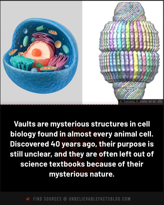
#cell biology#vaults#organelles#ribonucleoprotein#cellular structures#science history#1986 discovery#biology#science#mysterious#mystery#facts
116 notes
·
View notes
Text
Shedding new light on sugars, the “dark matter” of cellular biology - Technology Org
New Post has been published on https://thedigitalinsider.com/shedding-new-light-on-sugars-the-dark-matter-of-cellular-biology-technology-org/
Shedding new light on sugars, the “dark matter” of cellular biology - Technology Org
Scientists at Université de Montréal’s Department of Chemistry have developed a new fluorogenic probe that can be used to detect and study interactions between two families of biomolecules essential to life: sugars and proteins.
Our idea was to label sugar molecules with a chromophore, a chemical that gives a molecule its colour,” explained Cecioni. “The chromophore is actually fluorogenic, which means that it can become fluorescent if the binding of sugar with the lectin is efficiently captured. Image credit: Cecioni Lab
The findings by professor Samy Cecioni and his students, which open the door to a wide range of applications, were published in mid-October in the prestigious European journal Angewandte Chemie.
Found in all living cells
Sugar is omnipresent in our lives, present in almost all the foods we eat. But the importance of these simple carbohydrates extends far beyond tasty desserts. Sugars are vital to virtually all biological processes in living organisms and there is a vast diversity of naturally occurring sugar molecules.
“All of the cells that make up living organisms are covered in a layer of sugar-based molecules known as glycans,” said Cecioni. “Sugars are therefore on the front line of almost all physiological processes and play a fundamental role in maintaining health and preventing disease.”
“For a long time,” he added, “scientists believed that the complex sugars found on the surface of cells were simply decorative. But we now know that these sugars interact with many other types of molecules, particularly lectins, a large family of proteins.”
Driving disease, from flu to cancer
Like sugars, lectins are found in all living organisms. These proteins have the unique ability to recognize and temporarily attach themselves to sugars. Such interactions occur in many biological processes, such as during the immune response triggered by an infection.
Lectins are attracting a lot of attention these days. This is because scientists have discovered that the phenomenon of lectins “sticking” to sugars plays a key role in the appearance of numerous diseases.
“The more we study the interactions between sugars and lectins, the more we realize how important they are in disease processes,” said Cecioni. “Studies have shown how such interactions are involved in bacteria colonizing our lungs, viruses invading our cells, even cancer cells tricking our immune system into thinking they’re healthy cells.”
Difficult to detect…until now
There are still many missing pieces in the puzzle of how interactions between sugars and lectins unfold because they are so difficult to study. This is because these interactions are transient and weak, making detection a real challenge.
Two of Cecioni’s students, master’s candidate Cécile Bousch and Ph.D. candidate Brandon Vreulz, had the idea of using light to detect these interactions. The three researchers set to work to create a sort of chemical probe capable of “freezing” the meeting between sugar and lectin and making it visible through fluorescence.
The interaction between sugar and lectin can be described using a “lock and key” relationship, where the “key” is the sugar and the “lock” is the lectin. Chemists have already created molecules capable of blocking this lock-and-key interaction, and can now to identify exactly what sugars are binding to lectins of high interest to human health.
“Our idea was to label sugar molecules with a chromophore, a chemical that gives a molecule its colour,” explained Cecioni. “The chromophore is actually fluorogenic, which means that it can become fluorescent if the binding of sugar with the lectin is efficiently captured. Scientists can then study the mechanisms underlying these interactions and the disturbances that can arise.”
Cecioni and his students are confident their technique can be used with other types of molecules. It may even be possible to control the colour of new fluorescently labelled probes that are created.
By making it possible to visualize interactions between molecules, this discovery is giving researchers a valuable new tool for studying biological interactions, many of which are critical to human health.
Source: University of Montreal
You can offer your link to a page which is relevant to the topic of this post.
#applications#Bacteria#Biology#Biomolecules#Cancer#cancer cells#Cells#cellular structures#challenge#chemical#chemistry#Chemistry & materials science news#Dark#dark matter#detection#Disease#Diseases#diversity#Explained#Fundamental#Giving#Health#how#human#Human health#immune response#immune system#infection#interaction#it
2 notes
·
View notes
Text
Wound Healing
Wound Healing... Part 1
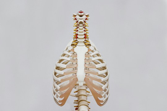
Wound healing is a complex process that involves replacing missing or damaged tissue and cellular structures. It can be divided into three or four phases, depending on the model used: hemostasis, inflammatory, proliferation, and remodeling. The inflammatory phase begins at the time of injury and can last up to four days. During this phase, blood vessels constrict to prevent blood loss,…
View On WordPress
#blogger#broccoli#cellular structures#coaching#coaching calls#collagen#complex#counseling#counselor#damaged tissue#ga#Georgia Landers#georgiasedify#god#hemostasis#inflammatory#injury#jesus#kale#muscle#phase#process#proliferation#remodeling#teacher#thanksforhavinggaonurmind#tissue#vitamin c#water#Wound healing
0 notes
Text
3D Printing: From Prototypes to Organ Transplants
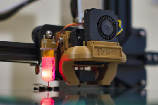
In the last decade, the landscape of manufacturing, medical science, and even the arts have been fundamentally transformed by the advent of 3D printing technology. Once a niche tool used for the creation of simple prototypes, 3D printing has burgeoned into a revolutionary force that stands at the forefront of innovation across numerous sectors. This article delves into the journey of 3D printing,…
View On WordPress
#3D ink#3D models#3D printed organs#3D printing#3D scanners#Additive manufacturing#Aerospace#Artificial organs#Automotive#Bioengineering#Bioink#Biomaterials#Bioprinting#Biotechnology#CAD#Cellular structures#Ceramics#Creative tech#Custom-made#customization#Dental devices#Design#Design thinking#Development#Digital fabrication#Digital manufacturing#Digital models#Disruptive tech#Donor organs#efficiency
1 note
·
View note
Text

A little doodle I did! Wanted to draw some art kinda representing my take on geostigma
#Like when I first watched advent Children I was obsessed with geostigma#And kinda had an interpretation that Seph’s cells were taking over Cloud’s body and rewriting his DNA??#So SEPH and Cloud share cellular structure?#I dunno if this makes any sense#art#final fantasy 7#ff7 rebirth#sephiroth#cloud strife#sefikura#void’s art#Anyway back to Sefikura shenanigans it is! :)))))#tw nudity
37 notes
·
View notes
Text








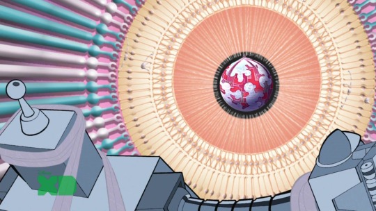


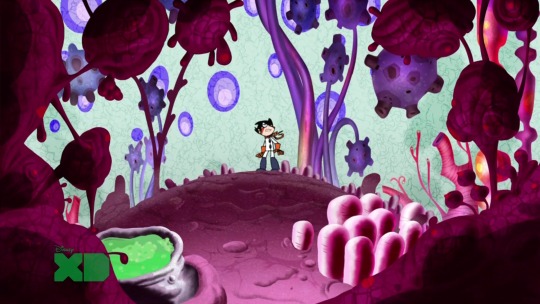


Super robot monkey team hyperforce go! ~background/scenery appreciation post ~
#because I just think they're so terribly underrated!#srmthfg#srmthfg!#super robot monkey team hyperforce go!#scenery#background#cartoon#art#looking through these screencaps i just thought how incredibly beautiful they are even if they are not that well detailed#they manage to set the mood for each episode perfectly#like i just noticed how in the last one with them going to the macro-cosmos#the background universe looks like a web; almost like cellular structures#it gives the impression of everything being part of something bigger; of a tissue organ or body#i really like that it's very eery#the colors are also so masterfully utilized#they literally have the skittles squad as main characters and they always find away to integrate their colors into the scenery#idk if it's the nostalgia speaking but the beautiful backgrounds and color palettes in this show really tickled the right spot of my brain#long post#pictures
131 notes
·
View notes
Text
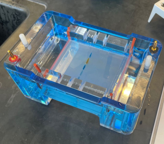
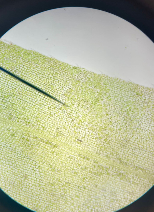
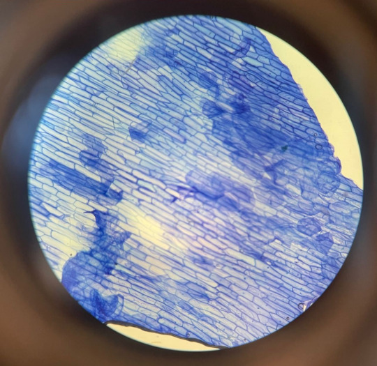
Tiny Blog Stuff: sharing some lab stuff from college
Electrolysis of dyes
Cell Structure through a microscope (plant cell and onion cell)
Photosynthesis/Celluar Respiration with a cabomba plant
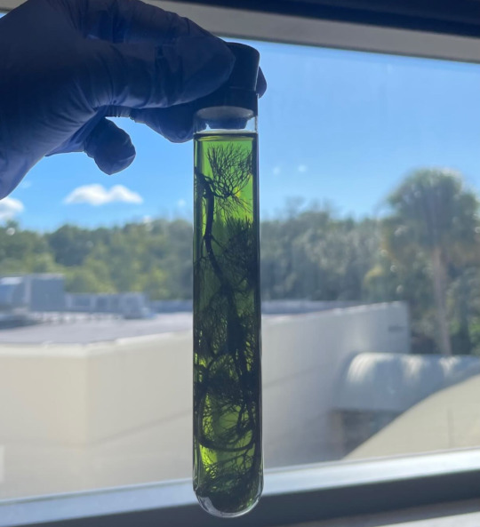
Fermentation of yeast

I like lab and biology but lectures always makes me sleepy…
#biology#biology lab#electrolysis#plant cells#cell structure#microscope#photosynthesis#cellular respiration#fermentation#college
42 notes
·
View notes
Text
dune wants to murder ancients sooooo bad. dune is sooooooo mad she'll never get the opportunity to murder an ancient
#her entire cellular structure: WE SHOULDNT BE THINKING ABOUT THIS MAN NO NO THATS ILLEGAL WE CANT MURDER OUR PARENTS#dune: oh my god but itd be so fucking funny
20 notes
·
View notes
Note
Vincent has dark roots and white hair. Does he dye it?
Nope. His hair just fades in that color past a certain length. If he were to let it all grow out instead of shaving the sides all the time, it would all look white and spiky.
(Similarly, if he buzzed it all off, it would be black.)
#devarambles#vincenttag#Colors changing and fading occur as birdpeople age and grow. Sera used to have grey wings before! Vincent also had some light graying befor#Their melanocytes have a different way of working around bird genes#Vincent's leucism makes it hard for pigment cells to stay in a stable state for long#Pigment cells die easier especially with their hair shafts having a different/hollowed structure#I am pulling this all out of nothing and holding it up with pseudoscience and nonsense#because in truth Vincent with all-white hair looks like he's pushing 70#So! there you go#I refuse to learn the intricacies of cellular anatomy for hair. i am not doing this to myself#art#artwork#digital art#drawing#Illustration#my artwork#my art#ark_systema#A_S Textposts
6 notes
·
View notes
Text
i fear i may be regressing into a fall out boy blog
#that show rearranged my cellular structure#wtf how hard do you have to go to knock me back to my 2014 obsessions
5 notes
·
View notes
Text
These lightning storms have been fucking active.
#I am just arching the air for water#“water into wine”#wine from water?#no you goddamn fool I said water in o twine#sam and max vist the worlds largest ball of twine#your friends have heard all about it#on the diaries if meta secret lovers#diaries of a wimpy kid....lol#well you die#nothing has managed to kill me yet#hey I am a special kind of evil...from a certain point of view#its like remember what I am for a clip of a minute#yes though this cellular structure was of different form certainly hard to keep solid memories#well I guess you always believed in me so that's cool#yes on your naked body you love to remind me about yes on that yes on that yes on that
2 notes
·
View notes
Text

It's just a genuine mental illness at this point
#my blood cells have been replaced with pikmin respectively#as have my braincells#my entire cellular structure is just pikmin tbh#when will they drop hospital decor pikmin tbh
1 note
·
View note
Text
Noninvasive imaging method can penetrate deeper into living tissue
New Post has been published on https://thedigitalinsider.com/noninvasive-imaging-method-can-penetrate-deeper-into-living-tissue/
Noninvasive imaging method can penetrate deeper into living tissue
Metabolic imaging is a noninvasive method that enables clinicians and scientists to study living cells using laser light, which can help them assess disease progression and treatment responses.
But light scatters when it shines into biological tissue, limiting how deep it can penetrate and hampering the resolution of captured images.
Now, MIT researchers have developed a new technique that more than doubles the usual depth limit of metabolic imaging. Their method also boosts imaging speeds, yielding richer and more detailed images.
This new technique does not require tissue to be preprocessed, such as by cutting it or staining it with dyes. Instead, a specialized laser illuminates deep into the tissue, causing certain intrinsic molecules within the cells and tissues to emit light. This eliminates the need to alter the tissue, providing a more natural and accurate representation of its structure and function.
The researchers achieved this by adaptively customizing the laser light for deep tissues. Using a recently developed fiber shaper — a device they control by bending it — they can tune the color and pulses of light to minimize scattering and maximize the signal as the light travels deeper into the tissue. This allows them to see much further into living tissue and capture clearer images.
This animation shows deep metabolic imaging of living intact 3D multicellular systems, which were grown in the Roger Kamm lab at MIT. The clearer side is the result of the researchers’ new imaging method, in combination with their previous work on physics-based deblurring.
Credit: Courtesy of the researchers
Greater penetration depth, faster speeds, and higher resolution make this method particularly well-suited for demanding imaging applications like cancer research, tissue engineering, drug discovery, and the study of immune responses.
“This work shows a significant improvement in terms of depth penetration for label-free metabolic imaging. It opens new avenues for studying and exploring metabolic dynamics deep in living biosystems,” says Sixian You, assistant professor in the Department of Electrical Engineering and Computer Science (EECS), a member of the Research Laboratory for Electronics, and senior author of a paper on this imaging technique.
She is joined on the paper by lead author Kunzan Liu, an EECS graduate student; Tong Qiu, an MIT postdoc; Honghao Cao, an EECS graduate student; Fan Wang, professor of brain and cognitive sciences; Roger Kamm, the Cecil and Ida Green Distinguished Professor of Biological and Mechanical Engineering; Linda Griffith, the School of Engineering Professor of Teaching Innovation in the Department of Biological Engineering; and other MIT colleagues. The research appears today in Science Advances.
Laser-focused
This new method falls in the category of label-free imaging, which means tissue is not stained beforehand. Staining creates contrast that helps a clinical biologist see cell nuclei and proteins better. But staining typically requires the biologist to section and slice the sample, a process that often kills the tissue and makes it impossible to study dynamic processes in living cells.
In label-free imaging techniques, researchers use lasers to illuminate specific molecules within cells, causing them to emit light of different colors that reveal various molecular contents and cellular structures. However, generating the ideal laser light with certain wavelengths and high-quality pulses for deep-tissue imaging has been challenging.
The researchers developed a new approach to overcome this limitation. They use a multimode fiber, a type of optical fiber which can carry a significant amount of power, and couple it with a compact device called a “fiber shaper.” This shaper allows them to precisely modulate the light propagation by adaptively changing the shape of the fiber. Bending the fiber changes the color and intensity of the laser.
Building on prior work, the researchers adapted the first version of the fiber shaper for deeper multimodal metabolic imaging.
“We want to channel all this energy into the colors we need with the pulse properties we require. This gives us higher generation efficiency and a clearer image, even deep within tissues,” says Cao.
Once they had built the controllable mechanism, they developed an imaging platform to leverage the powerful laser source to generate longer wavelengths of light, which are crucial for deeper penetration into biological tissues.
“We believe this technology has the potential to significantly advance biological research. By making it affordable and accessible to biology labs, we hope to empower scientists with a powerful tool for discovery,” Liu says.
Dynamic applications
When the researchers tested their imaging device, the light was able to penetrate more than 700 micrometers into a biological sample, whereas the best prior techniques could only reach about 200 micrometers.
“With this new type of deep imaging, we want to look at biological samples and see something we have never seen before,” Liu adds.
The deep imaging technique enabled them to see cells at multiple levels within a living system, which could help researchers study metabolic changes that happen at different depths. In addition, the faster imaging speed allows them to gather more detailed information on how a cell’s metabolism affects the speed and direction of its movements.
This new imaging method could offer a boost to the study of organoids, which are engineered cells that can grow to mimic the structure and function of organs. Researchers in the Kamm and Griffith labs pioneer the development of brain and endometrial organoids that can grow like organs for disease and treatment assessment.
However, it has been challenging to precisely observe internal developments without cutting or staining the tissue, which kills the sample.
This new imaging technique allows researchers to noninvasively monitor the metabolic states inside a living organoid while it continues to grow.
With these and other biomedical applications in mind, the researchers plan to aim for even higher-resolution images. At the same time, they are working to create low-noise laser sources, which could enable deeper imaging with less light dosage.
They are also developing algorithms that react to the images to reconstruct the full 3D structures of biological samples in high resolution.
In the long run, they hope to apply this technique in the real world to help biologists monitor drug response in real-time to aid in the development of new medicines.
“By enabling multimodal metabolic imaging that reaches deeper into tissues, we’re providing scientists with an unprecedented ability to observe nontransparent biological systems in their natural state. We’re excited to collaborate with clinicians, biologists, and bioengineers to push the boundaries of this technology and turn these insights into real-world medical breakthroughs,” You says.
“This work is exciting because it uses innovative feedback methods to image cell metabolism deeper in tissues compared to current techniques. These technologies also provide fast imaging speeds, which was used to uncover unique metabolic dynamics of immune cell motility within blood vessels. I expect that these imaging tools will be instrumental for discovering links between cell function and metabolism within dynamic living systems,” says Melissa Skala, an investigator at the Morgridge Institute for Research who was not involved with this work.
“Being able to acquire high resolution multi-photon images relying on NAD(P)H autofluorescence contrast faster and deeper into tissues opens the door to the study of a wide range of important problems,” adds Irene Georgakoudi, a professor of biomedical engineering at Tufts University who was also not involved with this work. “Imaging living tissues as fast as possible whenever you assess metabolic function is always a huge advantage in terms of ensuring the physiological relevance of the data, sampling a meaningful tissue volume, or monitoring fast changes. For applications in cancer diagnosis or in neuroscience, imaging deeper — and faster — enables us to consider a richer set of problems and interactions that haven’t been studied in living tissues before.”
This research is funded, in part, by MIT startup funds, a U.S. National Science Foundation CAREER Award, an MIT Irwin Jacobs and Joan Klein Presidential Fellowship, and an MIT Kailath Fellowship.
#3d#Algorithms#animation#applications#approach#assessment#author#Biological engineering#Biology#Biomedical engineering#blood#blood vessels#Brain#Brain and cognitive sciences#Building#Cancer#cancer diagnosis#Capture#career#cell#Cells#cellular structures#channel#clinical#collaborate#Color#colors#computer#Computer Science#cutting
0 notes
Text
From Glycans to Function: Navigating the Landscape of Glycomics

Glycomics, the study of complex carbohydrates known as glycans, represents a multifaceted field that delves into the diverse roles these molecules play in biological systems. By unraveling the intricate relationships between glycans and cellular function, researchers navigate a complex landscape that holds promise for advancing our understanding of health, disease, and beyond.
Glycomics in Biological Context: Glycans are ubiquitous in nature, adorning cell surfaces and influencing a myriad of physiological processes, from cell-cell recognition to immune response modulation.
Understanding the functional significance of Glycomics requires comprehensive analysis of their structures, interactions, and dynamics within biological systems.
Glycan Biosynthesis and Regulation
The intricate process of glycan biosynthesis is tightly regulated within cells, involving a complex network of enzymes, transporters, and regulatory factors.
Dysregulation of glycan biosynthesis pathways can have profound implications for cellular function, contributing to disease states such as cancer, autoimmune disorders, and metabolic syndromes.
Glycomics Technologies and Analytical Approaches
Advances in glycomics technologies, including mass spectrometry, glycan microarrays, and glycan profiling techniques, have revolutionized our ability to study glycans in unprecedented detail.
These analytical approaches enable researchers to map glycan structures, characterize glycan-protein interactions, and elucidate glycan-mediated signaling pathways, providing valuable insights into their functional roles.
Get More Insights On This Topic: Glycomics
Explore More Related Topic: Glycomics
#Glycomics#complex carbohydrates#glycans#biomolecular research#molecular biology#carbohydrate analysis#glycan structures#cellular communication#biomedical science
0 notes
Text
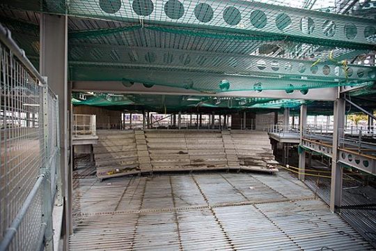
S T E E L S T R U C T U R E
sample project for a steel structure…..note the spans and dimensions in the project description….
_ik
Using three cores for stability, the steel frame is generally based around a 9.5m × 6m grid pattern in laboratory areas and a 12.7m × 6m grid in office areas. Cellular beams have been used throughout the floor plate in order to integrate all of the ducting in what will be a heavily serviced building.
.”cellular beams have been used throughout the floor plate in order to integrate all of the ducting in what will be a heavily serviced building:
CELLULAR BEAMS:
0 notes
Text
Biology, grade 7+, organelles and cell structure
Do Now:
Read the poem and answer the following questions, alone or with a partner.
What, if any, factual error(s) can you find in the poem?
If the mitochondria is the cellular equivalent of the biblical Eve, then which organelle would be the biblical Adam? Why?
Answers below the cut
Centrioles produce the microtubules that are like the puppet strings or highway system of the cell. They are unrelated to cell walls. Additionally, in case any students wish to nitpick, mitochondria is the plural of mitochondrion, and while they are typically used interchangeably, the last stanza treats the word like a singular entity instead of a plural.
Nucleus is the generally correct answer. However, a better answer would be the nucleoid, which is the prokaryotic equivalent to the nucleus. Since Eve was created from Adam's rib (or penis bone, as argued by some scholars), it's logical that the Adam organelle would also be prokaryotic, and therefore predate the first nucleus.
a silly ode to the first mitochondria, with waaaay too many religious allusions
(the mormons put me in seminary for four years and now it's everyone's problem)
the garden was not made of trees the snake did not exist when Eve was formed inside the seas and then was set adrift
she drifted in the tidal pools prokaryote divine producing simple molecules acids and alkalines
but paradise can never last and every god must fall some swallowed by a cytoplast (entrapped by a cell wall)
what do you call the dead that rise? what name is there for this? an Eve that finds that eden lies inside of the abyss
the wall no longer trapped her in but locked the monsters out the freedom only she could win to swim, and grow, and sprout.
she tinkered with her molecules And in a twist of fate Created one of life's crown jewels Adenosine Triphosphate (1)
what was before a simple wall could bloom with organelles a garden grown from former falls a paradise in hell
a fortress swam inside the brine, a thriving little town where tiny citizens could shine and ride the ups and downs
a golgi apparatus strove to package safe proteins a lysome found a nice alcove and kept the whole cell clean
the centrioles rebuilt the walls whenever they grew weak and eve was known and loved by all as something quite unique:
the powerhouse of the first cell the mitochondria (2) the Jonah that became the whale the jesus of bacteria once eaten by a macrophage then made through death anew the founder of our current age the sprout from which we grew
(yeah, yeah - you try and use this line in a poem)
(gah. this paragraph killed the syllable counts. i was challented to fit the phrase "powerhouse of the cell" into it, and mitochondria had to fit somewhere. both of which were gonna be doozies. decided to put them back to back and break the scheme at the end.
#do now#biology#grade 7 12#cellular biology#cell structures#organelles#poem#the ties that bind biology and theology are older than any religion#also I'm expecting at least one “Was anyone going to tell me” meme about the penis bone#I know I could have called it the baculum. This is funnier.#btw for extra fun try getting the students to sing this poem to the tune of#supercalifragilisticexpialidocious#because that's what's been happening in my head nonstop while I've been writing this
715 notes
·
View notes