#RNA-binding protein
Explore tagged Tumblr posts
Text
L'immunità che dialoga costantemente col cervello: il ruolo segreto di C1q nel cervello che matura ed invecchia
Uno studio innovativo condotto presso il laboratorio di Beth Stevens, PhD, presso il Boston Children’s Hospital, ha rivelato che una proteina immunitaria influisce sulla sintesi proteica neuronale nel cervello che invecchia. Un precedente lavoro del laboratorio Stevens aveva scoperto che le cellule immunitarie nel sistema nervoso centrale, la microglia, aiutano a sfoltire le sinapsi nel cervello…
#alternative splicing#complemento#neuroplasticità#proteina C1q#pruning sinaptico#ribonucleoproteina#RNA-binding protein#sinaptogenesi#sistema immunitario
0 notes
Text

TSRNOSS, Page 68.
#diabetes insipidus#immunity#RNA polymerase#error rate#antibodies to polysaccharides#glycosylation of proteins#alkaline tide#antigen binding site#allergens#blood urea#immune suppression#antibody#disulfide bonds#B-mercaptoethanol#salivary amylase#vitamin b12#cobalt#cursive#handwriting#manuscript#journal
0 notes
Photo

Multi-omics characterization of RNA binding proteins reveals disease comorbidities and potential drugs in COVID-19. Pan et al., Comput Biol Med. 2023 Mar; 155: 106651.
0 notes
Note
Hello! First of all, thank you for the wonderful content! It's a real joy, and an enrichment, food for both the brain and the heart! I was wondering if through your treasures, you could find some writing notes/words/concepts/vocabulary relating to genetic engineering? Like...creating a virus, and a vaccine for it, modifying the virus so it has certain specific effects.... Thank you in advance!
Writing Notes: Virus & Vaccine
References How Viruses Work; Replication Cycle; Mutation, Variants, Strains, Genetically Engineering Viruses; Writing Tips; Creating your Fictional Virus & Vaccine
Virus - an infectious microbe consisting of a segment of nucleic acid (either DNA or RNA) surrounded by a protein coat.
It is a tiny lifeform that is a collection of genes inside a protective shell. Viruses can invade body cells where they multiply, causing illnesses.
It cannot replicate alone; instead, it must infect cells and use components of the host cell to make copies of itself. Often, a virus ends up killing the host cell in the process, causing damage to the host organism.
Well-known examples of viruses causing human disease include AIDS, COVID-19, measles and smallpox. Examples of viruses:

Viruses are even smaller than bacteria and can invade living cells—including bacteria. They may interfere with the host genes, and when they move from host to host, they may take host genes with them.
Bacteriophages (also known as phages)—viruses that infect and kill bacteria.

Size differential between virus and bacterium
Viruses are measured in nanometers (nm).
They lack the cellular structure of bacteria, being just particles of protein and genetic material.
How Viruses Work
Viruses use an organism’s cells to survive and reproduce.
They travel from one organism to another.
Viruses can make themselves into a particle called a virion.
This allows the virus to survive temporarily outside of a host organism. When it enters the host, it attaches to a cell. A virus then takes over the cell’s reproductive mechanisms for its own use and creates more virions.
The virions destroy the cell as they burst out of it to infect more cells.
Viral shedding - when an infected person releases the virus into the environment by coughing, speaking, touching a surface, or shedding skin.
Viruses also can be shed through blood, feces, or bodily fluids.
Virus Replication Cycle
While the replication cycle of viruses can vary from virus to virus, there is a general pattern that can be described, consisting of 5 steps:
Attachment – the virion attaches to the correct host cell.
Penetration or Viral Entry – the virus or viral nucleic acid gains entrance into the cell.
Synthesis – the viral proteins and nucleic acid copies are manufactured by the cells’ machinery.
Assembly – viruses are produced from the viral components.
Release – newly formed virions are released from the cell.
Mutations, Variants, and Strains
Not all mutations cause variants and strains. Below are definitions that explain how mutations, variants, and strains differ.
Mutation - errors in the replication of the virus’s genetic code; can be beneficial to the virus, deleterious to the virus, or neutral
Variants - viruses with these mutations are called variants; the Delta and Omicron variants are examples of coronavirus mutations that cause different symptoms from the original infection
Strains - variants that have different physical properties are called strains; these strains may have different behaviors or mechanisms for infection or reproduction
Genetically Engineering Viruses
Using reverse genetics, the sequence of a viral genome can be identified, including that of its different strains and variants.
This enables scientists to identify sequences of the virus that enable it to bind to a receptor, as well as those regions that cause it to be so virulent.
Vaccine - a special preparation of substances that stimulate an immune response, used for inoculation
Vaccines & Fighting Viruses with Viruses
Common pathogenic viruses can be genetically modified to make them less pathogenic, such that their virulent properties are diminished but can still be recognized by the immune system to produce a robust immune response against. They are described as live attenuated.
This is the basis of many successful vaccines and is a better alternative than traditional vaccine development which typically includes heat-mediated disabling of viruses that tend to be poorer in terms of immunogenicity.
Viruses can also be genetically modified to ‘fight viruses’ by boosting immune cells to make more effective antibodies, especially where vaccines fail. Where vaccines fail, it is often due to the impaired antibody production by B-cells, even though antibodies can be raised against such viruses – including HIV, EBV, RSV & cold-viruses.
Related Articles: Modified virus used to kill cancer cells ⚜ Genetic Engineering ⚜ Engineering Bacterial Viruses ⚜ Benefits of Viruses
A Few Writing Tips
As more writers look to incorporate infectious diseases into their work, there are quite a few things writers should keep in mind:
Don’t anthropomorphize. Really easy to do, but scientifically wrong. Viruses don’t want to kill you; bacteria don’t want to infect you; parasites don’t want to make your blood curdle. None of these things are big enough to be sentient to want to do anything. They just do it (or don’t do it).
Personal protective equipment. This includes wearing gloves, lab coats, safety glasses, and tying your hair back if it’s long. It is the same as Edna Mode’s “no capes.” Flowing hair looks cool all the way to the explosive ball of flames that engulfs someone’s head.
Viruses are small. You can’t see viruses down a normal microscope—they need a special microscope called an electron microscope. These are highly specialized and take a long time to make the preparations to be able to see the virus. Normally viruses are detected by inference—measuring part of them using an assay that can amplify tiny amounts of material, for example PCR.
Viruses don’t really cause zombie apocalypses.
Vaccines work. But they take time. The best vaccine in the world will still only prevent infections two weeks after it is given. Drugs are quicker, but still take some time. But the good news is an infection is not going to kill you (or turn you into a zombie) quickly, so they both have time to work.
Scientists use viruses as a vector to introduce healthy genes into a patient’s cells:
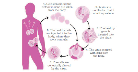
Your Fictional Virus & Vaccine
When creating your own fictional virus, research further on the topic and consider choosing a specific one as your basis/inspiration.
Here's one resource. For some of them, you'll need a subscription to access, but those that are available give you a good overview of the virus, as well as treatment options.
You can do the same for creating your fictional vaccine:
Here's one resource. And here's one on vaccine developments.
Sources: 1 2 3 4 5 6 7 8 9 10 11 12 13 ⚜ Writing Notes & References
Lastly, here's an interesting article on how science fiction can be a valuable tool to communicate widely around pandemic, whilst also acting as a creative space in which to anticipate how we may handle similar future events.
Thanks so much for your kind words, you're so lovely! Hope this helps with your writing. Would love to read your work if it does :)
#writing notes#virus#vaccine#writeblr#dark academia#spilled ink#writing reference#writing prompt#literature#science#writers on tumblr#creative writing#fiction#novel#light academia#lit#writing ideas#writing inspiration#writing tips#science fiction#writing advice#writing resources
80 notes
·
View notes
Note
I LOVE THIS BLOG… would you be able to explain the stuff you’re doing to someone who knows nothing about proteins? all I can remember is something to do with dna ..?
of course! ill do my best to give an entry level crash course here, but if any of this is unclear please leave a comment or send an ask so i can better explain.
DISCLAIMER: all of this has been simplified, and because biology is messy, there are exceptions to pretty much everything i've said. the point is not to give a perfect explanation, but rather a general understanding
the central dogma of molecular biology is pretty much our version of "the mitochondria is the powerhouse of the cell", and since you've alluded to it already, i'll start there. it states that genetic information goes from DNA to RNA to proteins. inside of almost any cell is DNA, which codes for all of the genetic information allowing the cell to function. for our purposes right now, just think of DNA as an instruction manual. when a protein is going to be made, the part of the DNA sequence encoding it is copied over to make a RNA sequence.
RNA is structurally similar to DNA, but while DNA is usually found as a double helix (with two complementary strands), RNA is more often single stranded. it is less stable than DNA, so it does not work as well for long term information storage, but is smaller and can cary out numerous crucial functions.
prokaryotes are things like bacteria, and are distinct from eukaryotes (which includes us) because they lack a nucleus. this means that their DNA is loose inside their cell, rather than sectioned away. in prokaryotes, transcription (which copies information from DNA -> RNA) and translation (which is the process of going from RNA -> proteins) can happen at the same time, while in eukaryotes these processes are separated, as DNA is too large to leave the nucleus. messenger RNA (mRNA) is the specific type of RNA used to code for proteins in all cells. inside a eukaryotic cell, mRNA must be processed to increase its stability and allow it to exit the nucleus.
now, getting to the part about proteins! proteins are made through a process called translation, which translates the information stored using a sequence of nucleic acids on RNA to a string of amino acids known as a protein. each set of three nucleic acids, which on mRNA can be A, U, C or G, makes up the codon for one amino acid. the code is referred to as 'degenerate', since there is a lot of redundancy built in and so some information is lost along the way. there are more possible codons than there are amino acids, and so there is a lot of overlap with several codons coding for the same amino acid.
translation is accomplished using organelles known as ribosomes. these bind to the relevant RNA sequence and help join together the amino acids that are encoded by their sequence, forming peptide bonds. this is done using another specialized type of RNA called a transfer RNA (tRNA), which sticks temporarily to the three-letter codon on the mRNA and carries the corresponding amino acid to the ribosome so that it can be joined with the others in the sequence. all proteins start with the same codon (AUG), and subsequent amino acids are added one at a time. RNA and proteins both have directionality, which means that the two different ends of these molecules are not the same, and the direction you read the sequence in matters.
as a protein is assembled, the N terminal end is put together first, and so this part exits the ribosome while the rest is still being built. at this point, it comes in contact with the liquid inside the cell, and starts to bend itself into different shapes in order to make the most thermodynamically stable structure. this happens spontaneously, and is an effort to minimize the free energy of the protein and the surrounding water molecules. basically, everything wants to be in a state that requires as little energy as possible, and will fold itself to get there. think of this as a similar process to getting home after a long day, and trying to make yourself comfortable as fast as possible. protein folding is the equivalent to you taking off your jeans and lying down on your couch.
the thing is, proteins are complicated, and they need to fold quickly, because the inside of a cell is crowded and chaotic. the way they fold is influenced by several different factors, including how fast translation takes place and whether anything else is nearby to help them fold correctly. proteins do countless different highly specific things in any given cell, and their ability to function is based entirely around their structure. just like how you probably have numerous different tools in your home made of plastic, but each one is a different shape and therefore does something unique. if someone came along and melted your plastic cups until they were completely deformed, they wouldn't be of much use.
the primary structure of a protein is its amino acid sequence, and the secondary structure is made by interactions between nearby backbone atoms, but the tertiary structure is the main thing you'll see looking at any real protein structure. it is the combination of interactions between all the atoms within one amino acid chain. if this gets damaged (which can happen with things like heat and strong chemicals), the protein is said to be denatured. some proteins also have a quaternary structure, which is formed as different folded chains of amino acids each making up one subunit assemble together to make a bigger, more complicated protein.
whether they folded wrong from the start (like your plastic cup getting made with a hole in the bottom at the factory) or they started off fine but then got broken (like your plastic cup melting after you leave it on the hot stove), misfolded proteins are the wrong shape and therefore cannot perform their function correctly. these can do a lot of damage in an organism, and are generally a waste of resources to keep around, so they get destroyed and their parts are recycled.
hope this helps!
letter sequence in this ask matching protein-coding amino acids:
ILVETHISLGwldyealeteplainthestffyredingtsmenewhknwsnthingatprteinsallIcanrememerissmethingtdwithdna
protein guy analysis:
this protein is strange, terrible and filled with holes! just like many of the other structures, the myriad of loops want nothing to do with each other, and everything is all over the place. this whole structure is disordered and likely wiggling around trying to find something else to stick to and mess with. just a toxic trainwreck that should never have existed.
predicted protein structure:

#science#biochemistry#biology#chemistry#stem#proteins#protein structure#science side of tumblr#protein asks
44 notes
·
View notes
Text
by Nicolas Hulscher, MPH
As the U.S. begins to escalate their response against H5N1 bird flu, we will likely see a push for more unsafe and ineffective countermeasures. The Biopharmaceutical Complex is currently preparing bird flu mRNA injections developed by Moderna, CEPI-funded H5N1 replicon (self-amplifying) shots, and Arcturus Therapeutics replicon ‘pandemic’ bird flu injections funded by BARDA and the Gates Foundation. Thus, it is of high priority to identify promising compounds with anti-avian influenza activity that don’t involve injection of modified genetic material.
A 2023 study revealed a comprehensive list of natural plants and bioactive compounds that have shown anti-avian influenza activity: A systemic review on medicinal plants and their bioactive constituents against avian influenza and further confirmation through in-silico analysis.
Methods: 33 plants and 4 natural compounds were identified and documented. Molecular docking was performed against the target viral protein neuraminidase (NA), with some plant based natural compounds and compared their results with standard drugs Oseltamivir and Zanamivir to obtain novel drug targets for influenza. Results: It was seen that most extracts exhibit their action by interacting with viral hemagglutinin or neuraminidase and inhibit viral entry or release from the host cell. Some plants also interacted with the viral RNA replication or by reducing proinflammatory cytokines. Ethanol was mostly used for extraction. Among all the plants Theobroma cacao, Capparis Sinaica Veil, Androgarphis paniculate, Thallasodendron cillatum, Sinularia candidula, Larcifomes officinalis, Lenzites betulina, Datronia molis, Trametes gibbose exhibited their activity with least concentration (below 10 μg/ ml). The docking results showed that some natural compounds (5,7- dimethoxyflavone, Aloe emodin, Anthocyanins, Quercetin, Hemanthamine, Lyocrine, Terpenoid EA showed satisfactory binding affinity and binding specificity with viral neuraminidase compared to the synthetic drugs.
13 notes
·
View notes
Text

Poliovirus is composed of an RNAgenome and a proteincapsid. The genome is a single-stranded positive-sense RNA (+ssRNA) genome that is about 7500 nucleotides long.[2] The viral particle is about 30 nm in diameter with icosahedral symmetry. Because of its short genome and its simple composition—only RNA and a nonenveloped icosahedral protein coat that encapsulates it—poliovirus is widely regarded as the simplest significant virus.[3]
7500 nucleotides is blowing my mind. did some more reading and found this

The MS2 genome is one of the smallest known, consisting of 3569 nucleotides of single-stranded RNA.[3] It encodes just four proteins: the maturation protein (A-protein), the lysis (lys) protein, the coat protein (cp), and the replicase (rep) protein.[1] The gene encoding lys overlaps both the 3'-end of the upstream gene (cp) and the 5'-end of the downstream gene (rep), and was one of the first known examples of overlapping genes. The positive-stranded RNA genome serves as a messenger RNA, and is translated upon viral uncoating within the host cell. Although the four proteins are encoded by the same messenger/viral RNA, they are not all expressed at the same levels.
only 4 genes!!! and the lysis one is fully contained in two other ones

Once the viral RNA has entered the cell, it begins to function as a messenger RNA for the production of phage proteins. The gene for the most abundant protein, the coat protein, can be immediately translated. The translation start of the replicase gene is normally hidden within RNA secondary structure, but can be transiently opened as ribosomes pass through the coat protein gene. Replicase translation is also shut down once large amounts of coat protein have been made; coat protein dimers bind and stabilize the RNA "operator hairpin", blocking the replicase start. The start of the maturation protein gene is accessible in RNA being replicated but hidden within RNA secondary structure in the completed MS2 RNA; this ensures translation of only a very few copies of maturation protein per RNA. Finally, the lysis protein gene can only be initiated by ribosomes that have completed translation of the coat protein gene and "slip back" to the start of the lysis protein gene, at about a 5% frequency.[1]
it had the first gene sequenced and the first full genome sequenced
43 notes
·
View notes
Note
Hello, Mr. Holmes! How are you?
So, long story short, I ended up with an optical microscope in my room more or less 4 months ago, with 200 previously made slides (secured in a proper box), and lots of new ones too, for me to prepare myself. I love microbiology (it's one of my hyperfixations, curse my neurodivergency) and now I love it even more (my mother has had to drag me away from the microscope - I named it Wesley - in the middle of the night multiple times now).
After much conversation, I finally convinced my mom to buy me the proper equipment to prepare the slides!
So, I'm sending this ask to you, as I know you also have a microscope and that you use it a lot: what kind of equipment do you recommend me buying (gloves, scalpel blades, tints, etc), while still remembering that all of the stuff needs to stay in my room (properly taken cared of by me, of course)?
For example, I'm unsure if different dyes are used for different smears and specimens due to it's affinity (I've noticed that on 'organic matter' slides, images are usually tinted purple or pink, while on plant-based slides, images are usually tinted green and blue, with a few red structures.) Considering that I don't have access to a mortuary, I will mostly make plant slides. There must be a difference in the dyes then, right?
Sorry for the long text! Hope this isn't too much of a bother.
- a 17-year-old :)
Congratulations on your new light microscope. I do hope you get the best out of it. I am overjoyed that someone else appreciates the art of microscopy and microbiology.
However, you need to be careful to not strain your eyes. It is recommended to take breaks every 15 minutes to close your eyes or focus on something in the distance to reaccommodate your eyes. And get up every 40 minutes, stretch and correct your posture. And it is recommended to not use a microscope more than 5 hours per day. John has to chase me away from my microscope sometimes to take a break when I sit there for hours, my posture like a Caridea.
Concerning equipment, you will obviously need a scalpel or other sharp blade to make very thin slices of your specimen, as thin as possible. And forceps to move your samples (best just get a whole dissection kit it has everything). Obviously slides and coverslips, pipettes for the stains or water, maybe some tubes. A pen to label your slides. In many staining procedures ethanol or acetone is also used. A waste jar to safely dispose of any chemicals, but be careful what you mix. A rack for staining and containers. I would recommend nitrile gloves, some people are sensitive to latex.
The dyes you use depend on the specimen. For example in histological slides of tissues hematoxylin and eosin are most commonly used (short HE-stain). That's what you most likely saw on your slides, it's blue, purple and pink. Hematoxylin is a basic compound extracted and oxidised from the logwood tree (Haematoxylum campechianum), and it stains acidic compounds in the cells (or basophilic because they have an affinity for basic substances). For example nucleic acids like DNA or RNA get stained by hematoxylin because they are basophillic. And where are lots of nucleic acids? In the nucleus and ribosomes, that is why they appear blue to purple in the staining because they bind hematoxylin. Eosin is an acidic compound, and stains basic or acidophilic compounds red or pinkish, like proteins, collagen, cytoplasm, extracellular matrix.

(Ductus epididymidis with HE-stain)
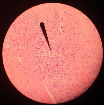
(Tongue HE-stain, pointer marking a ganglion; that is my picture)
Of course there are more specific stains for specific tissues like Golgi's silver staining for neurons.
For plants toluidine blue is often used, high affinity for acidic tissues, and can stain blue to green to purple. It is often combined with safranin, a basic azine, which is probably the red stain you saw. It stains polysaccharides and lignin, woody parts of the plant. Safranin and astrablue is also often combined, astrablue stains non-lignified parts of the plant.
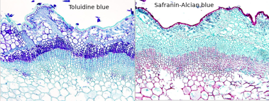
(Ulex europaeus stem; not my pictures I don't have any samples currently, source Atlas of plant and animal histology)
Safranin is also used in bacteriology, in the famous Gram staining. In Gram staining you use crystal violet (blue/purple), Lugol's iodine solution, then wash it with ethanol and add safranin (red) as a counter stain. Bacteria is gram-positive if the crystal violet stays in their thick murein cell wall, can't be washed out with the ethanol and the bacteria stays blue. Gram-negative appear red because of the counterstain.
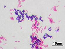
(Staphyloccocus aureus (violet, gram positive) & Escherichia coli (red, gram negative); not my picture, source Wikipedia)
However, I am not sure whether you have access to any of those substances, if they are too expensive for you or if they are too hazardous if used in your own room for a prolongued time. Of course those substances need to be stored properly, and your own room is probably not a good place, especially for ethanol or acetone. The fumes. I would recommend to ask your biology or chemistry teacher whether they can recommend anything further and where to buy said solutions in your area, and if they can't they are idiots. There are also many useful resources and tutorials on Youtube.
Another fascinating experiment for your microscope, that you can perform without buying any chemicals, is a hay infusion. You put hay into a container filled with water, and let it sit undisturbed for a week in a sunny area but not in direct harsh sunlight. During that time the microorganisms in the hay are reproducing in the solution, feeding on the polysaccharides of the hay. Protozoans also flourish in the hay infusion and eat the bacteria. It might get cloudy and a bit foul smelling (best not do it in your own room if you don't want to sleep next to a rotting smell). When you put a drop of the solution onto a slide and look at it in the microscope, you should see a variety of microorganisms like bacteria (like Bacillus subtilis), amoeba, ciliates, heliozoa, algae et cetera. At different depths of the liquid you should find different kinds of organisms, because of differing oxygen content. However, pathogens can also occur in the hay infusion so handle it carefully and work sterile, wash your hands properly.
And even if you don't work at a morgue you can still get tissue samples to experiment on, after all meat is sold in supermarkets, basically the same as a human body. And at the butchers they even sell organs like chicken hearts, pig kidney, liver, blood et cetera. Or observe your own hair under the microscope.
Which kind of samples and slides were included in your starter kit? Be careful to not leave them lying around in the sunlight, or the stain might fade. Always store them in the proper box.
#roleplay#rp#sherlock roleplay#sherlock rp#johnlock roleplay#johnlock rp#sherlock#bbc sherlock#sherlock holmes#sherlock holmes rp#sherlock holmes roleplay#science#scientist#research scientist#histology#microscope#microscopy#bacteria#bacteriology#pathology#anatomy#biology#chemistry#scientists#pictures#he stain#specimen#samples#slides#sherlock replies
47 notes
·
View notes
Text
You:
explain crispr mRNA
Ai:
CRISPR RNA (crRNA) is a sophisticated, programmable molecular tool that leverages the prokaryotic adaptive immune system's mechanism of defense against invasive genetic elements. This RNA-based component serves as a guide to direct the Cas9 nuclease enzyme toward specific genomic loci for targeted editing.
The crRNA structure consists of three distinct regions: the scaffold, the repeat sequence, and the protospacer. The scaffold provides structural support and facilitates binding with the Cas9 protein, while the repeat segment is derived from the CRISPR array in the bacterial genome and promotes crRNA-Cas9 complex formation.
The protospacer region, which constitutes the majority of the crRNA sequence, harbors the desired target specificity information. It possesses a complementarity to a predefined genomic sequence (the target site), thereby enabling the crRNA-guided Cas9 endonuclease to recognize and cleave the target DNA at the precise location.
crRNA synthesis typically involves in vitro transcription from a DNA template or ribosomal RNA processing in vivo. The resulting mature crRNAs are then processed into their functional forms through 5' and 3' trimming, which removes non-essential regions and generates a single-stranded structure that can base-pair with the target site.
Upon binding to Cas9, the crRNA acts as an adaptor, positioning the nuclease at its cognate genomic location. Upon encountering the target sequence, the Cas9-crRNA complex excises the DNA, creating a double-strand break (DSB). The DSB can then be repaired by the cell's endogenous pathways, which may result in precise gene editing via homology-directed repair (HDR) or introduce insertions/deletions (indels) through non-homologous end joining (NHEJ), effectively creating a mutation at the targeted site.
The programmable nature of crRNAs allows researchers to tailor them for specific applications, including gene knockouts, point mutations, and even precise base editing. Moreover, advances in RNA engineering have enabled the development of complex crRNA designs capable of targeting multiple genomic loci simultaneously or recognizing non-canonical target sites.
In summary, CRISPR RNA is a highly specialized, versatile molecular tool that empowers scientists to precisely manipulate genomes through its intricate interactions with Cas proteins and cellular repair machinery. Its unique properties have revolutionized the field of genome engineering, offering unprecedented control over genetic information.
You:
explain how a virus can be used as an mRNA encapsulate to deliver crisper RNA
Ai:
Viruses, particularly those in the families Lentiviridae (e.g., HIV-1), Adenoviridae (e.g., adenovirus serotype 5), and AAVidiae (adeno-associated viruses), can be engineered to function as mRNA encapsulation vectors for CRISPR RNA delivery.
The process typically involves:
Cloning the desired crRNA sequence into an expression plasmid, which is then transfected into virus-producing cells. The viral vector is assembled with its capsid protein and genomic RNA, replacing the original viral genetic material. The modified virus acquires the CRISPR RNA as a passenger payload, encapsulated within its lipid envelope or protein capsid. Upon infection of target host cells, the virus delivers its crRNA cargo into the cytoplasm via endocytosis and lysosomal escape. After entering the nucleus through nuclear pore complexes, the released crRNA can then interact with Cas proteins to initiate genome editing at the desired loci.
The choice of viral vector largely depends on factors such as host specificity, transduction efficiency, and immunogenicity concerns. Lentiviral vectors, for example, are capable of infecting non-dividing cells but may elicit stronger immune responses compared to AAV or adenoviral alternatives. AAVs, being less immunogenic, have become popular choices for gene therapy applications.
Incorporating CRISPR RNA into viral delivery systems offers several advantages over direct crRNA administration. It enables targeted and efficient transduction of cells, enhances the stability of crRNA in circulation, reduces off-target effects due to improved tissue specificity, and can facilitate simultaneous editing or multiplexed gene modifications.
However, challenges persist, including concerns over potential off-target effects from viral-mediated delivery itself, immune responses against the viral vector, and the need for further optimization of viral design and packaging strategies to ensure precise genome editing with minimal collateral damage. Continued research is necessary to refine this approach and maximize its therapeutic potential in various applications, ranging from treating genetic diseases to agricultural biotechnology.
————————————————————————
Virus AI describes as being the likely candidate for spreading crisper RNA

“A digital cryo-electron micrograph of the Adenovirus serotype 5 (Ad-5)/HVR5-33RGD vector. Adenoviruses are non-enveloped, double-stranded DNA viruses that cause cold-like infections of the upper respiratory tract.”
10 notes
·
View notes
Text
Exploring the Marvels of Biological Macromolecules: The Molecular Machinery of Life (Part 3)
Proteins and Enzymes: Catalysts of Molecular Reactions
Proteins are the central players in macromolecular interactions. Enzymes, a specialized class of proteins, catalyze biochemical reactions with remarkable specificity. They bind to substrates, facilitate reactions, and release products, ensuring that cellular processes occur with precision.
Protein-Protein Interactions: Orchestrating Cellular Functions
Proteins often interact with other proteins to form dynamic complexes. These interactions are pivotal in processes such as signal transduction, where cascades of protein-protein interactions transmit signals within cells, regulating diverse functions such as growth, metabolism, and immune responses.
Protein-Ligand Interactions: Molecular Recognition
Proteins can also interact with small molecules called ligands. Receptor proteins, for instance, bind to ligands such as hormones, neurotransmitters, or drugs, initiating cellular responses. These interactions rely on specific binding sites and molecular recognition.
Protein-DNA Interactions: Controlling Genetic Information
Transcription factors, a class of proteins, interact with DNA to regulate gene expression. They bind to specific DNA sequences, promoting or inhibiting transcription, thereby controlling RNA and protein synthesis.
Membrane Proteins: Regulating Cellular Transport
Integral membrane proteins participate in macromolecular interactions by regulating the transport of ions and molecules across cell membranes. Transport proteins, ion channels, and pumps interact precisely to maintain cellular homeostasis.
Cooperativity and Allosteric Regulation: Fine-Tuning Cellular Processes
Cooperativity and allosteric regulation are mechanisms that modulate protein function. In cooperativity, binding one ligand to a protein influences the binding of subsequent ligands, often amplifying the response. Allosteric regulation occurs when a molecule binds to a site other than the active site, altering the protein's conformation and activity.
Interactions in Signaling Pathways: Cellular Communication
Signal transduction pathways rely on cascades of macromolecular interactions to transmit extracellular signals into cellular responses. Kinases and phosphatases, enzymes that add or remove phosphate groups, play pivotal roles in these pathways.
Protein Folding and Misfolding: Disease Implications
Proteins must fold into specific three-dimensional shapes to function correctly. Misfolded proteins can lead to Alzheimer's, Parkinson's, and prion diseases. Chaperone proteins assist in proper protein folding and prevent aggregation.
References
Voet, D., Voet, J. G., & Pratt, C. W. (2016). Fundamentals of Biochemistry: Life at the Molecular Level. Wiley.
Lehninger, A. L., Nelson, D. L., & Cox, M. M. (2017). Lehninger Principles of Biochemistry. W. H. Freeman.
Berg, J. M., Tymoczko, J. L., & Stryer, L. (2002). Biochemistry. W. H. Freeman
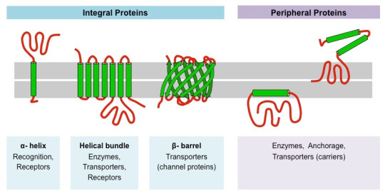
#science#college#biology#education#school#medicine#student#doctors#health#healthcare#proteins#molecular biology#molecular structure#chemestry#chemistry
98 notes
·
View notes
Text
SARS-CoV-2 can infect and replicate in human motor neurons differentiated from induced pluripotent stem cells - Published Jan 4, 2024
Numerous patients experience neurological and neuromuscular symptoms during and after infection with the severe acute respiratory syndrome coronavirus 2 (SARS-CoV-2). Apart from the central nervous system (CNS), the peripheral nervous system (PNS) is also affected. In this study, the Italian authors investigated whether SARS-CoV-2 can infect and replicate in human motor neurons (MNs) differentiated in vitro from induced pluripotent stem cells (iPSC-MNs). They also examined whether iPSC-MNs express the main receptors for SARS-CoV-2 entry and whether SARS-CoV-2 infection of iPSC-MNs changes the expression of 46 genes involved in cell survival, metabolism, inflammatory response, apoptotic and antiviral pathways.
SARS-CoV-2 is an enveloped, positive-sense, single-stranded RNA virus. Its genome encodes four structural proteins, namely the spike (S), envelope (E), nucleocapsid (N), and membrane (M) proteins. It seems that SARS-CoV-2 uses various neuroinvasive strategies and entry pathways to invade the nervous system, such as infection of the nasal olfactory epithelium and axonal transport along the olfactory nerve, retrograde axonal transport, invasion by compromising the blood-brain barrier, retrograde virus spread from the lungs to the CNS via the vagus nerve, and the use of infected hematopoietic cells as “Trojan horses” (hematogenous route).
It remains unknown whether observed neuromuscular manifestations of SARS-CoV-2 infection are caused by a direct viral invasion of motor neurons, and/or are a collateral injury resulting from an uncontrolled innate immune response. Damage to motor neurons leads to the deterioration of muscle function, manifested as muscle weakness, atrophy, or paralysis.
Two host-cell factors are important for SARS-CoV-2 viral entry into many cell types: angiotensin-converting enzyme 2 (ACE2), which is bound by the S protein, and transmembrane serine protease 2 (TMPRSS2), which cleaves the S protein, allowing this binding to take place. In addition to ACE2 and TMPRSS2, the S protein has been reported to engage other cell-surface factors proposed to serve as attachment factors promoting SARS-CoV-2 entry.
The expression level of ACE2 is low in the human brain. In contrast, neuropilin 1 (NRP1), CD147, TMPRSS2, and furin are higher and broader expressed than ACE2, indicating that they may be putative mediators of SARS-CoV-2 entry into human nervous system cells. According to the authors of this study, the infection of human motor neurons with SARS-CoV-2 mainly relies on CD147 and/or NRP1 binding. Previous studies have shown that NRP1, known to bind furin-cleaved substrates, potentiates SARS-CoV-2 infectivity and that the furin-cleaved S1 subunit of the S protein binds directly to cell surface NRP1. www.science.org/doi/10.1126/science.abd2985
About the study The authors used an in vitro model of human motoneurons (MNs) differentiated from induced pluripotent stem cells (iPSC-MNs) to investigate the infectability of these cells by SARS-CoV-2 and their expression of the main SARS-CoV-2 receptors. They also examined whether SARS-CoV-2 changes the expression of 46 genes involved in cell survival, metabolism, inflammatory response, and apoptotic and antiviral pathways.
The induced pluripotent stem cells (iPSCs) from three healthy donors (1 male and 2 females, aged between 37 and 49 years) differentiated into motor neurons that expressed both, neuronal (bIII-tubulin and SMI-312) and motoneuronal (ChAT, HB9) markers.
To verify the infectability of iPSC-MNs by SARS-CoV-2, VeroE6 cells were exposed to supernatants collected from iPSC-MNs infected with SARS-CoV-2. Reverse transcription polymerase chain reaction (rt-PCR) was used to analyze the expression of SARS-CoV-2-specific ORF7A, ORF3A, ORF8, RDRP, S, E, and N genes in uninfected (mock) and infected iPSC-MNs.
The expression of main SARS-CoV-2 receptors (ACE2, CD147, NRP1) and peptidases (TMPRSS2, furin) was assessed in iPSC-MNs infected with SARS-CoV-2 and the A549-hACE2 cells, as a positive control.
Results To validate the infectability of iPSC-MNs, VeroE6 cells were exposed to supernatants collected from infected iPSC-MNs at different time points. The results showed that supernatants collected from infected iPSC-MNs could re-infect VeroE6 cells. Furthermore, the SARS-CoV-2-specific genes (N, E, S, ORF3A, ORF8, and ORF7A) were detected exclusively in infected iPSC-MNs. The expression of SARS-CoV-2 N1, S1, S2, and E2 genes in infected iPSC-MNs was subsequently confirmed by rt-PCR.
The expression of SARS-CoV-2 specific N1 and N2 genes was identified in supernatants from infected iPSC-MN cultures by rt-PCR, confirming that SARS-CoV-2 can infect and replicate in human motor neurons. However, the viral replication level was lower in iPSC-MNs than in VeroE6 cells. In addition, SARS-CoV-2 replication in infected iPSC-MNs was not accompanied by a cytopathic effect as assessed by the crystal violet assay. In addition, the immunofluorescence assay detected N protein in infected iPSC-MNs, mainly at the perinuclear level, in the soma, and along the neurite extensions. However, the percentage of infected iPSC-MNs was very low.
Further analysis demonstrated that iPSC-MNs expressed the main entry receptors of SARS-CoV-2, including ACE2, CD147, NRP1, and TMPRSS2, but at different levels. ACE2 and furin were expressed at lower levels, whereas CD147 and TMPRSS2 were expressed at higher levels in infected iPSC-MNs compared to the control A549-hACE2 cell line. The NRP1 expression was comparable between iPSC-MNs and A549-hACE2 cells. The immunofluorescence assay for ACE2, CD147, and NRP1 confirmed these results.
In iPSC-MNs, SARS-CoV-2 infection changed the expression of 10 genes involved in cell survival, metabolism, antiviral, and inflammatory response. The virus up-regulated the expression of B-cell lymphoma-2 family protein (BCL2), BCL2-associated X protein (BAX), caspase 8, CD147, proinflammatory interleukin-6, and sphingosine-1-phosphate receptor 1, involved in the regulation of lymphocyte trafficking, brain and cardiac function, vascular permeability, and vascular and bronchial tone. The virus down-regulated the expression of human leukocyte antigen-A and endoplasmic reticulum aminopeptidase 1, involved in antigen processing and presentation, and angiogenin, which exerts neuroprotective functions and contributes to the systemic response to infection.
Interestingly, an increased ratio between the expression of anti-apoptotic BCL2 and pro-apoptotic BAX gene suggests that programmed cell death was somehow prevented in iPSC-MNs after the infection.
Conclusion The authors concluded that this study has shown, for the first time, that SARS-CoV-2 can infect and replicate in iPSC-derived human motor neurons. However, viral replication and the percentage of infected cells were lower than in VeroE6 cells, susceptible to SARS-CoV-2.
This article was published in Frontiers in Cellular Neuroscience.
Journal Reference Cappelletti G, Colombrita C, Limanaqi1 et al. Human motor neurons derived from induced pluripotent stem cells are susceptible to SARS-CoV-2 infection. Front. Cell. Neurosci, Sec. Cellular Neuropathology. 05 December 2023. Volume 17, 2023. (Open Access) doi.org/10.3389/fncel.2023.1285836 www.frontiersin.org/journals/cellular-neuroscience/articles/10.3389/fncel.2023.1285836/full
#mask up#public health#wear a mask#pandemic#covid#wear a respirator#covid 19#still coviding#coronavirus#sars cov 2
5 notes
·
View notes
Text
Antiviral Drugs: The Frontlines of Disease Prevention and Treatment

What are Antiviral Drugs? Antiviral drugs are medications for treating infections caused by viruses. Unlike antibiotics, which work against bacteria, antiviral drugs target virus infections such as influenza, hepatitis, herpes, and HIV/AIDS. These drugs do not cure viral infections but may help control symptoms and reduce the ability of viruses to multiply in the body. How do Antiviral Drugs Work? There are several ways that antiviral drugs work to fight viral infections: - Interfering with Viral Attachment: Antiviral Drugs is prevent viruses from attaching to and entering host cells in the body. This approach blocks the initial stage of viral infection. - Interrupting Viral Replication: Many antiviral medications interfere with the replication or reproduction of viruses already inside host cells. They target viral enzymes or proteins essential for viral replication and stop the virus from multiplying. - Incorporating Into Viral DNA/RNA: Some drugs incorporate themselves into the developing viral DNA or RNA during replication. This incorporation results in structural defects in the new viral particles that render them unable to function or infect other cells. - Mimicking Building Blocks of Viruses: Certain antiviral drugs resemble the natural building blocks viruses use during replication. The drugs get incorporated into new viral particles instead, resulting in non-functional or dead viral offspring. The Development of Antiviral Resistance Like bacteria, viruses can mutate and evolve resistance to medications over time. Repeated or prolonged use of antiviral drugs increases the likelihood that drug-resistant virus strains will emerge and spread. Some common mechanisms of antiviral resistance include: - Mutations in Viral Target Sites: Changes in the viral genes or proteins targeted by drugs can prevent the drugs from binding or having an effect. - Alterations in Cellular Target Sites: Mutations in host cell genes involved in viral infection processes may help viruses evade drug effects. - Enhanced Drug Efflux: Mutant viruses may efflux or pump out antiviral drugs more quickly, decreasing drug levels inside infected cells. - Enzymatic Drug Inactivation: Viruses resistant to some drugs produce enzymes that chemically inactivate the medications before they can take effect. To counter resistance, combination antiviral therapies targeting multiple parts of the viral life cycle are often prescribed. Constant monitoring of resistance remains important for treatment effectiveness. Newer antiviral medications continue being developed to stay ahead of evolving viral threats. The Clinical Significance of Antiviral Drugs
Discovery of effective antiviral drugs has transformed the medical management of many once-devastating viral illnesses: - Influenza: Neuraminidase inhibitor drugs like oseltamivir reduce flu severity and duration when taken early in infection. They helped minimise deaths during pandemics. - Hepatitis: Nucleoside and nucleotide analogues halt hepatitis B and C virus replication, are curative in many cases and prevent life-threatening complications like cirrhosis or liver cancer. - Herpes: Oral antivirals inhibit herpes simplex and varicella zoster virus replication, preventing recurrent outbreaks or severe infection in high-risk individuals. - HIV/AIDS: Potent combination antiretroviral therapy controls HIV replication and restores immune function, transforming AIDS from a fatal disease to a manageable chronic condition. - Ebola: Drugs like remdesivir proved effective against the recent Ebola outbreaks and research continues developing therapies before the next epidemic arises.
Get more insights on, Antiviral Drugs
For Deeper Insights, Find the Report in the Language that You want.
Japanese
About Author:
Vaagisha brings over three years of expertise as a content editor in the market research domain. Originally a creative writer, she discovered her passion for editing, combining her flair for writing with a meticulous eye for detail. Her ability to craft and refine compelling content makes her an invaluable asset in delivering polished and engaging write-ups.
(LinkedIn: https://www.linkedin.com/in/vaagisha-singh-8080b91)
#Coherent Market Insights#Combination Therapies#Increasing R&D Activities#Reverse Transcriptase Inhibitors#Hospital Pharmacies
3 notes
·
View notes
Text
the whole question of whether viruses are or are not really "alive" is kind of interesting to me because viruses are kind of like software if you think about it?
Like a virus on its own is a fairly inert thing - it's a packet of RNA code inside a protein casing and doesn't really do anything on its own except float around until they contact a matching cell membrane whereupon the casing gets hooked up and the RNA injected. In software terms, RNA is kind of like a segment of free-floating biological machine code, while the casing is the encapsulating data structure that allows it to interface with things, like the MZ EXE format of the DOS/Windows world or the ELF format of Unix and its relatives. What effectively makes biological viruses work at all is the fact that cells tend to absorb the contents of anything that can bind to their surface and have minimal if any protections in place against processing foreign RNA code. In computer security terms, cells are extremely vulnerable to remote code execution, and the antiviral protections that the body has is primarily a question of either identifying and eliminating viral particles before they get linked up to a cell, or murdering any cell that looks infected.
Okay that's interesting but what does that have to do with whether or not viruses are alive you may ask, so let me pull this analogy together a little by asking the following: Where does software exist?
This might seem like a silly question at first, but it's actually not as simple as it might seem. Consider this post you're reading - where is it? Well, on the servers of tumblr dot com you may say, but you're not looking at the servers right now, are you. Okay well a local copy on the device you're reading this on too then - and sure, there is definitely such a copy, but you're not looking at that either, not directly at least: that data only exists in memory as electrical signals and charges on a few microchips are not something we can see either. No, what you're looking at is an image most likely rendered on a screen through a complex interplay of code and data, of both hardware and software operating together, and only from that full stack of interoperating elements does the post you're reading emerge in a form that you can read.
Or, to go back to the main topic: viruses are code - code which comes alive when introduced to a living cell, but lies inert within the virus itself. They live in the sense that they have functional biological machine code, if you will, but lack the active process with which to execute said code themselves. Like software needs some kind of computer to run, a virus needs a living cell to run - they are alive but only when inside a cell, on their own in isolation they are just inert clumps of protein that depend on being picked up and executed.
A virus is kind of the ultimate expression of life as an emergent property: no one single part of the virus itself is really alive, and yet when introduced to a susceptible cell, its code will interact with the processes of that cell and real, living behaviour emerges. The life of viruses is not contained anywhere within the virus itself, it is something that only exists in interaction with other things outside of itself.
7 notes
·
View notes
Note
Hello! I am a molecular biologist, and I was wondering if I could get your opinion on some of my theories on Gallifreyans.
I haven't read through everything on your blog yet, but I'm working my way through it (lol). So some of this may not be quite accurate with what you have set up thus far.
Basically, I want to briefly discuss alternative splicing! Anyway, in metazoans, alternative splicing outcomes can be regulated in a time and tissue specific manner by legitimately hundreds of biomolecules such as RNA binding proteins, chromatin remodelers, hormones, etc etc. It is subject to epigenetic regulation as alternative splicing and transcription are coupled (and splicing largely occurs cotranscriptionally), so details such as DNA methylation, nucleosome positioning, histone modifications, etc can change the balance of different mRNA isoforms. This is largely because these factors will either help recruit splicing factors (or inhibit their recruitment) or because it will slow RNA Polymerase II elongation.
Onto my theories. I have been thinking for a little while that the lindos hormone can perhaps modulate splicing, triggering the production of regeneration-specific isoforms. Perhaps their bodies work so fast that isoforms promoting totipotency trigger a temporary transition away from the cells' differentiated states.
I also think it could be possible that they have some novel ability to, say, "unsplice," which humans cannot do. This could potentially allow them to use already made transcripts and then completely change them to produce unique proteins without needing to transcribe another mRNA. This could feasibly allow them to rapidly change what proteins are in each cell (perhaps quick enough that it occurs within the regeneration itself). Although, there would be some instability while now unused proteins get degraded or the splicing/unsplicing ratio stabilizes (the molding period). This would require intense regulation as well as unsplicing and resplicing would now be posttranscriptional, but I digress.
Sorry to bother you with the long post, I just had too many nerdy ideas going through my head. Thanks!
-gallifreyanhotfive
Molecular Biology: 'Unsplicing'
Oh, you thrill me with your biology talk! Molecular biology is not a speciality so apologies in advance for any limited response.
🔬 Lindos and Its Variations
Something to be covered in the new Anatomy and Physiology guide is a wider look at the role of Lindos in Time Lords, so we're hitting the nail on the head here.
Under stress, injury, or during the process of regeneration, the lindal gland significantly increases its production of the hormone Lindoneogen like a caffeine-fueled scientist, resulting in a corresponding surge in lindos cell production. There are several forms of lindos cells, including:
Lindopoetic Progenitor Cells (LPCs): Dormant cells that spring into action upon Lindoneogen stimulation.
Lindopoietic Stem Cells (LSCs): Residing in the yellow bone marrow, ready to differentiate under the guidance of Lindoneogen and the catalytic influence of artron, into ...
Lindoblasts and Phagolindotropes: Specialised cells responsible for regenerating tissue and recycling cellular components from the previous incarnation.
Haemolindocytes: Circulating cells that endow Gallifreyan blood with its regenerative properties.
💡Splicing and Lindoneogen
Lindoneogen could play a key role in alternative splicing, creating specific mRNA isoforms vital for regeneration. This implies that Lindoneogen is not just a cellular signal but also a molecular tool for crafting the necessary protein portfolio for regeneration. So Lindoneogen may trigger the production of specific mRNA isoforms that are vital for the regeneration process, which could lead to the expression of proteins that facilitate the transition of cells into a more pluripotent state.
🖇️Unsplicing
Love this idea. 'Unsplicing' as your concept presents would be particularly relevant during regeneration. It could allow cells to quickly alter their protein expression profiles without the lag of new mRNA transcription. This rapid adaptation would be pretty handy for the efficient transition of cells to suit the requirements of the new incarnation.
🔗Integrating with Lindos Cells
This concept of 'unsplicing' could be particularly prominent in the function of phagolindotropes. As these cells are responsible for consuming the previous incarnation's cells and replacing them with new ones, their ability to 'unsplice' and rapidly change protein expression would be pretty useful. This mechanism might also support the functions of lindoblasts and haemolindocytes in tissue regeneration and blood adaptability.
🏫 So ...
The addition of splicing and unsplicing mechanisms to the lindos theory suggests a more complex and dynamic process than simple cellular proliferation and differentiation, with dynamic genetic adaptations at the molecular level highlighting the advanced biological capabilities of Gallifreyans. Good work, Batman!
Any orange text is educated guesswork or theoretical. More content ... →📫Got a question? | 📚Complete list of Q+A and factoids →📢Announcements |🩻Biology |🗨️Language |🕰️Throwbacks |🤓Facts → Features:⭐Guest Posts | 🍜Chomp Chomp with Myishu →🫀Gallifreyan Anatomy and Physiology Guide (pending) →⚕️Gallifreyan Emergency Medicine Guides →📝Source list (WIP) →📜Masterpost If you're finding your happy place in this part of the internet, feel free to buy a coffee to help keep our exhausted human conscious. She works full-time in medicine and is so very tired 😴
#doctor who#gil#gallifrey institute for learning#dr who#dw eu#gallifrey#gil biology#gallifreyans#ask answered#GIL: Asks#gallifreyan biology#GIL: Biology#GIL: Biology/Foundations#GIL: Species/Gallifreyans
21 notes
·
View notes
Text

Exploring RNA Interference
Imagine a molecular switch within your cells, one that can selectively turn off the production of specific proteins. This isn't science fiction; it's the power of RNA interference (RNAi), a groundbreaking biological process that has revolutionized our understanding of gene expression and holds immense potential for medicine and beyond.
The discovery of RNAi, like many scientific breakthroughs, was serendipitous. In the 1990s, Andrew Fire and Craig Mello were studying gene expression in the humble roundworm, Caenorhabditis elegans (a tiny worm). While injecting worms with DNA to study a specific gene, they observed an unexpected silencing effect - not just in the injected cells, but throughout the organism. This puzzling phenomenon, initially named "co-suppression," was later recognized as a universal mechanism: RNAi.
Their groundbreaking work, awarded the Nobel Prize in 2006, sparked a scientific revolution. Researchers delved deeper, unveiling the intricate choreography of RNAi. Double-stranded RNA molecules, the key players, bind to a protein complex called RISC (RNA-induced silencing complex). RISC, equipped with an "Argonaut" enzyme, acts as a molecular matchmaker, pairing the incoming RNA with its target messenger RNA (mRNA) - the blueprint for protein production. This recognition triggers the cleavage of the target mRNA, effectively silencing the corresponding gene.
So, how exactly does RNAi silence genes? Imagine a bustling factory where DNA blueprints are used to build protein machines. RNAi acts like a tiny conductor, wielding double-stranded RNA molecules as batons. These batons bind to specific messenger RNA (mRNA) molecules, the blueprints for proteins. Now comes the clever part: with the mRNA "marked," special molecular machines chop it up, effectively preventing protein production. This targeted silencing allows scientists to turn down the volume of specific genes, observing the resulting effects and understanding their roles in health and disease.
The intricate dance of RNAi involves several key players:dsRNA: The conductor, a long molecule with two complementary strands. Dicer: The technician, an enzyme that chops dsRNA into small interfering RNAs (siRNAs), about 20-25 nucleotides long. RNA-induced silencing complex (RISC): The ensemble, containing Argonaute proteins and the siRNA. Target mRNA: The specific "instrument" to be silenced, carrying the genetic instructions for protein synthesis.
The siRNA within RISC identifies and binds to the complementary sequence on the target mRNA. This binding triggers either:Direct cleavage: Argonaute acts like a molecular scissors, severing the mRNA, preventing protein production. Translation inhibition: RISC recruits other proteins that block ribosomes from translating the mRNA into a protein.
From Labs to Life: The Diverse Applications of RNAi
The ability to silence genes with high specificity unlocks various applications across different fields:
Unlocking Gene Function: Researchers use RNAi to study gene function in various organisms, from model systems like fruit flies to complex human cells. Silencing specific genes reveals their roles in development, disease, and other biological processes.
Therapeutic Potential: RNAi holds immense promise for treating various diseases. siRNA-based drugs are being developed to target genes involved in cancer, viral infections, neurodegenerative diseases, and more. Several clinical trials are underway, showcasing the potential for personalized medicine.
Crop Improvement: In agriculture, RNAi offers sustainable solutions for pest control and crop development. Silencing genes in insects can create pest-resistant crops, while altering plant genes can improve yield, nutritional value, and stress tolerance.
Beyond the Obvious: RNAi applications extend beyond these core areas. It's being explored for gene therapy, stem cell research, and functional genomics, pushing the boundaries of scientific exploration.
Despite its exciting potential, RNAi raises ethical concerns. Off-target effects, unintended silencing of non-target genes, and potential environmental risks need careful consideration. Open and responsible research, coupled with public discourse, is crucial to ensure we harness this powerful tool for good.
RNAi, a testament to biological elegance, has revolutionized our understanding of gene regulation and holds immense potential for transforming various fields. As advancements continue, the future of RNAi seems bright, promising to silence not just genes, but also diseases, food insecurity, and limitations in scientific exploration. The symphony of life, once thought unchangeable, now echoes with the possibility of fine-tuning its notes, thanks to the power of RNA interference.
#science sculpt#life science#science#molecular biology#biology#biotechnology#dna#double helix#genetics#artists on tumblr#rna#rna sequencing#RNA interference#cell biology#cells#biomolecules#illustrates#scientific illustration#illustration#illustrative art#scientific research
12 notes
·
View notes
Text
The holdout method does not exhibit statistical or adaptive overfitting. This is not an “always” statement. But a long list of work led by Becca Roelofs, Ludwig Schmidt, and Vaishaal Shankar has shown that it’s true. There isn’t overfitting on Imagenet, in the sense that the better the models on a test set, the better they are on new data sets. There isn’t overfitting in NLP question answering. There isn’t overfitting on Kaggle Leaderboards. If train and test are iid. (Or nearly iid as in MNIST), we do not witness overfitting. The holdout method works better than statistics suggest it should. All of the theorems we prove to justify the holdout method (usually doing some sort of union bound over possible tests) are laughably conservative about what happens in practice. You can even write theory papers digging into this conservatism (this one or that one). Let me be as clear: it’s possible for ML engineers to initially “fit the training set too quickly,” so the test error goes up. This certainly happens, and then they have to deploy tricks to “fit the training set more slowly” or whatever. Add weight decay or dropout or batch norm or whatever is trendy today. Go for it. I don’t even know the best ones anymore because you all are writing tens of thousands of machine learning papers every year. I’m not arguing against the art and skill of machine learning engineering.
You motherfucker is image generation the only application of machine learning you know of? One thing I've had to do in the past is to look for things in the genome, protein binding sites, RNA structure, whatever. if there aren't a ton of them to measure then it's dead easy to build a model that will learn the features based on totally irrelevant pieces of the genome, the genome is fucking huge and there are so many things you can incorrectly use as predictors.
6 notes
·
View notes