#scientific microscopes
Explore tagged Tumblr posts
Text
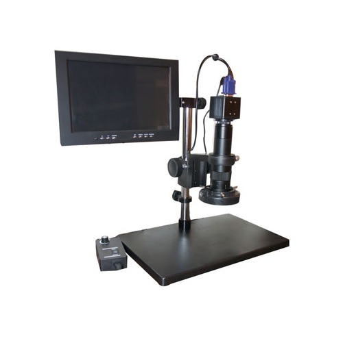
LabgoIndia is premier manufacturer of Binocular, Dissecting, Fluorescent, Gemology, Inclined, Inverted, Medical, Metallurgical, Penta Head and other scientific microscopes.
Visit at:- https://labgoindia.com/product-category/microscope
#Video-Zoom-Microscope#Microscope#LabgoIndia#scientific microscopes#scientific microscopes manufacturer
0 notes
Text
43 notes
·
View notes
Text
streddit is such an interesting place because I’ve never seen a community that was so active in their fandom that so adamantly rejects what is happening on screen. Like it’s not a case of GA who watch the show casually so they may not pick up on certain things— it’s fans who frequently engage with the show, the supplemental books and comics, fan material, but they also so adamently ignore surface level canon material if it doesn’t match the version of the show they built in their head. They are too involved to be GA and let they know less than actual GA
#i wanna observe streddit under a microscope#theyre like a scientific research group#how the hell did we get here#it is not even just byler#if you suggest will has any importance to the show at all they grill you alive#st reddit#byler#<- target audience
213 notes
·
View notes
Text

// not to be predictable. but how mad would y'all be,
#raine is not allowed to comment on this post /j and its sheerly because she knows ive been pondering him like an orb for months BHGKTRBKHGRB#LOOK. I AM EASILY SWAYED. I THINK HE'S A FUN BUG TO STUDY UNDER A MICROSCOPE AND AFTER OBSERVING MULTIPLE RAD ASS PEOPLE -#- IN THEIR PRETTY SCIENTIFIC LABCOATS DO EXPERIMENTS ON HIM I AM NOT IMMUNE TO BEING THE TODDLER THAT PRETENDS TO BE AN EQUAL GTBHRGKBRHB#i dont think i'd be able to write him anywhere near as well as harerazor and inverteds and maskfetisch but what am i but the clown -#- that gets sooo interested in the blorbos my friends are interested in BHGKTRBGHKR#this is also a shoutout to tewwors' siwoo i fucking LOOOOVE him he's not a small part of my humming and hawing#im not tagging him bc idk. popular vote i guess BKHGTBR#��� ♔ your slightly mad sea-captain : ooc.#━ ♔ the new green of spring is shimmering : dash.#jjk //
7 notes
·
View notes
Text
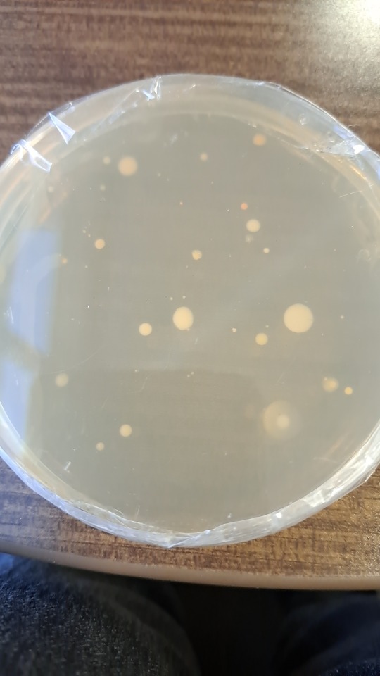
Here, you can see the fungus I've been growing. If you look close enough, you can see some pink growth aswell, this is most likely bacteria that ended up in my dish, which is super cool because I wasn't expecting to also be growing bacteria!
#bachelor of science#bacteria#biology#biology student#college#i love biology#microbiology#post secondary education#science#stem#fungi#fungus#microscope#the scientific method#undergrad life#student life#undergrad student#university student#studyblr#study motivation#studying#studyspo#petri dish#stem studyblr#stem student#biologist#studying biology#undergraduate#university#student
12 notes
·
View notes
Text

Pelomyxa is a genus of large amoebae with distinctive biological features that make them significant in the study of protists. These organisms possess multiple nuclei that exist at various stages throughout their life cycle, allowing them to adapt to specific environmental conditions.
Although their surface is covered by numerous flagella, these structures are not functional for movement, as they have undergone evolutionary reduction and lost their role in locomotion. Rather than utilizing flagella for locomotion, Pelomyxa employs a slow crawling mechanism along the bottom of lakes and ponds, moving at a gradual pace.
Pelomyxa is specialized for life in low-oxygen zones found in the bottom sediments of freshwater environments. These amoebae are typically located in the sediment-covered bottoms of ponds and small lakes, where the soil is rich in decaying organic matter, especially decomposed plant material such as broken-down leaves. These conditions provide an ideal environment for Pelomyxa to develop in relative isolation, hidden from most other aquatic organisms.
Currently, 14 species of Pelomyxa are recognized, while historical records mention about 20 additional species that have not been observed since their initial descriptions. Some of these species may have gone extinct, while others may still exist in unexplored habitats, awaiting rediscovery.
An interesting example is Amoeba quarta, first described in 1884 by the researcher August Gruber. After its initial observation, this species seemingly disappeared from scientific knowledge until 2024, when researchers from St. Petersburg rediscovered it during a study of the sediments of Lake Osinovskoe in northwest Russia. Subsequent investigations revealed that this organism belonged to the genus Pelomyxa, and the species was renamed Pelomyxa quarta.
These rediscoveries emphasize the importance of studying ecosystems that often remain overlooked but may conceal organisms critical for understanding biodiversity. The slow-moving, inconspicuous Pelomyxa offers valuable insights into ecological processes in the depths of freshwater ecosystems, which are frequently underexplored. This also serves as a reminder that many unknown species may still be waiting to be discovered and included in contemporary biological research.
For the curious and the scientifically minded, you can read more in the full research paper here: https://doi.org/10.21685/1680-0826-2024-18-3-5
#nature#biology#microbiology#microorganisms#protists#zoology#amoeba#life science#science#sciencenews#scientific research#scientific discovery#scientificresearch#biodiversity#amoebae#microscopic organisms#microscopic#microscope#microscopy#microscopic life
4 notes
·
View notes
Text
rip rhinedottir taking middle school biology probably could’ve saved you
#she’d go fucking insane over all of that#I always forget they don’t have the same scientific tools and knowledge in teyvat#-> she’d foam at the mouth over gel electrophoresis. dare I say#rip rhinedottir you would’ve loved dna transcription#do they even know about dna structure in teyvat. Rhinedottir do you know about this be honest#I don’t know enough about alchemy to know if that’s required knowledge#imagine creating life and not knowing what RNA is#do they even have advanced microscopes????? do they know what cells look like????????#rip Rhine would’ve loved organelles :/#rhine hcs
10 notes
·
View notes
Text
species don’t exist — i mean, obviously, they do. but they aren’t objective. species are (as most things are) a cultural construction, a coalition of humans deciding where and when to draw what lines. constantly in debate: did you know paleoanthropologists are unintentionally incentivized to claim to have discovered entire new genera along the path of human evolution because they are more likely to generate media buzz and gain desperately needed funding. thousands of plants may be categorized together but a centimeter’s difference in skull thickness warrants an entire new genus name. we are more genetically similar to chimpanzees than they are to their fellow non-human primates, but due to the rules of Linnaean taxonomy humanity will never be collapsed into the same genus as them because the rules dictate that the older genus name prevails: humanity would never accept becoming Pan sapiens, especially not after it took decades for it even to be accepted that humans were a part of the taxonomy in the first place. even the most basic of criteria we’ve used in the past to decide where a species stops and starts continues to be debunked - fish from entire opposites of the world can produce fertile offspring. analogous evolution can find lines that split millions of years back creating critters that would be side by side in a disney cartoon. categorization is a eternal battleground of western scientific standards requiring universalized objective qualifiers vs. the futile efforts to recognize the unmeasurable amounts of nuance held in traditional ecological knowledge — versus the fact that, inevitably, it all boils down to a vast continuum contained within only a few percentage points of variation in the squiggly lines that tell the cells of everything on the entire globe how to eat
#PONDERING . i should cite some of these with sources but i’m pondering pop science and genuinely curious how many people have got the full#‘the way we categorize living beings into species isn’t an innate trackable quality in dna but a constructed system of assigning names to#certain observable traits - be those visible to the eye or the microscope on a chromosome#<- has had no less than five evolution lectures at varying levels of complexity in the last three years <- anthropology student#i am approaching this both from a scientific perspective (genetic variation is so vast that there are many cases in which species distincti#ons boil down to two creatures or plants just being considered different by the people who interacted with them)#AND an anthropological linguistic one (those ways of categorizing animals or plants are inherently cultural and there is no objective#inherent quality of those plants/animals/fungi/whatever that would say it’s the binomial name or the colloquial name or anything at all)#text✨#idk what i’m doing. species don’t exist much like gender it’s biologically ambiguous and culturally valuable
17 notes
·
View notes
Text
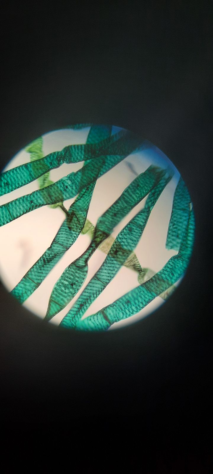
Gotta love science class... Especially when you show up early and the class before you was looking at spirogyra samples. I can't lie though these things are pretty damn cool to look at!
#science#ontario#original photographers#spirogyra#gotta love science class#photography#scientific photography#plants#plant photography#pond#pond plant#microscope
20 notes
·
View notes
Note
YOURE THE REALEST PERSON EVER FOR TRANTHARRY POSTING!!!!!! ive been saying this since day 1. You get me. Thank you. Cheering and screaming.
omg thank you so so so much 😳😭🫡 i almost felt bad for the spam at first but idk trantharry make me crazy!!! like it started with me like “i think they could kiss. for the laughs.” but then i gave it some thought and like… i think they could be really compatible! they have several common interests, they’re both SOOOOO divorced, trant would know how to help harry get and stay sober, harry could maybe become the father that stepped up (emphasis on maybe), i think they’d both be total total freaks, trant would actually be interested in how harry’s mind works, they could be so annoying together, etc etc etc. 🥰
#it did start as me like ‘i think trant’s scientific curiosity in harry is sexy actually i too want to examine harry under the microscope’#but i gave it too much thought and now i want them to kiss for real. oops!#i also think the implications of their relationship before/after harry’s memory loss would be so interesting!!!!! and a little bittersweet?#kfkekfkefj SORRY I WENT OFF I JUST LIKE TO THINK ABOUT THEM!!! i’m smashing them together repeatedly!#this was very very kind again thank you this ask made my day ☺️☺️☺️ TRANTHARRIES… OUR VISION… I’M GIVING US ALL A ROUND OF APPLAUSE
7 notes
·
View notes
Text
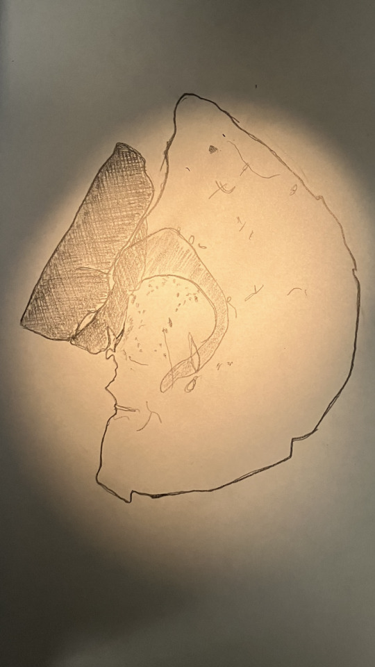
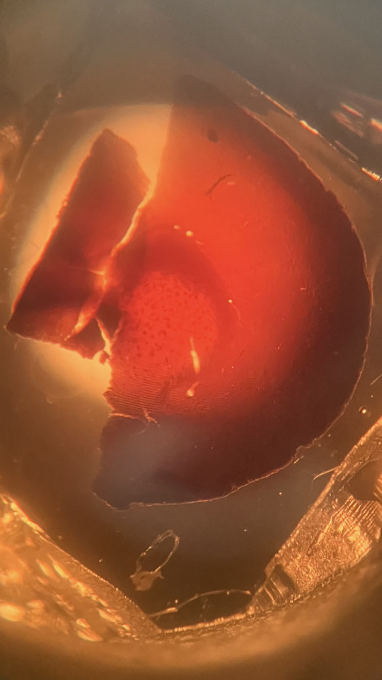
More brainy sketches
#brain anatomy#anatomical drawing#human anatomy#anatomy#neural science#science#microscope#biology#scientific illustration#brain#stuydblr#study aesthetic#stem student#studyinspo#stem studyblr#study blog#chaotic studyblr#studyblr#studyspo#study motivation#laboratory
8 notes
·
View notes
Note
icarus you are a study to everyone, legit you are in a green test tube filled with goo to me
thbis is the nicest thing anyone’s ever said to me

#muse talk#sadgeish#hope this is true…… i hope u all see me on the dash and break out the microscopes and test tubes……#i yearn to be a scientific anomaly
11 notes
·
View notes
Text
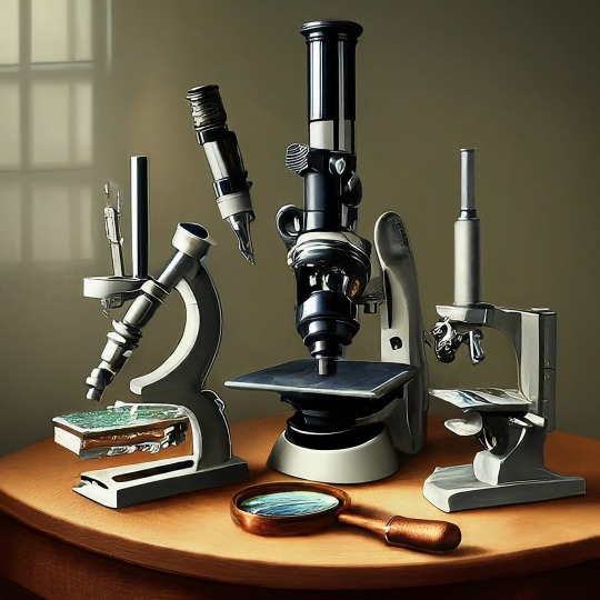
A Journey into the World of Microscopy: From Humble Beginnings to High-Tech Magnification
The science of looking into the hidden invisible Microscopy has transformed our understanding of the world around us. It can explore the universe beyond the reach of our naked eyes, with complex cellular structures, red blood cells, viruses and other viruses and microorganisms taking on amazing perspectives
The history of the microscope is a fascinating story of human curiosity, scientific genius, and relentless exploration. From the humble beginnings of simple magnifying glasses to the sophistication of modern electronic microscopes, the invention of microscopes has shaped our understanding of the microscopic world
In the 1600s, Dutch opticians such as Hans and Zachary Janssen are credited with inventing the first microscope. Known for this hybrid microscope, many lenses were used to magnify objects up to 30 times.At the end of the 17th century, Antony van Leeuwenhoek, Dutch draper some changed our perception of thumbnails. Armed with a well-made single-lens microscope, and explored the hidden reaches of nature. In 1674, Leeuwenhoek discovered microorganisms in lake water, which he aptly named “animalcules”. His discovery laid the foundations of biology and inspired generations of scientists. This incredible feat allowed him to uncover a hidden universe – the first sightings of bacteria, red blood cells, and other microorganisms.
Formation of the scientific environment (17th-19th centuries): Leeuwenhoek’s discoveries boosted scientific research. Robert Hooke, an English scientist, established these developments. In 1665, his book "Micrographia" recorded his observations with a compound microscope. Notably, the term "cell" was coined by Hooke when he examined cork tissue, laying the foundation for cell biology.Microscope systems flourished throughout the 18th and 19th centuries Joseph Lister and other scientists addressed the limitations of the early lenses, introducing improvements that reduced image distortion.
Beyond the Limits of Light: The Beginning of the New Age (19th-20th century): As the 19th century progressed, the limitations of optical microscopy became apparent and scientists yearned for a tool which can go deeper into cells. This research culminated in the development of the electron microscope in the 1930s. The 20th century was revolutionary with the invention of the electron microscope. Unlike light microscopes, which use visible light, electron microscopes use electron beams to achieve much higher magnification.Formation of the scientific environment (17th-19th centuries): Leeuwenhoek’s discoveries boosted scientific research. Robert Hooke, an English scientist, established these developments. In 1665, his book "Micrographia" recorded his observations with a compound microscope. Notably, the term "cell" was coined by Hooke when he examined cork tissue, laying the foundation for cell biology.Microscope systems flourished throughout the 18th and 19th centuries Joseph Lister and other scientists addressed the limitations of the early lenses, introducing improvements that reduced image distortion.
Beyond the Limits of Light: The Beginning of the New Age (19th-20th century): As the 19th century progressed, the limitations of optical microscopy became apparent and scientists yearned for a tool which can go deeper into cells. This research culminated in the development of the electron microscope in the 1930s. The 20th century was revolutionary with the invention of the electron microscope. Unlike light microscopes, which use visible light, electron microscopes use electron beams to achieve much higher magnification.
In the 1930s, German experts Max Knoll and Ernst Ruska made the first electron microscope. This tool let us see tiny things like cells and even atoms by using electron beams, not light, getting images many times bigger. This cool invention showed us the tiny parts inside cells, viruses, and stuff too small to see before. The 1900s brought even more cool microscopes. New kinds like phase-contrast and confocal microscopy let scientists look at live cells without using stuff that could hurt them. Now, the world of looking at tiny things is getting even better. Today, we have high-tech microscopes that use computers and lasers. These let us see and even change tiny things in ways we never could before.
Modern Microscopy's Diverse Arsenal - Today, the field of microscopy boasts a diverse range of specialized instruments, each tailored to address specific scientific needs. Here's a glimpse into some remarkable examples:
Scanning Electron Microscope (SEM): Imagine a high-tech camera that captures images using a beam of electrons instead of light. That's the essence of a SEM. By scanning the surface of a sample with a focused electron beam, SEMs generate detailed information about its topography and composition. This makes them ideal for studying the intricate structures of materials like insect wings, microchips, and even pollen grains.
Transmission Electron Microscope (TEM): While SEMs provide exceptional surface detail, TEMs take us a step further. They function by transmitting a beam of electrons through a very thin sample, allowing us to observe its internal structure. TEMs are the go-to instruments for visualizing the intricate world of viruses, organelles within cells, and macromolecules like proteins.
Confocal Microscopy: Ever wished to focus on a specific layer within a thick biological sample and blur out the rest? Confocal microscopy makes this possible. It utilizes a laser beam to precisely illuminate a chosen plane within the sample, effectively eliminating information from out-of-focus regions. This allows researchers to create sharp, three-dimensional images of cells, tissues, and even small organisms.
Atomic Force Microscopy (AFM): This technique takes a completely different approach, venturing into the realm of physical interaction. AFM employs a tiny cantilever, akin to a microscopic feeler, to physically scan the surface of a sample. By measuring the minute forces between the cantilever and the sample's surface, AFM can map its topography at an atomic level. This provides invaluable insights into the properties of materials at an unimaginable scale, making it crucial for research in fields like nanotechnology and surface science.
Fluorescence Microscopy: Imagine illuminating a sample with specific wavelengths of light and observing it glowing in response. That's the essence of fluorescence microscopy. This technique utilizes fluorescent molecules or tags that bind to specific structures within a cell or tissue. When excited by light, these tags emit their own light, highlighting the target structures with remarkable clarity. This allows researchers to visualize specific proteins, DNA, or even pathogens within biological samples.
Super-resolution Microscopy (SRM): Overcoming the limitations imposed by the wavelength of light, SRM techniques like STED (Stimulated Emission Depletion) and PALM (Photoactivated Localization Microscopy) achieve resolutions surpassing the diffraction limit. This allows researchers to visualize structures as small as 20 nanometers, enabling the observation of intricate cellular machinery and the dynamics of individual molecules within living cells.
Cryo-Electron Microscopy (Cryo-EM): This powerful technique takes a snapshot of biological samples in their near-life state. Samples are rapidly frozen at ultra-low temperatures, preserving their native structure and minimizing damage caused by traditional fixation methods. Cryo-EM has been instrumental in determining the three-dimensional structures of complex molecules like proteins and viruses, providing crucial insights into their function and potential drug targets.
Correlative Microscopy: Combining the strengths of multiple microscopy techniques, correlative microscopy offers a comprehensive view of biological samples. For instance, researchers can utilize fluorescence microscopy to identify specific structures within a cell and then switch to electron microscopy to examine those structures in high detail. This integrated approach provides a deeper understanding of cellular processes and their underlying mechanisms.
Light Sheet Microscopy (LSM): Imagine illuminating a thin slice of a sample within a living organism. LSM achieves this feat by focusing a laser beam into a thin sheet of light, minimizing photobleaching and phototoxicity – damaging effects caused by prolonged exposure to light. This allows researchers to observe dynamic processes within living organisms over extended periods, providing valuable insights into cellular behavior and development.
Expansion Microscopy (ExM): This innovative technique physically expands biological samples by several folds while preserving their structural integrity. This expansion allows for better resolution and visualization of intricate cellular structures that would otherwise be difficult to distinguish using traditional microscopy methods. ExM holds immense potential for studying the organization and function of organelles within cells.
Scanning Near-Field Optical Microscopy (SNOM): This innovative technique pushes the boundaries of resolution by utilizing a tiny probe that interacts with the sample at an extremely close range. SNOM can not only image the surface features of a sample with exceptional detail but also probe its optical properties at the nanoscale. This opens doors for research in areas like material science and photonics, allowing scientists to study the behavior of light at the interface between materials.
X-ray Microscopy: Stepping outside the realm of light and electrons, X-ray microscopy offers unique capabilities. By utilizing high-energy X-rays, this technique can penetrate deep into samples, making it ideal for studying the internal structure of dense materials like bones and minerals. Additionally, it allows for the visualization of elements within a sample, providing valuable information about their distribution and composition.
From revealing the building blocks of life to aiding in the development of new medicines, the microscope has played an undeniable role in shaping our scientific understanding. As technology continues to evolve, one can only imagine the future breakthroughs this remarkable invention holds in unveiling the secrets of our universe, both seen and unseen. These advancements hold the potential to revolutionize our understanding of biological processes, develop new materials with extraordinary properties, and ultimately pave the way for breakthroughs in medicine, nanotechnology, and countless other fields. As we continue to refine and develop novel microscopy techniques and the future holds immense promise for further groundbreaking discoveries that will undoubtedly revolutionize our perception of the world around us.
#science sculpt#life science#science#molecular biology#biology#biotechnology#artists on tumblr#microscopy#microscope#Scanning Electron Microscope#Transmission Electron Microscope#Confocal Microscopy#Atomic Force Microscopy#Fluorescence Microscopy#Expansion Microscopy#X-ray Microscopy#Super-resolution Microscopy#Light Sheet Microscopy#illustration#illustrator#illustrative art#education#educate yourself#techniques in biotechnology#scientific research#the glass scientists#scientific illustration#scientific advancements
7 notes
·
View notes
Text
youtube
The confocal microscope at Imperial College's Sir Alexander Fleming Building lab is used for imaging the interior of living plant and animal cells.
During my PhD project, I used the confocal microscope to view the interior of Nicotiana benthamiana plant cells which were expressing Green Fluorescent Protein (GFP) tagged genes of interest. I aimed to find out where the proteins encoded by the genes of interest were localised in the plant cell, which turned out to be in the cytoplasm.
From Wikipedia's entry on Confocal Microscopy: "Confocal microscopy, most frequently confocal laser scanning microscopy (CLSM) or laser scanning confocal microscopy (LSCM), is an optical imaging technique for increasing optical resolution and contrast of a micrograph by means of using a spatial pinhole to block out-of-focus light in image formation. Capturing multiple two-dimensional images at different depths in a sample enables the reconstruction of three-dimensional structures (a process known as optical sectioning) within an object. This technique is used extensively in the scientific and industrial communities and typical applications are in life sciences, semiconductor inspection and materials science. Light travels through the sample under a conventional microscope as far into the specimen as it can penetrate, while a confocal microscope only focuses a smaller beam of light at one narrow depth level at a time. The CLSM achieves a controlled and highly limited depth of field."
Music by the Fiechter Brothers
Images by Katia Hougaard & the Facility for Imaging by Light Microscopy at Imperial College London
#katia plant scientist#botany#plant biology#plants#plant science#biology#science#science and technology#confocal microscopy#microscopy#microscope#scientific instruments#laboratory#research#phdblr#phd#phd life#molecular biology#cell biology#green fluorescent protein#plant scientist#Youtube
4 notes
·
View notes
Text

AP SOUTH PARK YAOI REAL
#no unfortunately these are just notes for my ap lang class#but boy would i like to Analyze sp style..#put them under a microscope..conduct various scientific experiments..#analyze and dissect some of the classics (god tier style fics)..#i would get a 5 on that exam guys im so serious..#ap south park yaoi#sp style#south park
6 notes
·
View notes
Text
im getting my bacteria back from the lab this thursday, im so excited :3
i swabbed the bottom of my shoe (super basic i know😭💔)
ill post a photo asap!
#bachelor of science#bacteria#biology#biology student#college#i love biology#microbiology#post secondary education#science#stem#hatsune miku#microscope#studying biology#biologist#growing bacteria#undergrad student#university student#student life#studyblr#studying#stem studyblr#studyspo#study motivation#education#stem student#university#undergrad life#undergraduate#the scientific method#scientist
6 notes
·
View notes