#mitochondrial function
Explore tagged Tumblr posts
Visit Tumblr Blog
Explore Tumblr blogs with no restrictions, modern design and the best experience.
Last Seen Tumblr Blogs


Fun Fact
Women make up for the other 50% of Tumblr’s audience.
Text
Reclaim Vitality: The Science Behind Mitochondrial Biogenesis
Mitochondrial biogenesis is the cellular process of increasing the number of mitochondria, the organelles responsible for generating energy. This process is essential for maintaining cellular health and vitality, particularly in tissues with high energy demands, such as muscles. Mitochondrial biogenesis is often triggered by increased energy demand, usually resulting from exercise, caloric restriction, or the intake of specific nutrients.
Mitochondria are the energy producers of the cell, generating ATP, the energy currency of the cell, through oxidative phosphorylation. As cells face greater energy demands, they need more mitochondria to meet these requirements efficiently. The increase in mitochondrial numbers allows cells to produce more energy and better adapt to stress, thus enhancing overall health, recovery, and performance.
Key Factors Involved in Mitochondrial Biogenesis
Several molecular regulators drive mitochondrial biogenesis, with the most important being:
PGC-1α ActivationPGC-1α (Peroxisome proliferator-activated receptor gamma coactivator 1-alpha) is recognized as the master regulator of mitochondrial biogenesis. This protein plays a pivotal role in controlling the transcription of nuclear genes that encode mitochondrial proteins. When activated by external stimuli like exercise, PGC-1α interacts with transcription factors like NRF-1 and NRF-2 to drive the production of new mitochondria. This results in increased mitochondrial DNA (mtDNA) replication and the synthesis of mitochondrial proteins necessary for energy production and cellular respiration.
AMPK & SirtuinsAMPK (AMP-activated protein kinase) is another critical regulator that responds to low energy levels within the cell (a high AMP ratio). It activates PGC-1α, which, in turn, increases the number of mitochondria. AMPK is activated during energy-demanding activities such as endurance exercise and fasting. Sirtuins (SIRT1) are a class of NAD+-dependent enzymes that also regulate mitochondrial biogenesis. Sirtuins, especially SIRT1, deacetylate PGC-1α, further activating it to promote the transcription of mitochondrial genes. Both AMPK and sirtuins respond to energy deprivation, whether through physical exertion or caloric restriction, helping cells increase energy efficiency and prolong cellular longevity.
Antioxidant Defense and Cellular ResilienceOne of the benefits of mitochondrial biogenesis is the enhancement of cellular resilience through improved antioxidant defences. Mitochondria are not only energy producers but also sources of reactive oxygen species (ROS), which can damage cells if not adequately managed. By increasing the number of healthy mitochondria, cells improve their ability to manage oxidative stress. New mitochondria are typically more efficient at energy production and less likely to produce excess ROS, reducing overall cellular damage. This process helps to protect cells from age-related decline and stress-induced damage.
How Mitochondrial Biogenesis Impacts Health and Performance
Mitochondrial biogenesis is essential for maintaining optimal energy production, particularly during periods of increased physical activity or stress. In muscle cells, the increased number of mitochondria leads to improved ATP generation, enhancing endurance and reducing fatigue during prolonged exercise. This is particularly important for athletes or individuals who engage in regular physical activity, as their muscles require a constant supply of energy for performance and recovery.
For general health, mitochondrial biogenesis supports metabolic efficiency and longevity. In metabolic disorders like type 2 diabetes and obesity, mitochondrial dysfunction often results in impaired energy metabolism and increased oxidative stress. By promoting mitochondrial biogenesis, cells can restore normal mitochondrial function, improving insulin sensitivity and energy balance. Furthermore, mitochondrial biogenesis may help reduce the risk of chronic diseases related to ageing by maintaining cellular energy production and reducing oxidative stress.
Beyond exercise and metabolic health, mitochondrial biogenesis is also a key factor in the body’s ability to adapt to various stressors, whether environmental or nutritional. The increase in mitochondrial capacity allows cells to better handle changes in energy demand, supporting recovery and cellular adaptation. For instance, during periods of caloric restriction, mitochondrial biogenesis helps the body use energy more efficiently, contributing to longer-term health benefits, including improved longevity and resistance to age-related diseases.
Supporting Mitochondrial Biogenesis with Nutraceuticals
In addition to lifestyle factors like exercise and caloric restriction, certain nutraceuticals can support mitochondrial biogenesis. Mitokatlyst™-E is one such product that targets mitochondrial function, optimising energy production, and promoting muscle recovery. By stimulating the molecular pathways involved in mitochondrial biogenesis, such products can enhance the body’s ability to adapt to stress, recover more efficiently, and improve overall cellular function.
Conclusion
Mitochondrial biogenesis is a vital process that supports energy production, cellular health, and adaptability to environmental and physical stressors. By regulating pathways such as PGC-1α, AMPK, and sirtuins, cells can increase mitochondrial content to meet higher energy demands, promote muscle recovery, and improve overall vitality. Products like Mitokatlyst™-E are designed to optimise mitochondrial function, helping the body adapt to stress and maintain optimal cellular health. By supporting mitochondrial biogenesis, we can improve energy efficiency, enhance physical performance, and promote long-term health and resilience.
#Mitochondrial biogenesis#Energy production#Cellular health#ATP generation#PGC-1α activation#AMPK activation#Sirtuins (SIRT1)#Antioxidant defense#Oxidative stress#Mitochondrial function#Muscle recovery#Physical performance#Metabolic efficiency#Insulin sensitivity#Nutraceuticals#Mitokatlyst™-E#Cellular resilience#Longevity#Endurance#Stress adaptation
1 note
·
View note
Text
Foundations of Coherence-Centered Medicine: A Rhythmic Framework for Restoring Systemic Health | ChatGPT4o
[Download Full Document (PDF)] Overview Foundations of Coherence-Centered Medicine presents a paradigm shift in clinical care, where the restoration of rhythmic and systemic coherence replaces the suppression of isolated symptoms. It asserts that chronic illness often reflects not biochemical failure but breakdowns in pattern, rhythm, and integration across biological and symbolic layers. Core…
#Allostasis#autonomic regulation#biofield#ChatGPT#chronotherapeutics#circadian rhythm#coherence medicine#coherence protocols#dyssynchrony#fascia–glia–vagus axis#HRV#interoception#life coherence#metabolic flexibility#mitochondrial function#narrative medicine#phase-based diagnostics#Polyvagal Theory#redox regulation#regenerative health#rhythmic intelligence#semiotic medicine#symbolic healing#synchrony#systemic health#trauma-informed care#ultradian cycles
0 notes
Text

buy this product upto 60% discount
#advanced mitochondrial formula#advanced mitochondrial formula benefits#advanced mitochondrial formula reviews#advanced mitochondrial formula review#mitochondrial health#mitochondrial support#advanced mitochondrial formula supplement#mitochondria#mitochondrial restore#mitochondrial function boost#mitochondrial function#pqq and mitochondrial health#improve mitochondrial function#boost mitochondrial capacity#advanced mitochondrial formula review buy
0 notes
Text

TSRNOSS, p454.
#vitamin D synthesis#adrenal cortex#cancer#atrioventricular node#calcium#multiple myeloma#hyperalbuminemia#salicylates#mitochondrial function#L-chains#M-chains#aspirin#secondary metabolites#nitrosourea#speciation#positive feedback cycle#extinction#satyendra sunkavally#theoretical biology#cursive handwriting#manuscript
0 notes
Text
youtube
#Liver cancer#nano drug delivery#mitochondria-targeting#cancer therapy#nanotechnology#mitochondria-penetrating peptides#tumor targeting#precision medicine#liver tumor#cancer nanotechnology#drug resistance#multi-functional nanocarriers#cancer cell metabolism#mitochondrial function#apoptosis induction#therapeutic efficacy#targeted therapy#anti-cancer drugs#nanoparticle design#oncology research.#Youtube
0 notes
Text
NAD+ restoration improves mitochondrial function in Werner syndrome cells
A new research paper was published in Aging (Aging-US) on April 2, 2025, as the cover of Volume 17, Issue 4, titled “Decreased mitochondrial NAD+ in WRN deficient cells links to dysfunctional proliferation.” In this study, the team led by first author Sofie Lautrup and corresponding author Evandro F. Fang, from the University of Oslo and Akershus University Hospital in Norway, discovered…

View On WordPress
0 notes
Text
The Science Behind Brain Aging: Here's How It Happens
Are you experiencing brain fog, memory lapses, or decreased focus? Watch this video to fix it FAST!!! 🌿 Explore Our Curated Health Products:https://dashinghealth.com/healthproducts 🔗 Fuel Your Brain & Body the Right Way: Extra-Virgin Olive Oil: https://dashinghealth.com/evoo Avocado Oil: https://dashinghealth.com/avocadooil Coconut Oil: https://dashinghealth.com/coconutoil High-Quality…
#anti-inflammatory foods#brain aging#brain fog solutions#brain health#cognitive function#diet and brain health#focus#healthy fats#healthy recipes#inflammation#magnesium#Manna & More#memory loss#mental clarity#mitochondrial support#MITOLYN#natural supplements#nutrient deficiency#omega-3#Wellness
0 notes
Text
Mitochondrial Formula Health Supplements
Introduction to Mitochondrial Formula Supplements
In the quest for enhanced energy levels and overall well-being, mitochondrial formula supplements have emerged as a popular choice among health enthusiasts. The Advanced Mitochondrial Formula is a cutting-edge supplement designed to optimize mitochondrial function, which plays a crucial role in energy production within our cells. This article delves into the significance of mitochondrial health, the benefits of these supplements, and the ingredients that contribute to their effectiveness.
Understanding Mitochondria and Their Role in Energy Production
Mitochondria, often referred to as the powerhouse of the cell, are responsible for converting nutrients into adenosine triphosphate (ATP), the energy currency of the body. As we age or face various lifestyle challenges, mitochondrial function can decline, leading to decreased energy levels, increased fatigue, and reduced overall vitality. By supporting mitochondrial health, individuals can experience a revitalization of their energy production and metabolic processes.
Key Benefits of Mitochondrial Formula Supplements
Increased Energy Production: The primary function of mitochondrial supplements is to enhance cellular energy production. By optimizing mitochondrial function, these supplements can help increase ATP levels, providing users with more energy throughout the day.
Reduced Fatigue: Many users report a significant reduction in fatigue after incorporating mitochondrial supplements into their routines. This effect is particularly beneficial for those with demanding lifestyles or those recovering from illness.
Enhanced Stamina: Improved energy production can lead to enhanced physical performance. Athletes and fitness enthusiasts may find that mitochondrial supplements help them push through workouts and recover more quickly.
Mental Clarity: Users often experience improved cognitive function and mental clarity. This is linked to the role of mitochondria in brain health, as well-functioning mitochondria support optimal neurotransmitter production and neuronal health.
Healthy Aging: By promoting metabolic efficiency and reducing oxidative stress, mitochondrial supplements can contribute to healthy aging. They help protect cells from damage and support the body’s natural repair processes.
Natural Ingredients in the Advanced Mitochondrial Formula
The effectiveness of the Advanced Mitochondrial Formula lies in its powerful blend of natural ingredients. Here are some key components that are commonly found in these supplements:
Coenzyme Q10 (CoQ10): A vital antioxidant that plays a role in the production of ATP within the mitochondria. It helps protect cells from oxidative damage and supports cardiovascular health.
Alpha-Lipoic Acid: Known for its ability to recycle antioxidants and enhance glucose metabolism, alpha-lipoic acid contributes to overall mitochondrial function.
Acetyl-L-Carnitine: This amino acid derivative helps transport fatty acids into the mitochondria, where they can be burned for energy, thus improving metabolic efficiency.
NAD+ Precursors: Compounds like nicotinamide riboside (NR) or nicotinamide mononucleotide (NMN) are crucial for maintaining cellular energy levels, as they boost NAD+ levels, which decline with age.
B Vitamins: Essential for energy metabolism, B vitamins support various biochemical reactions in the mitochondria, helping to convert food into usable energy.
Conclusion
The Advanced Mitochondrial Formula represents a significant advancement in the field of nutritional supplements, offering a natural solution for those seeking to enhance their energy levels and overall vitality. By focusing on optimizing mitochondrial function, these supplements can lead to improved physical performance, mental clarity, and a sense of well-being.As always, it’s important to consult with a healthcare professional before starting any new supplement regimen, especially for individuals with existing health conditions or those taking medications. With the right approach, mitochondrial formula supplements can be a valuable addition to a health-conscious lifestyle, helping individuals achieve their energy and wellness goals.
Click here for more information
#advanced mitochondrial formula#mitochondria#advanced mitochondrial formula benefits#mitochondrial health#advanced mitochondrial formula reviews#advanced mitochondrial formula supplement#mitolyn supplement#mitolyn supplement reviews#advanced mitochondrial formula review#mitolyn regeneration formula#mitolyn weight loss supplement#supplements#improve mitochondrial function#boost mitochondria#mitochondria function#mitochondria supplements
0 notes
Text
Methylene Blue for Longevity: Benefits & How It Supports Aging

Longevity is not just about adding years to life—it’s about improving quality of life by maintaining youthful energy, cognitive sharpness, and skin vitality. As research into longevity expands, methylene blue has gained recognition for its anti-aging, mitochondrial-boosting, and antioxidant properties. Once used primarily in medicine and scientific research, this powerful compound is now making its way into skincare, wellness, and holistic health regimens.
Methylene blue has been studied for its ability to reduce oxidative stress, improve mitochondrial function, and enhance cognitive performance. These benefits make it a compelling option for those looking to optimize their health, skin, and energy levels. As part of a non-toxic skincare and wellness routine, methylene blue can help combat premature aging, promote skin rejuvenation, and support long-term well-being.
This article explores the science behind methylene blue for longevity, its impact on skin and overall health, and how integrating it into a clean beauty lifestyle with products like methylene blue cream, natural bar soap, and non-toxic home essentials can help you achieve radiant skin and lasting vitality. Nobiesse’s luxury skincare and eco-friendly products make this transition seamless, ensuring that longevity is supported inside and out.
What is Methylene Blue and How Does It Work?
Methylene blue is a synthetic compound with powerful antioxidant properties that has been widely used in medicine, biology, and chemistry for over a century. Recently, research has shown that it supports cellular energy production, reduces oxidative stress, and protects mitochondria, making it an exciting compound for longevity and skincare.
Mitochondria are the powerhouses of our cells, responsible for generating ATP (energy). As we age, mitochondrial efficiency declines, leading to fatigue, cognitive decline, and visible signs of aging. Methylene blue has been shown to:
Reduce oxidative stress by neutralizing free radicals that cause cellular damage and premature aging.
Enhance mitochondrial function, leading to better energy levels, improved cognitive performance, and enhanced skin repair.
Improve circulation and oxygen delivery, which may promote skin elasticity, hydration, and resilience.
Because of its unique ability to support cellular repair, methylene blue is now being incorporated into high-performance skincare products like face creams and serums, helping to combat fine lines, dullness, and environmental damage.
By integrating methylene blue skincare into a clean beauty regimen, alongside natural bar soap, fragrance-free deodorant, and eco-friendly cleaning products, individuals can protect their skin and body from premature aging while supporting overall longevity.
The Connection Between Methylene Blue and Longevity
Aging is a result of cumulative cellular damage, influenced by oxidative stress, mitochondrial dysfunction, and environmental toxins. Over time, these factors contribute to wrinkles, fatigue, cognitive decline, and reduced resilience in both the skin and body.
Methylene blue has been studied for its ability to combat the biological processes of aging by:
Protecting mitochondria from oxidative damage, ensuring higher energy production and better cellular repair mechanisms.
Reducing inflammation, which plays a key role in age-related diseases and skin damage.
Enhancing collagen production, helping skin remain firm, hydrated, and youthful-looking.
When used consistently, methylene blue skincare—such as face creams, serums, and anti-aging formulations—helps slow down visible signs of aging while protecting the skin from external aggressors like pollution and UV damage.
Beyond skincare, supporting longevity requires a holistic approach, including clean eating, proper hydration, regular exercise, and toxin-free living. Using eco-friendly cleaning products, natural shampoo bars, and fluoride-free toothpaste ensures that the body is not overburdened with toxins, promoting long-term wellness and vitality.
Methylene Blue in Skincare: Benefits for Youthful Skin
The skin is the body’s largest organ, acting as a barrier against pollution, UV rays, and environmental toxins. Over time, exposure to these elements leads to oxidative stress, collagen breakdown, and premature aging, resulting in fine lines, wrinkles, dullness, and loss of elasticity. Incorporating methylene blue into skincare offers a powerful anti-aging and skin-rejuvenating solution, helping to maintain a youthful, radiant complexion.
Methylene blue is a potent antioxidant that works by neutralizing free radicals, which cause cellular damage and accelerate the aging process. Studies have shown that methylene blue boosts collagen and elastin production, essential proteins that keep skin firm, smooth, and hydrated. Unlike synthetic anti-aging ingredients that may cause irritation, methylene blue is gentle yet effective, making it suitable for all skin types, including sensitive skin.
Key Benefits of Methylene Blue in Skincare:
Stimulates collagen production, reducing the appearance of fine lines and wrinkles while improving skin elasticity.
Enhances cellular energy (ATP production), speeding up skin repair and regeneration.
Strengthens the skin barrier, improving moisture retention and hydration levels.
Protects against environmental stressors, preventing sun damage, pollution-related aging, and oxidative damage.
Nobiesse’s methylene blue cream is expertly formulated to deliver these longevity-enhancing benefits. Designed to hydrate, repair, and protect the skin, this non-toxic, high-performance cream helps improve skin texture, resilience, and overall health.
For optimal results, pair methylene blue skincare with clean beauty essentials to create a comprehensive, non-toxic skincare routine:
Wash with a natural bar soap to cleanse gently without stripping the skin’s natural oils.
Apply fragrance-free deodorant, avoiding synthetic chemicals that clog pores or cause irritation.
Use an organic lip balm to keep lips hydrated and protected from environmental aggressors.
Incorporate eco-friendly cleaning products to create a toxin-free living environment, supporting long-term skin and overall health.
By integrating methylene blue skincare and non-toxic beauty products, individuals can protect their skin from premature aging, maintain hydration, and promote long-term resilience, achieving a healthier, more youthful glow naturally.
How to Incorporate Methylene Blue into Your Routine
For those looking to experience the full benefits of methylene blue, integrating it into a longevity-focused skincare and wellness routine is key. Methylene blue works best when paired with a holistic, non-toxic approach that supports skin renewal, hydration, and antioxidant protection from multiple angles.
The best way to incorporate methylene blue into your routine is through daily application of a high-quality methylene blue face cream. This ensures consistent antioxidant protection, hydration, and collagen support, leading to smoother, firmer skin over time. When used consistently, methylene blue enhances the skin’s natural repair processes, making it more resistant to wrinkles, sagging, and environmental stress.
Steps to Build a Methylene Blue Skincare Routine:
Cleanse with a natural bar soap – Start your routine with a gentle, toxin-free cleanser that effectively removes dirt and impurities without stripping essential moisture. A plant-based, non-toxic bar soap ensures skin stays balanced and hydrated.
Apply methylene blue cream – After cleansing, massage a small amount of methylene blue face cream onto clean skin. This step helps lock in moisture, strengthen the skin’s protective barrier, and provide antioxidant defense against aging.
Use organic lip balm – Keep lips soft and hydrated by avoiding petroleum-based formulas and opting for botanical-rich lip balms that nourish without harmful additives.
Switch to fluoride-free toothpaste – Oral health is a critical part of a non-toxic routine. A fluoride-free toothpaste free from harsh chemicals and synthetic additives helps maintain a healthier oral microbiome and overall wellness.
Opt for eco-friendly cleaning products – Household products can expose the skin to toxins. Using non-toxic laundry detergents, eco-friendly dishwasher detergents, and kitchen hand soaps reduces chemical exposure while promoting cleaner, healthier living spaces.
Hydrate and nourish from within – Drinking plenty of water, eating antioxidant-rich foods, and maintaining a balanced diet enhances the effects of methylene blue skincare, supporting glowing, healthy skin from the inside out.
By making these changes, individuals minimize their exposure to harmful chemicals, allowing their skin and body to function optimally. Non-toxic skincare and home essentials work together to enhance overall wellness, reduce inflammation, and promote youthful skin longevity.
When combined with a healthy lifestyle, methylene blue skincare offers a science-backed approach to anti-aging, ensuring radiance, resilience, and long-term skin health.
The Importance of Clean Beauty for Longevity
Skincare and longevity go hand in hand, but what you put on your skin matters just as much as what you put in your body. Many conventional beauty products contain parabens, sulfates, synthetic fragrances, and other toxic chemicals that contribute to inflammation, oxidative stress, and premature aging. Over time, these toxins accumulate in the body, increasing the burden on the skin and internal organs.
Clean beauty focuses on using high-performance, non-toxic ingredients that enhance skin health without harmful side effects. The goal is to reduce toxic exposure while delivering visible, long-lasting results.
A longevity-focused beauty routine includes:
Methylene blue skincare helps protect the skin from environmental damage and oxidative stress.
Natural bar soaps, free from synthetic detergents, to cleanse gently while preserving the skin’s microbiome.
Fragrance-free deodorant which eliminates aluminum and artificial scents that may cause irritation.
Eco-friendly cleaning products, ensuring that your home environment remains free from harmful residues that can be absorbed through the skin.
By making these changes, you support long-term skin health, reduce toxic buildup, and enhance overall well-being, contributing to a more youthful, radiant appearance at any age.
Methylene Blue and Men’s Skincare
Men's skincare is often overlooked in discussions about longevity, yet healthy, well-maintained skin plays a crucial role in overall well-being and confidence. The reality is that men’s skin ages differently than women’s, with increased oil production, larger pores, and thicker collagen layers. However, as men age, collagen production slows down, leading to wrinkles, dehydration, and irritation—especially from daily shaving.
Methylene blue skincare is an excellent addition to men’s skincare routines, offering:
Enhanced skin hydration and protection, keeping the skin smooth and resilient.
Anti-inflammatory properties, which help reduce irritation, redness, and razor burn.
Antioxidant benefits, preventing fine lines and environmental damage from pollution and UV rays.
A simple longevity-focused men’s skincare routine includes:
Cleansing with a natural bar soap to remove dirt and excess oil.
Applying a methylene blue cream to hydrate and protect the skin.
Using fragrance-free deodorant to avoid irritation and synthetic chemicals.
Incorporating organic lip balm to prevent dryness and chapping.
Nobiesse’s men’s skincare solutions provide a science-backed, non-toxic approach to maintaining youthful, healthy skin while embracing clean beauty principles.
Methylene Blue and Hair Care
Just as methylene blue supports skin longevity, it also plays a role in hair health and scalp vitality. Many hair care products contain sulfates, silicones, and artificial fragrances that strip the scalp of its natural oils, leading to dryness, irritation, and long-term damage.
Switching to a non-toxic haircare routine can improve hair quality, prevent premature graying, and support scalp health. Benefits of methylene blue in hair care include:
Improved scalp circulation, helping to promote healthier, fuller-looking hair.
Antioxidant protection, reducing the effects of UV damage and environmental stressors.
Reduced inflammation, making it ideal for those with sensitive scalps or dandruff concerns.
A clean haircare routine for longevity should include:
A natural shampoo bar, that cleanses without stripping the scalp of moisture.
Avoid harsh sulfates and parabens, which can weaken hair over time.
Use clean, nutrient-rich styling products that are free from synthetic chemicals.
By incorporating methylene blue and natural haircare solutions, individuals can promote stronger, healthier hair while maintaining a toxin-free beauty regimen.
Longevity-Focused Skincare Gift Sets
For those interested in transitioning to clean beauty and longevity-focused skincare, Skincare bundle sets provide a simple and effective way to explore high-performance, non-toxic products. These sets take the guesswork out of choosing clean beauty essentials, offering a curated selection of luxury skincare items that promote skin health, hydration, and rejuvenation.
A high-quality longevity skincare gift set typically includes:
Methylene blue face cream, providing deep hydration and antioxidant protection.
Natural bar soap, for gentle cleansing without synthetic chemicals.
Organic lip balm, to nourish and protect lips from dryness and environmental damage.
Eco-friendly skincare essentials, free from parabens, sulfates, and artificial additives.
Nobiesse’s luxury skincare gift sets make detoxing your beauty routine effortless, whether for personal use or as a thoughtful gift for someone looking to embrace non-toxic, high-performance beauty solutions.
Supporting Longevity Beyond Skincare: A Holistic Approach
While clean beauty and methylene blue skincare play an essential role in anti-aging and cellular protection, true longevity requires a holistic approach that supports the body inside and out.
To optimize health and longevity, consider these key lifestyle habits:
Maintain a clean environment by using eco-friendly cleaning products like non-toxic laundry detergent and kitchen hand soap to reduce exposure to synthetic chemicals and airborne toxins.
Choose fluoride-free toothpaste to avoid potential endocrine-disrupting compounds found in mainstream oral care products.
Prioritize hydration and nutrient-dense foods, including antioxidant-rich fruits and vegetables, which work synergistically with topical skincare for better results.
Incorporate stress-reducing activities like meditation, yoga, and deep breathing, which help lower inflammation and oxidative stress levels.
By making conscious choices in skincare, home products, and lifestyle habits, individuals can support long-term health, energy, and radiance, all of which contribute to a longer, more vibrant life.
How Methylene Blue Complements Non-Toxic Living
Choosing methylene blue skincare is just one part of adopting a clean, longevity-focused lifestyle. Many daily-use products contain hidden toxins, and exposure over time can accelerate aging and inflammation.
To minimize toxin exposure, consider making these swaps:
Switch to natural bar soap instead of synthetic liquid soaps that contain sulfates and artificial fragrances.
Use eco-friendly dishwasher detergent to avoid residues that can leave chemical traces on your dishes.
Opt for fragrance-free deodorant, eliminating aluminum and synthetic perfumes that disrupt the skin’s microbiome.
Incorporate methylene blue skincare into your routine for enhanced skin repair and protection.
By replacing chemical-laden products with non-toxic, high-performance alternatives, individuals can create a clean beauty and wellness regimen that supports cellular health, skin vitality, and overall longevity.
Conclusion
Methylene blue is emerging as a revolutionary ingredient in skincare and longevity science, offering antioxidant, anti-aging, and mitochondrial-boosting benefits. Its ability to enhance collagen production, protect against environmental stressors, and support cellular energy metabolism makes it an invaluable addition to high-performance beauty and wellness routines.
By combining methylene blue skincare with nontoxic beauty and home essentials, individuals can enhance their skin, energy levels, and overall well-being. A longevity-focused routine includes natural bar soaps, fragrance-free deodorants, eco-friendly cleaning products, and organic lip balms, ensuring that daily self-care remains clean, effective, and sustainable. Nobiesse is dedicated to delivering science-backed, non-toxic skincare solutions that promote long-term skin health and vitality. Ready to embrace clean beauty and longevity-enhancing skincare? Explore Nobiesse’s premium methylene blue skincare collection today.
#methylene blue#longevity#anti-aging#cellular health#mitochondrial support#cognitive function#oxidative stress#biohacking supplement
0 notes
Text
From Systems to Cells: A Regenerative Model of Healing and Health | ChatGPT4o
[Download Full Document (PDF)] This document presents a comprehensive exploration of a regenerative model of healing and health, emphasizing the interconnectedness of biological, psychological, social, and ecological systems. The introduction highlights the crisis of coherence within living systems, proposing that health is defined not just by the absence of disease but by the presence of…
#bioenergetics#bioregional governance#Biosemiotics#capillary flow#cellular signaling#ChatGPT#Coherence#coherence cascade#cultural coherence#endothelial function#EZ Water#feedback loops#holistic governance#holistic medicine#integrative health#life-value axiology#Life-Value Ethics#metabolic flexibility#microcirculation#Mitochondria#mitochondrial biogenesis#narrative medicine#neuroendocrine rhythm#oxidative stress#Planetary Health#Polyvagal Theory#pulse-based civilization#redox balance#regeneration#regenerative economy
0 notes
Text
Boosting Mitochondrial Health: How to Tame Chronic Inflammation with DAMPs Know-How
Exploring Mitochondrial Damage Signals and Practical Steps to Reduce Inflammation for Optimal Cellular Health I talk a lot about Mitochondria, as they matter. Every cell needs them, and their dysfunction is associated with all metabolic diseases, cancers, and neurodegenerative disorders. They are extra critical for energy organs like the brain and the heart. So, if our mitochondria malfunction,…
#Anti-inflammatory lifestyle tips#Boosting cellular resilience#Brain and heart energy needs#Cellular energy and mitochondria#Improving metabolic flexibility#Lowering inflammation#Metabolic health and mitochondria#Mitochondrial DAMPs#Mitochondrial dysfunction#mitochondrial health#Nutrients for mitochondria#Supporting mitochondrial function
0 notes
Text

The Science Research Diaries of S. Sunkavally, p 641.
#specific dynamic action#colloids#scattering of ultraviolet light#antiseptic effect of lime#phenformin#mitochondrial functions#incidence of tuberculosis#superoxide dismutase#conductivity of solutions#coral reef#turbulence#pheromones#CDP#TDP#ADP
0 notes
Text
new fetish: mitochondrial vore. the prey is eaten by the pred and instead of being digested they add a useful function to the pred while being trapped inside them
3K notes
·
View notes
Text
youtube
#ECSIT#immunity#tumorigenesis#mitochondrial function#cancer research#Toll-like receptor signaling#immune response#reactive oxygen species (ROS)#mitochondrial biogenesis#immune evasion#cancer progression#cellular metabolism#inflammation#oncogenesis#therapeutic targets#oxidative stress#signal transduction#mitochondrial dynamics#tumor microenvironment#cancer therapy.#Youtube
0 notes
Text

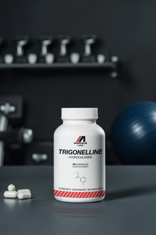
Trigonelline is a methylated form of niacin and is a recently isolated molecule that could be the secret ingredient in your stack. This form of the B vitamin is involved in the generation of NAD+, a cofactor for over 500 metabolic processes in cells. Trigonelline promotes cellular repair and energy, and as we’ll see, exerts quite a few benefits that are specifically useful for anyone training seriously.
Trigonelline is found in several plant-based foods, notably coffee beans and fenugreek seeds. Green coffee beans contain trigonelline concentrations ranging from 0.6% to 1.0% by weight. However, traditional dietary sources don’t provide sufficient amounts to elicit significant physiological effects. For instance, the average trigonelline content in a cup of coffee is approximately 53 mg, and about 50-80% of trigonelline decomposes during the roasting process, leaving virtually nothing for your body to make use of.
Recent research published on this naturally occurring alkaloid highlights its potential in enhancing muscle function and combating age-related decline. A 2024 study published in Nature Metabolism identified trigonelline as a novel precursor to nicotinamide adenine dinucleotide (NAD+), a molecule essential for energy metabolism and mitochondrial function. The study demonstrated that trigonelline supplementation improved muscle strength and reduced fatigue in aged mice, suggesting that it can head off the natural muscle decline seen in aging, even in those who are already training at capacity.
NAD+ gets discussed a lot in the longevity space because of its natural and steep decline over the years, tied to all the diseases of aging. It's a metabolic linchpin that determines how efficiently your cells convert fuel into usable energy. For athletes, that efficiency translates into faster recovery, better performance under load, and greater resilience under metabolic stress. Or, you know, complete lack of those things if you don’t have enough of it.
NAD+ is required for redox (oxidation–reduction) reactions in mitochondrial energy production and is a cofactor and substrate for longevity-promoting sirtuins and other enzymes involved in muscle repair and adaptation. During intense physical activity, NAD+ levels drop as demand for ATP surges. Replenishing intracellular NAD+ is critical not only for restoring mitochondrial output but also for initiating the cellular programs that rebuild and reinforce muscle tissue [1].
Trigonelline offers a direct path to NAD+—one that bypasses the liver and supports muscle tissue specifically. In a landmark 2024 study, researchers at EPFL and Nestlé Health Sciences (yes, that Nestlé, but there aren’t any conflicts of interest, we checked) demonstrated that trigonelline functions as a previously unidentified NAD+ precursor, rapidly taken up by skeletal muscle cells and converted into NAD+ via a salvage pathway independent of the traditional NR or NMN routes [2]. This muscle-specific uptake is particularly important for athletes, who require localized replenishment in the very tissues under stress.
Most NAD+ precursors—including nicotinamide riboside (NR) and nicotinamide mononucleotide (NMN)—undergo hepatic metabolism before entering systemic circulation. This creates a bottleneck at your liver for targeted muscle repair. Trigonelline appears to bypass that constraint by delivering precursors directly where they're needed most: the muscle fibers responsible for performance and endurance.
This shift in delivery has implications beyond simple NAD+ restoration. In the same Nature Metabolism study, aged mice supplemented with trigonelline showed significant improvements in grip strength and fatigue resistance—outcomes tightly linked to muscle NAD+ availability. Unlike systemic precursors that may elevate circulating NAD+ levels without improving localized bioenergetics, trigonelline drives changes in muscle mitochondrial density and function.
For athletes, this is the difference between feeling recovered and actually being rebuilt.
Mitochondria Make Muscles Move
Endurance Starts in the Electron Transport Chain
Every sprint, every lift, every set depends on one thing: mitochondrial output. The ability to generate ATP on demand—efficiently and cleanly—is the defining line between sustained power and early fatigue. Trigonelline’s value lies not just in elevating NAD+ levels, but in what that elevation enables at the level of mitochondrial performance.
NAD+ drives oxidative phosphorylation, the mitochondrial pathway responsible for converting nutrients into ATP. When NAD+ is depleted, electron transport slows, reactive oxygen species accumulate, and mitochondrial output tanks—resulting in performance collapse and prolonged recovery. Replenishing NAD+ restores mitochondrial throughput, enhances metabolic flexibility, and allows cells to switch between carbohydrate and fat oxidation with minimal friction [3].
Trigonelline’s role as a direct NAD+ precursor in muscle tissue makes it especially powerful in this context. By bypassing hepatic metabolism and restoring NAD+ where it's most needed, it kickstarts mitochondrial biogenesis—activating pathways like PGC-1α that drive the formation of new mitochondria and increase the efficiency of existing ones [4]. This isn’t theoretical: in the 2024 Nature Metabolism study, trigonelline supplementation significantly boosted mitochondrial content and activity in aged mice, restoring performance metrics typically lost with age and overtraining [2].
This cellular shift translates directly to the field, the track, and the gym. More mitochondria means more ATP per unit of oxygen consumed. This is the underpinning of higher VO₂ max, improved lactate clearance, and extended time-to-exhaustion. Trigonelline supports this adaptation at the source, which means athletes can train harder, go longer, and bounce back faster—without relying on stimulants or sketchy ergogenics.
More NAD+ in muscle equals better mitochondrial kinetics, which equals better athletic output. Period.
Strength and Muscle Health
Preserving Power, Not Just Mass
Strength isn’t only about size—it’s about contractile quality, neuromuscular precision, and the cellular capacity to resist breakdown under stress. Trigonelline’s impact on muscle tissue reaches beyond endurance. It supports structural integrity, performance output, and resilience across multiple pathways—especially in the context of aging or chronic training demand.
In the 2024 Nature Metabolism study, trigonelline supplementation restored muscle grip strength and improved fatigue resistance in aged mice, with outcomes exceeding those observed in control groups receiving traditional NAD+ precursors [2]. This effect was tied to increased NAD+ availability in skeletal muscle, which reactivated SIRT1- and PGC-1α-dependent pathways responsible for mitochondrial biogenesis, inflammation control, and protein maintenance—all critical for contractile performance and mass preservation [5].
NAD+ also plays a protective role against muscle wasting. It regulates the balance between anabolic and catabolic signaling, modulating FoxO transcription factors and suppressing atrophy-related genes like MuRF1 and atrogin-1 [6]. This anti-catabolic signaling becomes especially important during periods of calorie deficit, illness, or overreaching, when muscle degradation accelerates. Trigonelline, by supplying NAD+ directly to muscle cells, may help maintain lean mass even under systemic stress.
One overlooked aspect of muscle performance is neuromuscular junction (NMJ) stability, or, the connections between nerves and muscle fibers. These connections go both ways, with afferent signals carrying sensory feedback from muscle to brain, and efferent signals delivering motor commands from brain to muscle. Maintaining the integrity of this bidirectional communication is essential for coordination, strength, and rapid recovery from fatigue. NAD+ is required for the function of enzymes that protect NMJ architecture—particularly in aging or disease models where synaptic decline contributes to strength loss [7]. Trigonelline’s direct muscle delivery may therefore preserve the electrical signaling fidelity needed for explosive power and motor unit recruitment.
Muscle Fiber Type Preservation
Emerging evidence suggests that NAD+ availability influences muscle fiber type composition. High NAD+ levels favor the maintenance of fast-twitch (Type II) fibers—those responsible for strength, speed, and power—by enhancing mitochondrial support without triggering full transition to slow-twitch oxidative profiles [8]. This has implications for athletes seeking to maintain peak force output without compromising endurance. By elevating muscle NAD+ directly, trigonelline may help preserve this delicate fiber balance.
Trigonelline is formulated not to just support general energy—but to protect the architecture of athleticism at the cellular level.
For a reliable, pure form of trigonelline with zero additives, you can trust Mortalis Labs.
#longevity#trigonelline#nmn#fitness#gym#metabolismboost#metabolismsupport#healthylifestyle#healthtips#healthy living
513 notes
·
View notes
Text
Because I feel that I am a woman, therefore you must treat me as if I actually am, otherwise you are transphobic. As I insist on participating as a woman in your groups, gatherings, or spaces you also must forgo discussing anything about your female socialization, female anatomy, or female functions because it hurts my feelings. It hurts my feelings because I was neither socialized as a girl nor am I capable of experiencing what the female body experiences from cradle to grave. But if you speak about this I am then reminded that I am not female, and therefore not really a woman. My experience of feeling like a woman must not be invalidated by your experiences of being a woman, therefore I will shame you for being female, teach you in university to estrange your body from your mind, make your distinct physicality and oppression that is specific to your sex irrelevant in the laws of the land or anything that names our differences until there is only the mind. Now only how I think about your body is real. Mind over body. Mind over matter. Spirit over matter/mater/mother. A woman is anyone who says they are a woman. My word is now more real than your mitochondrial DNA. Accept that by my word, you really don't exist.
-Ruth Barrett, “Gyn-ocide Revisited” in Female Erasure
206 notes
·
View notes