#ChestRadiology
Explore tagged Tumblr posts
Text


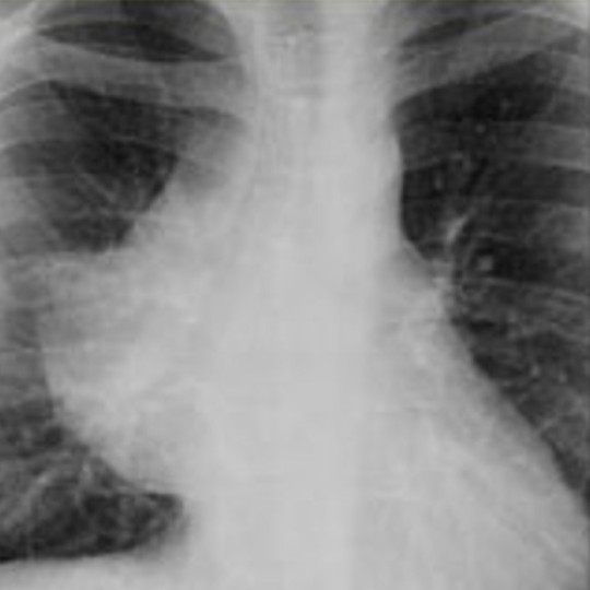
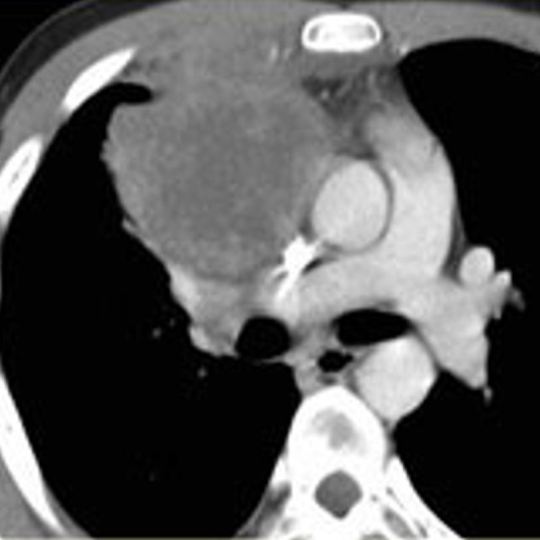
The differential diagnosis of a middle mediastinal mass includes lymph nodes, foregut duplication cysts, tracheal/bronchial lesions, lipoma, esophageal lesions (benign or malignant) and vascular lesions (aortic aneurysm, varices).
This case is a 70-year-old man who presented with cough and hemoptysis. Chest radiograph reveals a right mediastinal mass that silhouettes the right hilar vessels, localizing the lesion to the middle mediastinum. There is also mass effect on the trachea and widening of the right paratracheal stripe. CT reveals a mass along the right tracheal border, which also encases the right mainstem bronchus. Biopsy revealed small cell bronchial carcinoma.
Case courtesy of Townsville radiology training, Radiopaedia.org, rID: 18285
#TeachingRounds#FOAMEd#FOAMRad#Anatomy#mediastinum#radiology#chestradiology#chestrad#cardiothoracicradiology#chestxray#CTRad
6 notes
·
View notes
Photo

- “ Na #retrospectiva2019 é muito importante lembrar que o padrão #piu encontrado na #tcar não é igual a #fpi ( #fibrosepulmonaridiopatica ) É comum a várias outras doenças fibrosantes pulmonares como #pneumonitedehipersensibilidade crônica, #dtc, #ssc ,#pneumoconioses, etc • • #annualreview #pneumologia #radiologia #chestradiology #patologia #reumatologia #fibrosepulmonar #hrct (em DAPI Diagnóstico Avançado Por Imagem) https://www.instagram.com/p/B5nAyTPlI_j/?igshid=1ky661iykpjbx
#retrospectiva2019#piu#tcar#fpi#fibrosepulmonaridiopatica#pneumonitedehipersensibilidade#dtc#ssc#pneumoconioses#annualreview#pneumologia#radiologia#chestradiology#patologia#reumatologia#fibrosepulmonar#hrct
0 notes
Text


The differential diagnosis for posterior mediastinal masses is primarily neurogenic tumors (such as neurofibromas and schwannomas) and extramedullary hematopoiesis.
This is a case of a 25-year-old man with a history of thalassemia. Bilateral mediastinal masses demonstrate a hilum overlay sign at the right hilum (hilar vessels are visible) and silhouette the descending aorta. This localizes the masses to the posterior mediastinum. There is subtle widening of some posterior ribs. In addition, there is splenomegaly (which can be difficult to spot on CXR!). At CT, bilateral paraspinous masses are confirmed. This is an example of extramedullary hematopoiesis.
Case courtesy of Ian Bickle, Radiopaedia.org, rID: 39630
#TeachingRounds#FOAMEd#FOAMRad#Anatomy#mediastinum#radiology#chestradiology#chestrad#cardiothoracicradiology#chestxray#CTRad#hematology
0 notes
Text


Here is another mass overlying the right hilum on chest radiograph. Obtuse angles with the lung localize this to the mediastinum. You can see the hilar vessels through the mass, so it is not in the middle mediastinum (this is known as the hilum overlay sign - if it were in the middle mediastinum it would obscure the vessels). The majority of the right heart border is obscured by the mass so this must be in the anterior mediastinum (this is the silhouette sign - loss of a border due to an adjacent finding). The anterior location is confirmed at CT.
So what is the differential diagnosis for an anterior mediastinal mass? A popular mnemonic is the 4 T's - thymoma (or other thymic mass), teratoma (or other germ cell tumor), thyroid carcinoma (or goiter), and (terrible) lymphoma. Vascular lesions are also possible, but these tend to arise from the middle mediastinum.
At biopsy, this mass proved to be a lymphoma.
Source: S. Balla. Masses Differential Diagnosis. RadiologyAssistant.nl
#TeachingRounds#FOAMEd#FOAMRad#Anatomy#mediastinum#radiology#chestrad#chestradiology#cardiothoracicradiology#xray#chestxray
0 notes
Text

Let's break down a chest radiograph of an anterior mediastinal mass.
On the PA view, there is a mass overlying the right hilum. How can we tell if this is in the lung or mediastinum? Lung masses tend to have acute angles where they abut the mediastinal border. This mass has an obtuse angle at the superior border with the mediastinum (yellow) and a right angle at the inferior border (red), favoring mediastinum. At lateral CXR, there is no retrosternal fat, confirming this is an anterior mediastinal mass (but you won't always have a lateral CXR!).
Further evaluation with CT confirms the anterior mediastinal mass. Stay tuned for a discussion of the differential diagnosis of an anterior mediastinal mass (or if you know it, dro#cardiothoracicradiology
Edit to add: This is a lymphoma.
Case courtesy of Arkadi Tadevosyan, Radiopaedia.org, rID: 53664
#TeachingRounds#FOAMEd#FOAMRad#Anatomy#mediastinum#radiology#chestrad#chestradiology#cardiothoracicradiology
0 notes
Text

The mediastinum is typically divided into the anterior (purple), middle (blue), and posterior (yellow) compartments, shown here on sagittal (a) and axial (b-d) CT. These divisions assist in forming a differential diagnosis for lesions arising in each compartment.
Source: B.W. Carter, et al. Radiographics 2017, 37, 2, 413-436.
#TeachingRounds#FOAMed#FOAMRad#Anatomy#mediastinum#radiology#chestradiology#chestrad#cardiothoracicradiology
0 notes
Link
This week we are shifting gears to highlight #onlineresources for adult radiology. Today's case is from Radiopaedia.org. Whether you follow them on Tumblr, FB, Instagram, or Twitter, they consistently post great content and the articles on their site are highly educational. Here is an interesting case of Kartegener syndrome: Complete situs inversus with lower lobe predominant scarring and bronchiectasis due to chronic infection. Note the right-sided gastric air bubble and left sided liver. Read full case:https://radiopaedia.org/cases/kartagener-syndrome-23?lang=us Case courtesy of Dr Issac Yang, rID: 77896 #TeachingRounds #FOAMed #FOAMRad #Radiology #XRay #ChestRadiology #ChestRad #Genetics #Syndromes
2 notes
·
View notes
Photo

- " 🤔 #anamnese: história de #quilotorax, #asma? #4anosdeidade; #TCAR: Discreto espessamento de septos #pulmonares intralobulares: #linfoangiomatose #pulmonar ? ¤ ¤ #pneumo🎯 #pneumologia #radiology #chestradiology #residentrounds #hcpr #pulmonology #pulmão (em Complexo Hospital de Clínicas UFPR) https://www.instagram.com/p/Bu_O-SdFb2_/?utm_source=ig_tumblr_share&igshid=k40018lrq54d
#anamnese#quilotorax#asma#4anosdeidade#tcar#pulmonares#linfoangiomatose#pulmonar#pneumo🎯#pneumologia#radiology#chestradiology#residentrounds#hcpr#pulmonology#pulmão
0 notes
Photo

- " Remembering your #bestfriends that #dogs #🐶 can't operate #mri #scanners, but #catscan #🐱 !" - • • #radiology #chestradiology #pulmonology #pneumologia #cartoon #charge #joke #docjokes #😂 #cartoonoftheday #jokepic #medicina #cat #dog #bestfriends #bff #piada #medjoke (em DAPI Diagnóstico Avançado Por Imagem) https://www.instagram.com/p/BsLG280ABAT/?utm_source=ig_tumblr_share&igshid=ka86wc7v2z8r
#bestfriends#dogs#🐶#mri#scanners#catscan#🐱#radiology#chestradiology#pulmonology#pneumologia#cartoon#charge#joke#docjokes#😂#cartoonoftheday#jokepic#medicina#cat#dog#bff#piada#medjoke
0 notes
Photo

Pulmonary CTA can also diagnose chronic pulmonary embolism. This is a case of a woman who presented with shortness of breath. There are peripheral filling defects in the left lower lobar and segmental arteries, consistent with chronic pulmonary thromboembolism (filling defects from acute PE are in the center of the vessel, as in yesterday's case). Check out the full case: Case courtesy of Dr Julien Pagniez, Radiopaedia.org, rID: 25993 https://radiopaedia.org/.../chronic-pulmonary-embolism...
1 note
·
View note
Photo


CTA of the pulmonary arteries is the preferred imaging study to order for suspected acute pulmonary embolism. Even if negative for PE, CTA can evaluate for other causes of the patient's symptoms.
This is a case of a 28 year-old man who presented with chest pain, SOB, and tachycardia. CTA shows central filling defects in segmental left lower lobe arteries, likely acute pulmonary thromboembolism although the differential diagnosis includes tumor thrombus. Left basal consolidation (2nd image) is concerning for lung infarct. Ratio of right ventricle to left ventricle was >1, a poor prognostic indicator (image not shown).
Case courtesy of Dr Goran Mitrovic, Radiopaedia.org, rID: 37422 https://radiopaedia.org/cases/pulmonary-embolism-21?lang=us
1 note
·
View note
Photo

A #machinelearning algorithm that could b used in place of #surgical #lung #biopsy 2 confirm the idiopathic #pulmonaryfibrosis #fpi & #cpfe diagnosis with high + predictive value? #Pleasure 2 meet you: #Envisia #genomic classifier by Veracyte https://lnkd.in/dwBzUrn #pneumologia #fpi #cfpe ¤ ¤ #ai #lungbiopsy #pneumo🎯 #dpi #dips #ild #biópsia #biópsiapulmonar #pulmão #lung #fibrosepulmonar #pneumologia #pneumologista #chestradiology #🔬 https://www.instagram.com/p/Bwx3qxMF8ks/?igshid=efx30rw0zskp
#machinelearning#surgical#lung#biopsy#pulmonaryfibrosis#fpi#cpfe#pleasure#envisia#genomic#pneumologia#cfpe#ai#lungbiopsy#pneumo🎯#dpi#dips#ild#biópsia#biópsiapulmonar#pulmão#fibrosepulmonar#pneumologista#chestradiology#🔬
0 notes
Photo

#TipofTheDay in #pneumologia: chama a atenção a #tcar evidenciando #bronquiectasias e #bronquioloectasias de #tração em todos os #lobos #pulmonares #vidro fosco de permeio notadamente em #ápice direito, e #faveolamento incipiente subpleural. Todavia, procure lembrar ao auscultar o pulmão da alta correlação de #estertores em #velcro com a existência de #fibrosepulmonar / #doençasintersticiais na #TCAR! Logo, a ausculta do torax pode ser a chave de um diagnóstico precoce em #dips / #fibrosepulmonardiopática #fpi #cpfe #ssc #ar #ILDs #ipfwhywait ¤ #pneumologia #pneumo🎯 #tipoftheday #reumatologia #rheumatology #fisio #radiologist #chestradiology #reumato🎯 #reumatologiaemfoco #pulmonology #pneumologista #fisioterapia #semiologia #chestrehab #simplesassim (em Associação Paranaense de Pneumologia) https://www.instagram.com/p/BwMfND4lGTI/?utm_source=ig_tumblr_share&igshid=1llbq50zcx7vk
#tipoftheday#pneumologia#tcar#bronquiectasias#bronquioloectasias#tração#lobos#pulmonares#vidro#ápice#faveolamento#estertores#velcro#fibrosepulmonar#doençasintersticiais#dips#fibrosepulmonardiopática#fpi#cpfe#ssc#ar#ilds#ipfwhywait#pneumo🎯#reumatologia#rheumatology#fisio#radiologist
0 notes
Photo

-" #2nd #TipofTheday in #pulmonology🎯 #Velcrocrackles are abnormal #lung sounds. So, remember: #velcrosounds it's equal 2 #HRCT lung #imaging! #CPFE is prevalent & accurate & earliest diagnosis has a significant impact in #IPF / #ILD " - #Dicadodia em #pneumologia 🎯: #estertores em #velcro #CFPE #FPI " - #pneumologistas #cardiologistas #geriatras #cardiology #geriatry #radiology #chestradiology (em Hospital Pavilhão Pereira Filho-Santa Casa) https://www.instagram.com/p/Bp5Pky8ADTN/?utm_source=ig_tumblr_share&igshid=t3qe2phjisgi
#2nd#tipoftheday#pulmonology🎯#velcrocrackles#lung#velcrosounds#hrct#imaging#cpfe#ipf#ild#dicadodia#pneumologia#estertores#velcro#cfpe#fpi#pneumologistas#cardiologistas#geriatras#cardiology#geriatry#radiology#chestradiology
0 notes
Photo

Here is the image for our last post - apparently Tumblr doesn’t allow us to use the “Link” option AND have an image in the post. This is a case of Kartagener syndrome. Read the full case over at Radiopaedia: https://radiopaedia.org/cases/kartagener-syndrome-23?lang=us
3 notes
·
View notes