#chestrad
Explore tagged Tumblr posts
Text


The differential diagnosis of a middle mediastinal mass includes lymph nodes, foregut duplication cysts, tracheal/bronchial lesions, lipoma, esophageal lesions (benign or malignant) and vascular lesions (aortic aneurysm, varices).
This case is a 70-year-old man who presented with cough and hemoptysis. Chest radiograph reveals a right mediastinal mass that silhouettes the right hilar vessels, localizing the lesion to the middle mediastinum. There is also mass effect on the trachea and widening of the right paratracheal stripe. CT reveals a mass along the right tracheal border, which also encases the right mainstem bronchus. Biopsy revealed small cell bronchial carcinoma.
Case courtesy of Townsville radiology training, Radiopaedia.org, rID: 18285
#TeachingRounds#FOAMEd#FOAMRad#Anatomy#mediastinum#radiology#chestradiology#chestrad#cardiothoracicradiology#chestxray#CTRad
6 notes
·
View notes
Text
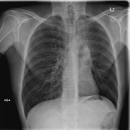
This week we will be showing a case series of chest radiographs. Take a minute to look at the image before you read on. Can you spot the problem?
...
...
...
...
This case demonstrates a mass at the left hilum. In addition, the left hemidiaphragm is elevated, indicating volume loss, and there is air outlining the aortic arch and proximal descending aorta (the so-called "Luftsichel sign," or "air sickle" due to its shape). Findings are classic for left upper lobe collapse (in this case post-obstructive due to the mass). The hyperinflated superior segment of the left lower lobe is what creates the air sickle, as it is situated betwixt the collapsed lung and the aorta.
Case courtesy of Dr Charlie Chia-Tsong Hsu, Radiopaedia.org, rID: 35263
3 notes
·
View notes
Text
Spotters Set 44 - Aunt Minnie Radiology Cases 3

#RGCOD #10 What is your diagnosis for this #chestrad case?
More radiology cases: https://buff.ly/2OAvQVn
#FOAMed #FOAMrad #radiology #radiogyan #chestrad #radres #raded #frcr #frcr2b #MedEd #XRAY #radiologia #radiologyassistant
Read the full article
0 notes
Photo

From: I miss my old buddy! @Chestrade
0 notes
Photo

Volunteers at the Centre for Health Sciences Training, Research and Development (CHESTRAD) International http://fornaija.tk/volunteers-at-the-centre-for-health-sciences-training-research-and-development-chestrad-international/
0 notes
Text
Expert wants parents, caregivers to handle Down syndrome children with love, care
Expert wants parents, caregivers to handle Down syndrome children with love, care
A medical researcher, Dr Olufunmilola Lola-Dare, has advised parents, caregivers and communities to always show love and give adequate care to children living with Down syndrome.
Lola-Dare, also the President, Centre for Health Services Training, Research and Development (CHESTRAD), gave the advice on Tuesday in an interview in Ibadan.
According to her, let us condemn a situation whereby Down…
View On WordPress
#care#caregivers to handle Down syndrome children with love#Expert wants parents#happenings#news#updates
0 notes
Text


The differential diagnosis for posterior mediastinal masses is primarily neurogenic tumors (such as neurofibromas and schwannomas) and extramedullary hematopoiesis.
This is a case of a 25-year-old man with a history of thalassemia. Bilateral mediastinal masses demonstrate a hilum overlay sign at the right hilum (hilar vessels are visible) and silhouette the descending aorta. This localizes the masses to the posterior mediastinum. There is subtle widening of some posterior ribs. In addition, there is splenomegaly (which can be difficult to spot on CXR!). At CT, bilateral paraspinous masses are confirmed. This is an example of extramedullary hematopoiesis.
Case courtesy of Ian Bickle, Radiopaedia.org, rID: 39630
#TeachingRounds#FOAMEd#FOAMRad#Anatomy#mediastinum#radiology#chestradiology#chestrad#cardiothoracicradiology#chestxray#CTRad#hematology
0 notes
Text
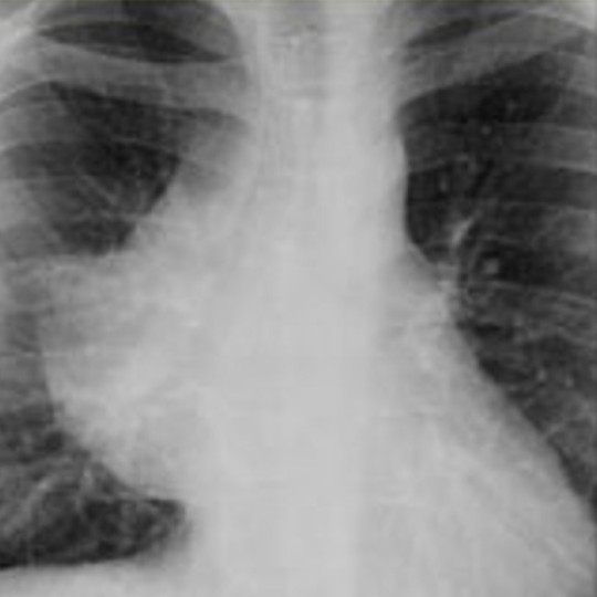
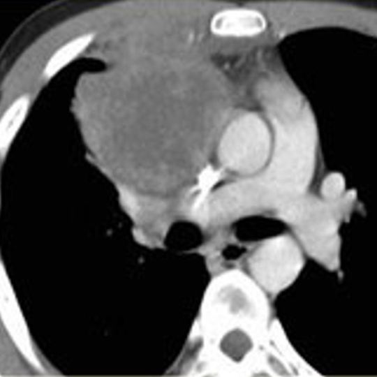
Here is another mass overlying the right hilum on chest radiograph. Obtuse angles with the lung localize this to the mediastinum. You can see the hilar vessels through the mass, so it is not in the middle mediastinum (this is known as the hilum overlay sign - if it were in the middle mediastinum it would obscure the vessels). The majority of the right heart border is obscured by the mass so this must be in the anterior mediastinum (this is the silhouette sign - loss of a border due to an adjacent finding). The anterior location is confirmed at CT.
So what is the differential diagnosis for an anterior mediastinal mass? A popular mnemonic is the 4 T's - thymoma (or other thymic mass), teratoma (or other germ cell tumor), thyroid carcinoma (or goiter), and (terrible) lymphoma. Vascular lesions are also possible, but these tend to arise from the middle mediastinum.
At biopsy, this mass proved to be a lymphoma.
Source: S. Balla. Masses Differential Diagnosis. RadiologyAssistant.nl
#TeachingRounds#FOAMEd#FOAMRad#Anatomy#mediastinum#radiology#chestrad#chestradiology#cardiothoracicradiology#xray#chestxray
0 notes
Text

Let's break down a chest radiograph of an anterior mediastinal mass.
On the PA view, there is a mass overlying the right hilum. How can we tell if this is in the lung or mediastinum? Lung masses tend to have acute angles where they abut the mediastinal border. This mass has an obtuse angle at the superior border with the mediastinum (yellow) and a right angle at the inferior border (red), favoring mediastinum. At lateral CXR, there is no retrosternal fat, confirming this is an anterior mediastinal mass (but you won't always have a lateral CXR!).
Further evaluation with CT confirms the anterior mediastinal mass. Stay tuned for a discussion of the differential diagnosis of an anterior mediastinal mass (or if you know it, dro#cardiothoracicradiology
Edit to add: This is a lymphoma.
Case courtesy of Arkadi Tadevosyan, Radiopaedia.org, rID: 53664
#TeachingRounds#FOAMEd#FOAMRad#Anatomy#mediastinum#radiology#chestrad#chestradiology#cardiothoracicradiology
0 notes
Text

The mediastinum is typically divided into the anterior (purple), middle (blue), and posterior (yellow) compartments, shown here on sagittal (a) and axial (b-d) CT. These divisions assist in forming a differential diagnosis for lesions arising in each compartment.
Source: B.W. Carter, et al. Radiographics 2017, 37, 2, 413-436.
#TeachingRounds#FOAMed#FOAMRad#Anatomy#mediastinum#radiology#chestradiology#chestrad#cardiothoracicradiology
0 notes
Text
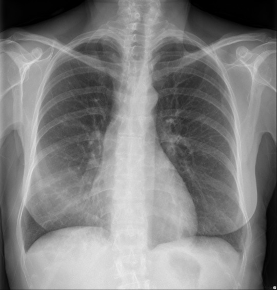
Can you spot the finding that gives this case away?
...
...
...
...
...
...
Indistinct right heart border is right middle lobe pneumonia / right middle lobe collapse until proven otherwise. This is easily confirmed on the lateral view, where the finding is usually obvious. (lateral view posted in the comments).
Case courtesy of A/Prof Stefan Heinze, Radiopaedia.org, rID: 37865
4 notes
·
View notes
Text

Take a look at today's chest radiology case.
...
...
...
...
...
There is rightward shift of the mediastinum, including the trachea, indicating volume loss in the right hemithorax. The right hemidiaphragm is obscured. These are the classic findings of right lower lobe collapse.
Case courtesy of Assoc Prof Frank Gaillard, Radiopaedia.org, rID: 15328
4 notes
·
View notes
Text

This week we will be showing a case series of chest radiographs. Take a minute to look at the image before you read on. Can you spot the problem?
...
...
...
...
This case demonstrates a mass at the left hilum. In addition, the left hemidiaphragm is elevated, indicating volume loss, and there is air outlining the aortic arch and proximal descending aorta (the so-called "Luftsichel sign," or "air sickle" due to its shape). Findings are classic for left upper lobe collapse (in this case post-obstructive due to the mass). The hyperinflated superior segment of the left lower lobe is what creates the air sickle, as it is situated betwixt the collapsed lung and the aorta.
Case courtesy of Dr Charlie Chia-Tsong Hsu, Radiopaedia.org, rID: 35263
5 notes
·
View notes
Photo
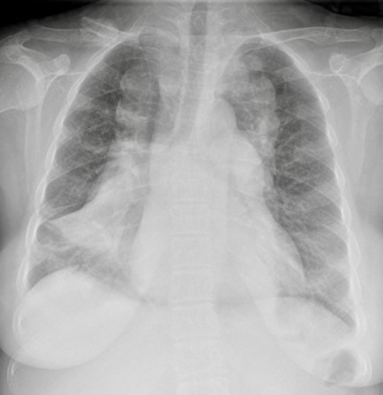
This chest radiograph shows bilateral paraspinal masses throughout the posterior mediastinum. In addition, there is medullary expansion of several posterior ribs (esp the left 4th and 5th ribs).
The combination of rib expansion and paraspinal masses is essentially diagnostic of extramedullary hematopoiesis. Differential diagnosis includes hemoglobinopathies and myeloproliferative disorders. This patient had beta-thalassemia major.
Case courtesy of Dr. Eric Stern, University of Washington.
4 notes
·
View notes
Photo

Today's #onlineresource is LearningRadiology.com. This is a great site for medical students and other non-radiologists, with lots of cases for beginners, as well as more advanced content for advanced readers.
This is a case of Swyer-James syndrome: post-infectious obliterative bronchiolitis leading to a hyper-expanded, hypovascular left lung. Read the full case here: http://learningradiology.com/.../chestn.../swyer%20james.htm
3 notes
·
View notes
Link
This week we are shifting gears to highlight #onlineresources for adult radiology. Today's case is from Radiopaedia.org. Whether you follow them on Tumblr, FB, Instagram, or Twitter, they consistently post great content and the articles on their site are highly educational. Here is an interesting case of Kartegener syndrome: Complete situs inversus with lower lobe predominant scarring and bronchiectasis due to chronic infection. Note the right-sided gastric air bubble and left sided liver. Read full case:https://radiopaedia.org/cases/kartagener-syndrome-23?lang=us Case courtesy of Dr Issac Yang, rID: 77896 #TeachingRounds #FOAMed #FOAMRad #Radiology #XRay #ChestRadiology #ChestRad #Genetics #Syndromes
2 notes
·
View notes