#medical imaging
Explore tagged Tumblr posts
Text

"Normal lumbar spine and pelvis." Radiography and radio-therapeutics. 1917.
Internet Archive
2K notes
·
View notes
Text

He looks like Harry du bois
#get well soon#bunnunny#bunny#dumb bunny#vet#radiography#animals#at the vet#medical imaging#animal memes#animalsgettingmedicalimaging#under the weather#pets#disco elysium#harry du bois
263 notes
·
View notes
Text
By popular (???) request, based on the outcome of this poll.
A WARNING: you guys really did pick the most complex one. This is loooooong. A DISCLAIMER. This is a silly little lesson aimed at folks who know sod-all about MRI. There are memes. There is (arguably) overuse of the term ‘big chungus’. If you are looking to delve deeper into the mysteries of K-Space, this is not the Tumblr post for you.
So, without further ado...
Today I am introducing you to my one true love. The legend. The icon.
Ferromagnetic material loves him. Claustrophobic people fear him.
Yeah, that’s right – we’re talking about the big boom-boom sexyboy magnet machine, hereby known as Big Chungus.

Aka...
MAGNETIC RESONANCE IMAGING
First off, though? Let’s start small.
Very, very small.
Meet HYDROGEN.

The nucleus of this element is made up of a single proton, which has a magnetic dipole – i.e., it acts like a tiny bar magnet.
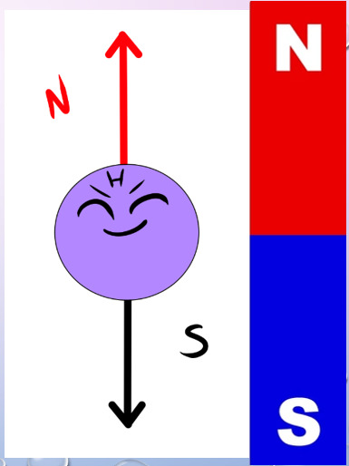
Hydrogen is also a component of water. As we all know, we’re basically walking sacks of goop – meaning that Hydrogen is abundant throughout our bodies.
Therefore, when we stick you in a strong magnetic field… say, within our friend Big Chungus… we can manipulate all those tiny Hydrogen atoms in a variety of fun ways.

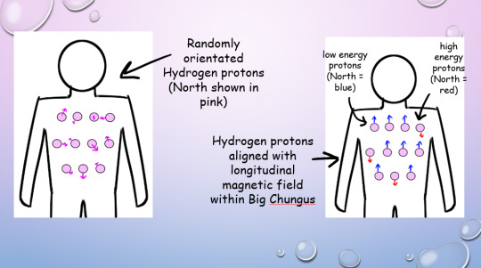
Under normal conditions, all your Hydrogen protons are pointing every-which-way.
But in Big Chungus, there is a strong longitudinal magnetic field that travels along the Z-axis of the machine. So, all your teeny tiny Hydrogen protons swivel to align with that field!
If a proton’s energy is LOWER than that of the longitudinal magnetic field (a majority), they will align PARALLEL with the field. If their energy is HIGHER (a minority) they will align ANTI-PARALLEL.
As most of the protons align with the longitudinal magnetic field, the net magnetisation vector within the human body is also longitudinal! This is called the thermodynamic equilibrium – the resting state for all those li’l protons when your body is within Big Chungus.
(You won’t feel any different, btw! We’re flipping a bunch of teeny-tiny bits inside you, but you won’t feel a thing!) (You might do later, when we activate the Gradient coils. We’ll….. get to that)
But, while all of this is very cool, it gives us no actual information. We gotta play some more with your protons - which brings us to arguably the most important concept in MRI. I mean, it’s literally in the name!

Let’s go back to our Hydrogen protons.

We’ve established that they’re all pointing in different directions. But they’re not just sitting still. They’re spinning and wobbling all over the shop.
We call this rotational wobbly movement precession.
In their natural state, these protons all precess at different speeds. When we subject them to Big Chungus, as well as all lining up neatly with the magnetic field, they all start to precess at the same speed.
However, their magnetic North will be pointing to different points at any given moment. Imagine two clocks, both of which are ticking at the same rate, but which have been set to read different times.
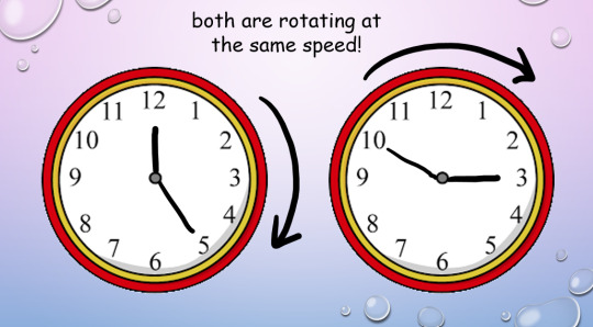
This is where magnetic resonance comes in.
In addition to the homogenous longitudinal magnetic field provided by Big Chungus, we also create an oscillating magnetic field in the transverse plane by using a radiofrequency (RF) pulse. We can tune that oscillation to the ‘resonant frequency’ of Hydrogen atoms.
Every molecule capable of resonance has its own specific frequency. We use a funky equation called the Larmor Equation to work this out, or, as I like to call it, W, BOY!!!
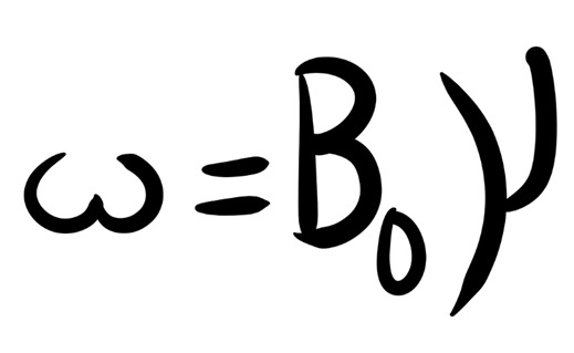
(The weird ‘w’ is the resonance frequency; the weird ‘Bo’ is the magnetic field strength, and the weird ‘Y’ is the gyromagnetic ratio of each particular element.)
So, we know exactly at what frequency to apply that RF pulse to your protons, to achieve resonance!
But what is resonance?

In acoustics, a ‘resonant frequency’ is the frequency an external wave needs to be applied at in order to create the maximum amplitude of vibrations within the object. Like when opera singers shatter glass with their voice! They’re singing at the resonant frequency of the glass, which makes it vibrate to the point where it compromises its structural integrity.
A similar concept applies in magnetic precession, with, uh, less destructive results. We’re not exploding anything inside of you, don’t worry!
(We do explode your innards accidentally in Ultrasound sometimes, via a different mechanism. But you’ll have to ask me more about that later. >:3)
To put it simply, magnetic resonance is the final step in getting those protons to BEHAVE. Now, the clocks have been corrected so their hands move at exactly the same time, in the same position. The protons are precessing ‘in phase’. Yay!
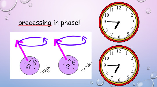
This creates transverse magnetisation, as the magnetic vectors of all those protons (which, remember, act as bar magnets) will swing around to point in one direction at the same time.
But the cool thing about resonance? It also allows the protons to absorb energy from the RF pulse.
(Do NOT ask me how. Do NOT. I will cry.)
And remember how the higher-energy protons flip anti-parallel to the longitudinal magnetic vector of Big Chungus, while the lower-energy protons are aligned parallel? And because we have more low-energy protons than high-energy protons, our body gains a longitudinal magnetic vector to match Big Chungus?
Zapping those protons at their resonant frequency gives 'em energy (a process known as ‘excitation’, which I love, because I get to imagine them putting little party hats on and having a rave).
So, loads of them flip anti-parallel! Enough to cancel out the net longitudinal magnetic vector of our bodies – despite the best efforts of good ol’ Chungus!
(Keep trying, Chungus. We love you.)

Our protons are as far from our happy equilibrium as they can possibly be. We’ve lost longitudinal magnetisation, and gained transverse magnetisation. Oh noooo however can we fix this ohhhh noooooo
Simple. We turn off the RF pulse.
Everything returns to that sweet, sweet thermodynamic equilibrium.
Longitudinal magnetisation is regained. I.e., the protons realign with Big Chungus’s longitudinal magnetic field, with the majority aligned parallel rather than anti-parallel.
This is called SPIN-LATTICE RELAXATION.
‘T1 time’ is the point by which 63% of longitudinal magnetisation has been regained after application of the RF pulse. A T1-weighted image shows the difference between T1 relaxation times of different tissues.
And, without that oscillating RF pulse, we lose resonance – the protons fall out of phase randomly, due to the delightful unpredictable nature of entropy, and Transverse magnetisation reduces.
This is called SPIN-SPIN RELAXATION.
Or, if we’re feeling dramatic…

‘T2 time’ is the point by which 37% of the transverse magnetisation has been lost. A T2-weighted image shows the difference between T2 relaxation times of different tissues.
(Spin-spin is objectively a hilarious phrase to say in full seriousness when surrounded by important physics-y people. However, a word to the wise: do not make a moon-moon joke. They are not on Tumblr (present company excluded). They will not understand. You will get strange looks.)

But remember how resonance lets our protons shlorp up that sweet, sweet energy from the RF pulse? Well, in order to get back to thermodynamic equilibrium and line up with Big Chungus again, they have to splort that energy back out.
This is why we stick a cage over the body part we’re imaging. That cage isn’t a magnet, or a way of keeping you still – it’s a receiver coil.
It picks up the RF signal that’s given off by your innards as they relax from the intense work-out we just put them through. How cool is that??

The amount of time we wait between applying the RF pulse and measuring the ‘echo’ from within your body is called the ‘ECHO TIME’, or ‘TE’ (because we didn’t want to call it ET).
(yes, we’re cowards. Sorry.)
We also have ‘REPETITION TIME’ or ‘TR’ – the amount of time we leave between RF pulses! This determines how much longitudinal magnetisation can recover between each pulse.

By manipulating TE and TR, we can alter the contrast (i.e., the blacks and whites) on our image.
Areas of high received signal (hyperintense) are shown as white, while areas of low received signal (hypointense) are shown as black. Different sorts of tissue will have different ratios of Hydrogen-to-other-shit, and different densities of Hydrogen-and-other-shit – ergo, some tissue blasts out all of its stored energy SUPER QUICK. Others give it off slower.
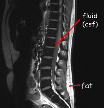
A T1-weighted image has a short TR and TE time.
Fat realigns its longitudinal magnetisation with Big Chungus SUPER QUICK. This means, on a T1-weighted image, it looks hyperintense. However, water realigns its longitudinal magnetisation with Big Chungus slooooowly. Therefore, on a T1-weighted image, fluid looks hypointense! Ya see?
A T2-weighted image has a long TR and TE time.
The precession of protons in fat decays relatively slow, so it will look quite bright on a T2-scan. But water decays slower, and therefore, by the time we take the T2 image, fluids within the body will be giving off comparatively ‘more’ signal than fat – meaning they’ll appear more hyperintense!
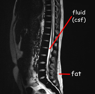
If we have a substance with intrinsically long T1 and T2 values, it will appear dark on a T1-weighted image and bright on a T2-weighted image, and the same in reverse. If a substance has a short T1 value and a long T2 value, it will appear relatively ‘bright’ on both T1 and T2-weighted images – i.e., fat and intervertebral discs.
As every tissue has its own distinct T1 and T2 property… we can work out precisely what sort of tissue we’re looking at.
When we build in all our additional sequences, this becomes even clearer! This is why your MRI scan takes sooooo long – we’re running SO MANY sequences, manipulating TR and TE to determine the exact T1 and T2 properties of various tissues within your bod.


There is, however, a problem.
The RF signal given off by each proton doesn’t shoot out in a handy-dandy straight line. Meaning, we have no idea where the signal is coming from within your body.
Enter our lord and saviour:
THE GRADIENT COILS.
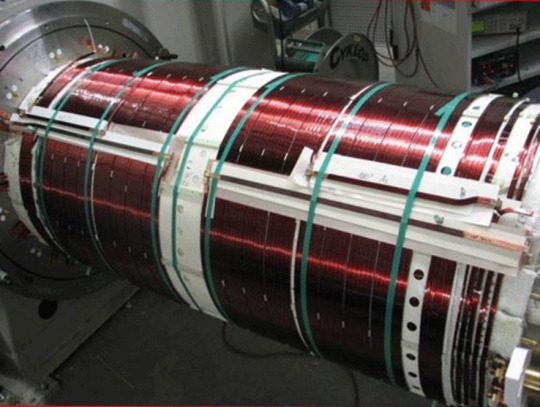
(Shim coils are also very important – they maintain field homogeneity across the whole of Big Chungus. While Big Chungus wouldn’t need them in a perfect theoretical scenario… reality ain’t that. Big Chungus’s magnetic field is all wibbly-wobbly, so we use Shims to keep everything smooth! That’s all you need to know about them. BACK TO THE GRADIENTS.)
There are three of them, wrapping around each of the three planes of your body. When these activate, they cause those epicly eerie booming noises, characteristic of a Big Chungus ExperienceTM.
youtube
The Gradient coils are also what causes those weird tingling sensations you get in an MRI machine – which, don’t worry, aren’t permanent! Your nerves just go ‘WOAHG. THASSALOT OF MAGNET SHIT. HM. DON’T LIKE THAT.’ But they’ll calm down again once you’re freed from Big Chungus.
The gradient coils cause constant fluctuations in the magnetic field across all three dimensions. They activate sequentially, isolating one chunk of your body after the next.
As these fluctuations cause variation within the signal received, we can look at how much THAT particular signal, received at THAT particular number of milliseconds after an RF pulse, varied when THAT particular gradient was activated, in comparison to when THAT OTHER gradient was activated.
For every single bit of signal output.
That gives us A WHOLE LOTTA DATA.

^ imagine this, but the cupboard contents is just. data.
Way too much data, in fact, for our puny human brains to comprehend – so obviously, we feed it to an algorithm.
K-space is a funky computational matrix where all this info gets compiled during data acquisition. Once we’ve finished the scan sequence and have all that yummy raw data, it can be mathematically processed to create a final image!
Just like that. Simple, right?

TL;DR
You are full of Hydrogen.
Hydrogen nuclei (protons) are basically tiny magnets
These tiny magnets are orientated completely randomly, with ‘North’ pointing in all directions
We stick billions of these tiny magnets (i.e., you) into a mahoosive magnet (i.e., Big Chungus)
All the tiny magnets flip around to align with the longitudinal magnetic field of Big Chungus
High energy protons = antiparallel Low energy protons = parallel
As you have more low energy protons than high energy protons in your body, the net magnetic vector of your body is longitudinal – just like Big Chungus!
All your protons are spinning and wobbling (precessing) at random rates
We use an RF pulse, tuned to the Resonance Frequency of Hydrogen, to make ‘em precess in phase (wobble at the same time, all pointing in the same direction at once). This creates a Transverse magnetic vector.
This in-phase precession is ‘Magnetic Resonance’
Magnetic Resonance means the protons can absorb energy from the RF pulse
Now there are more high energy protons within your body! They flip antiparallel, and the net longitudinal magnetic vector of your body decreases.
We measure the time it takes for the high-energy protons to release that energy and return to alignment with the net magnetic vector of Big Chungus (Spin-Lattice Relaxation / T1 recovery)
And the time it takes for the precessing-in-phase protons to Quit That Nonsense and all start wobbling in random directions again (Spin-Spin Decay / T2 recovery)
Each tissue within your body has a different composition & density of Hydrogen atoms – which means each tissue within your body has a unique T1 & T2 recovery time
By measuring the signal at different times (TE) and by varying the frequency with which we apply RF pulses (TR), we ‘take pictures’ that show variations in the amount of signal these tissues are giving off. The signal is caught by the large radiofrequency receiver coils we put over you when you enter the machine.
Because the signal given off during recovery/decay blasts out in all directions, we don’t know exactly where it originated within your body.
Gradient coils are arranged across X, Y, and Z axes throughout the gantry of Big Chungus. They cause tiny fluctuations in the magnetic field, in sequential chunks throughout space. This is the booming noise you hear when you’re in the machine.
These tiny fluctuations cause variations in the signal we receive, depending on how close the signal is to the activated gradient coil. All this data is compiled in a magical computational matrix called K-space. A funky algorithm then decodes those variations and couples them up with the strength of the signal to give us 1) How much signal is being blasted out at that particular moment 2) Where exactly that signal comes from within your body, according to the 3D map produced by the gradient coils
It then represents these values with a pretty picture!
Tl;dr tl;dr:

74 notes
·
View notes
Text
“And this is when we open the patient file.”
Radiologist when they have no idea what is going on in an image.
60 notes
·
View notes
Text

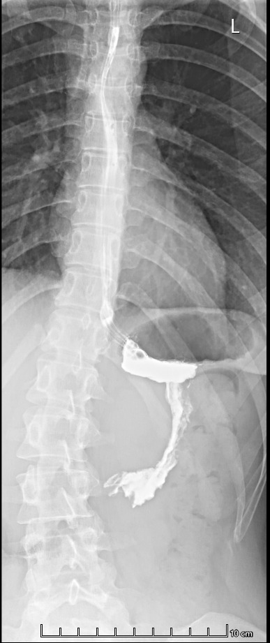
chat am i cooked
#xrays#medical imaging#i sorta just want to know if my spine is supposed to look like that....#it me#ignore the goo in my stomach#it tasted feral#also i was standing against a flat surface like pressed into it
12 notes
·
View notes
Text

Scan Do Attitude
Being able to monitor brain changes in mild cognitive impairment or Alzheimer's disease at point-of-care is desirable but, because of their necessarily lower magnetic field strength, portable MRI scanners have resolution limitations due to a lower signal-to-noise ratio. Here, combination with a machine learning pipeline overcomes disadvantages and renders the portable system more freely available and cost-effective
Read the published research article here
Selection from an image from work by Annabel J. Sorby-Adams and colleagues
Department of Neurology, Massachusetts General Hospital and Harvard Medical School, Boston, MA, USA
Image originally published with a Creative Commons Attribution – NonCommercial – NoDerivs (CC BY-NC-ND 4.0)
Published in Nature Communications, December 2024
You can also follow BPoD on Instagram, Twitter and Facebook
#science#biomedicine#biology#brain#neuroscience#neurodegeneration#alzheimer's#mri#brain scan#medical imaging
12 notes
·
View notes
Text
Ouuhhhh the medical tape wrap, you just KNOW he wriggles
#the wriggler#animals#get well soon#at the vet#medical imaging#animal memes#vet#radiography#animalsgettingmedicalimaging#under the weather#pets#cat#silly cat#cat memes#cats#cats of tumblr
44 notes
·
View notes
Text
I wanna make a fun little online lesson about radiography!
if a 'tell me in the comments' option wins, I'll pick the most popular answer from the comments! If anyone actually replies lololol
70 notes
·
View notes
Text
Harrison.ai raises $112 million to expand AI-powered medical diagnostics globally

- By InnoNurse Staff -
Harrison.ai, a Sydney, Australia-based startup, develops AI-powered diagnostic software for radiology and pathology to enhance disease detection and streamline workflows for healthcare professionals.
The company recently secured $112 million in Series C funding to expand internationally, with plans to establish a presence in Boston.
Founded in 2018 by brothers Dr. Aengus Tran and Dimitry Tran, Harrison.ai has launched Annalise.ai for radiology and Franklin.ai for pathology, aiming to address global clinician shortages and improve patient outcomes. Its products are deployed in over 1,000 healthcare facilities across 15 countries, with regulatory clearance in 40 markets, including 12 FDA approvals in the U.S.
The startup differentiates itself from competitors like Aidoc and Rad AI through its broader diagnostic capabilities and extensive datasets. Its AI models detect lung cancer earlier and outperform standard radiology exams. With its latest funding, Harrison.ai plans to expand AI automation beyond radiology and pathology.
Read more at TechCrunch
///
Other recent news and insights
Advanced imaging and AI detect smoking-related toxins in placenta samples (Rice University/Medical Xpress)
Study suggests AI may outperform humans in analyzing long-term ECG recordings (Lund University)
#ai#radiology#medtech#harrison ai#imaging#medical imaging#australia#health tech#pathology#automation#diagnostics
5 notes
·
View notes
Text

source
#op#medcore#medical aesthetic#hospitalcore#operating room#medical imaging#radiology#medical equipment#c arm#hospital
2 notes
·
View notes
Text
Revelation Revolution
Whole-body positron emission tomography (PET) one hour after injection of radioactive glucose highlights rapidly metabolising tissue such as the liver metastases of a colorectal tumour seen here within the abdomen, along with normal accumulation of the tracer in the heart, bladder, kidneys and brain
Edward J Hoffman – born on this day, January 1st in 1942 – along with Michel Ter-Pogossian and Michael E. Phelps – developed the first human Positron Emission Tomography scanner in 1973
Movie adapted from PET scan GIF created by Jens Maus
Video in the Public Domain
You can also follow BPoD on Instagram, Twitter and Facebook
#science#biomedicine#biology#medical imaging#colorectal cancer#metastasis#liver#PET scan#positron emission tomography#born on this day
20 notes
·
View notes
Text
Laparoscopic exploratory surgery pics below the cut!

Wish they'd given me an actual CD with the pictures but pics of the print out is the best I can do for now. Can you spot the problem?

There she is.

It takes up more space than I thought. But it's still wild how something seemingly so small can be disabling. Gotta have a coping game plan going forward, because it's gonna probably take 6+ months until surgery #2 to get this bitch out of me for good. :') Just made a medical binder for all future appointments, still have a lot more to print out and organize.
#adenomyosis#endometriosis#laparoscopy#chronic illness#chronic pain#teku#medical imaging#new’s first surgery
8 notes
·
View notes
Text
Applications of GenAI in Healthcare

Generative AI in Medical Imaging revolutionizes healthcare practices by streamlining various processes. In diagnostics, Generative AI enhances the accuracy of disease identification through detailed image analysis, leading to quicker treatment decisions and improved patient outcomes.
Moreover, this technology facilitates personalized treatment plans tailored to individual patients based on comprehensive imaging data. Beyond diagnostics, Generative AI assists in drug discovery by simulating molecular structures, accelerating research efforts to develop new treatments. Its applications extend to improving operational efficiency in healthcare settings, optimizing resource allocation, and workflow management.
By integrating Generative AI into healthcare systems, providers can enhance patient care, drive innovation, and pave the way for transformative advancements in medical practices.
For more in-depth insights on the transformative applications of Generative AI in healthcare, explore:
#generative ai#healthcare#generative ai in healthcare#medical imaging#medical technology#future of healthcare#technology#technologies#healthcare technology
4 notes
·
View notes
Text

The tightly taped totem
#hes being very brave#radiography#get well soon#at the vet#cats#kittens#kitten#animals#vet#medical imaging#animal memes#animalsgettingmedicalimaging#under the weather#pets
32 notes
·
View notes
Text
memes for the tired rad student





34 notes
·
View notes
Text
Researchers use AI to uncover sex-specific risk factors for aggressive brain cancer

- By InnoNurse Staff -
Researchers from the University of Wisconsin–Madison, led by Professor Pallavi Tiwari, are using AI to uncover risk factors for glioblastoma, a highly aggressive brain cancer, and how they vary between men and women.
Glioblastoma is more common and aggressive in men, but identifying patterns that predict tumor behavior has been challenging. Tiwari and her team have developed an AI model that analyzes pathology slides to identify subtle tumor characteristics linked to patient survival, while accounting for sex differences.
The study found that for women, tumors infiltrating healthy tissue were linked to worse outcomes, while in men, the presence of certain cells (pseudopalisading cells) indicated more aggressive tumors. This AI-driven research aims to enable more personalized cancer care and inspire new treatment approaches. Tiwari’s work extends beyond glioblastoma, with AI applications in analyzing pancreatic and breast cancers, and she is helping shape UW–Madison’s AI and health initiatives.

Image: These images display regions of glioblastoma tumors in females (top) and males (bottom) where the AI models predict the presence of higher and lower risk characteristics. Red areas indicate higher risk, while blue areas represent lower risk. Credit: Tiwari Lab / UW–Madison.
Read more at UW–Madison
2 notes
·
View notes