#peptidoglycan
Explore tagged Tumblr posts
Text
Watch what happens to Germs when you wash your hands with Soap at microscopic level. 🔬 The Soap molecules surround germ cells and disrupt their cell walls, causing them to burst.
Germ cells are surrounded by a cell wall that protects them from the environment. This cell wall is made up of a layer of peptidoglycan, which is a polymer of amino acids and sugars. Soap molecules are made up of two parts: a hydrophobic (water-fearing) tail and a hydrophilic (water-loving) head. When soap is added to water, the hydrophobic tails group together and the hydrophilic heads face outward, forming micelles. These micelles can surround germ cells and the hydrophobic tails can then disrupt the cell walls, causing the cells to burst.
The hydrophobic tails of the soap molecules can disrupt the cell wall in two ways. First, they can bind to the peptidoglycan molecules and weaken the bonds between them. Second, they can create holes in the cell wall. Once the cell wall is disrupted, the germ cells lose their internal contents and die.
It is important to note that soap only works to kill germ cells that are surrounded by a cell wall. Germ cells that do not have a cell wall, such as viruses, are not affected by soap.
The size of the soap micelles is important. Micelles that are too small will not be able to surround the germ cells. Micelles that are too large will not be able to penetrate the cell walls.
The concentration of soap is also important. A higher concentration of soap will be more effective at killing germ cells.
The temperature of the water can also affect the effectiveness of soap. Soap is more effective at killing germ cells in warm water than in cold water.
I hope this post has helped you understand the importance of handwashing and why doctors always ask you to do it regularly. Washing your hands with soap and water for at least 20 seconds is one of the best ways to prevent the spread of germs and stay healthy. So please, wash your hands often and help keep yourself and others safe!
Thank you for reading this post. I hope you found it informative and helpful. Please share it with your friends and family so they can learn about the importance of handwashing too. 😊🙏
#soap#germs#cellwall#science#biology#micelles#peptidoglycan#mechanism#explanation#education#health#safety
1K notes
·
View notes
Text

TSRNOSS. Page 240.
#blood testosterone level#resistance weight training#protein intake#calcium deficiency#vitiligo#psoralen#gut bacteria#gram-negative bacteria#viscosity#peptidoglycan#lysozyme#histones#toxicity#satyendra sunkavally#vitamin A overdose#ultrasound#effect on bacteria#Le Chatelier's Principle#fructose#diabetes#growth hormone#herbivory#vitamin D synthesis#anti-herbivore defence
0 notes
Text
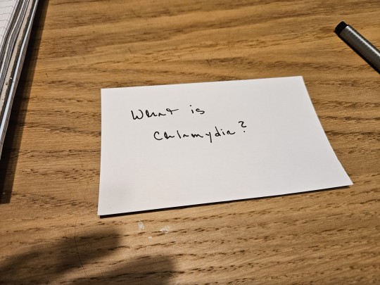
Asking the real questions
#i'm making flash cards for my microbiology test lol#for those curious: it's an obligate intracellular parasite#gram negative#and has no peptidoglycan#there is nothing else anyone needs to know about chlamydia#oscar talks to himself#school blogging
5 notes
·
View notes
Note
68, 82
satan's last name? i didn't know that was a thing people wondered about (aka: i have no clue)
favorite word? right now i think its smithereens or peptidoglycan. they're just very fun words to say
#peptidoglycan is pretty much waht helps differentiate between gram + and gram - cells fyi#heard it in epidemiology and have never stopped saying it#em rambles#chaos !!#thank you for the ask :)#ask game
0 notes
Text
VOTE BACILLOTA!
Bacillota vs. Fusobacteriota
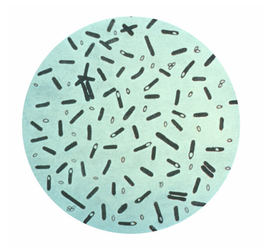

Bacillota propaganda here
Fusobacteriota propaganda here
#bacillota definitely has peptidoglycan btw#the confusion might be that their spores don't?#but regular vegetative cells do#bacteria showdown
36 notes
·
View notes
Text
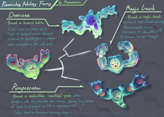
when i saw @n0rtist's ability forms i had to do reuniclus.....i hope it's not too late to be featured in the video! :0 we may have crossed from ability form into actual alternate form territory (see the original colours below the cut to see how crazy different they could have been)
as someone in evo microbio this was insane amounts of fun to do and i was able to pull inspo from places i'd never think to reference in my regular art!
itemized essay explaining the science and my design choices in more detail below the cut:
Ability Form 1: Overcoat
The science
Based on bacterial biofilm, which is basically when a bunch of bacteria get together in the same spot and start excreting this sticky slime stuff that structurally keeps the bacteria together (and also act as a medium for sharing useful resources between the bacteria, like enzymes, nutrients, etc.).
The design
The strings connecting the bacteria is based on what the slime stuff looks like on a microscopic level, specifically in electron micrographs like this one
The violet colour of the (rod) bacteria in the biofilm is a reference to Gram staining, which is a type of bacterial stain used to classify bacteria into two groups: bacteria that contain peptidoglycan in their cell walls (which stain violet) and bacteria that don't (these stain pink). Peptidoglycan is this pretty chonky polymer compound that's used to strengthen bacterial cell walls, so I thought it fit the role of Overcoat (which protects your Pokémon from things like weather)
Ability Form 2: Magic Guard
The science
Based on the myelin sheath, which are these segments of tube-like insulation that surrounds your neurons (see picture here). Mostly people talk about how it makes your neural signals propagate faster, which is true, but this ability form was more of a reference to its general protective role; it physically and electrically insulates your neurons. (Surprisingly I could not find a super good primary source for myelin providing physical protection, so don't cite me on that, but given it's literally a physical barrier this seems like a pretty safe assumption.)
The design
The entire body is based on a pseudounipolar neuron, which just means it only has one part extend out of the main body but shortly after it splits into two long parts (axons). I could have made it a bipolar neuron I guess (two parts extend out of the main body), but having a neck made it look a little closer to the base form's body
I wanted to give it dendrite fingers but it looked too creepy. I'm not sure if the three long fingers I gave it in the final design made it less creepy.
Since Overcoat and Magic Guard are both shield-type abilities, they're drawn as more closely related on the the phylogenetic tree (white thing in the center).
Ability Form 3: Regenerator
The science
This one is the most hype imo. In 2015 Dr. Will Ratcliff did this pretty sick experiment about the evolution of multicellularity (since at some point a long time ago life was single-celled) where he kept propagating the same single-celled yeast for a mega long time, and eventually the yeast evolves a multi-cellular "snowflake" form where after undergoing mitosis, the resulting daughter cells don't split, but stay attached, resulting in the yeast forming these clusters that create these cute little branches. I don't know where I was going with this. Oh right, the branching out reminded me a lot of regenerating limbs, so that was the inspiration for this one.
Anyway this is like one of my favourite experiments ever, there's some pretty good news articles out there about if you want to learn more about it!
The design
The segments are yeast cells, and the balls within are the nuclei.
The colour scheme was based on this fluorescent microscopy photo of the snowflake yeast. Originally I had the nuclei be bright orange in reference to this other microscopy picture but I thought the colour scheme was deviating too much from the base form for an ability form lol.
Speaking of here's the original unhinged colour drafts:
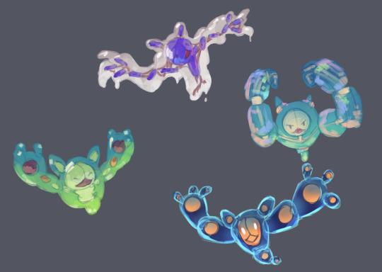
if i did commit to the full alternate form i think the biofilm one is poison, myelin one is fighting, yeast one is uhh...dude idek, i mean the fluorescent microscopy vibe is pretty strong so maybe electric lol? it's giving ghost vibe too though
i was originally planning on citing stuff but it's a tumblr post and I've already linked Ratcliff's work and i want to go to bed lol. If any of the science is wrong just call me out i'll fix it. otherwise, i hope someone out there appreciates the science references !! it's 8am good Night
#art tag#biology#evolutionary biology#microbiology#reuniclus#pokemon#ability forms#biology art#i guess#bacteria#evolution#nintendo#pokemon fanart#pokemon gen 5
371 notes
·
View notes
Text
Yapping about leprosy's biology because I've yapped about everything BUT the bacteria itself
-
Leprosy is caused by 2 bacteria in humans; Mycobacterium leprae and Mycobacterium lepromatosis. The mycobacterium genus is a group of actinomycetota bacteria, the latter being known for their high guanine + cytosine content in their DNA and the former for their mycolic acid.
Bacteria are usually separated in two groups; gram positive and gram negative, positive bacteria have a thicker peptidoglycan layer, which is kind of like.. the 'skin' of a bacteria, while negative bacteria have a thinner peptidoglycan layer but they have an outer lipid wall that has lipopolyssacharides (LPS). LPS aids bacteria to recognize and bind to specific receptors, usually, a pathogen will be a gram-negative bacterias they are more adapted to recognize other cells but this isn't set in stone.
Mycobacteria however are neither of those, they have an outer wall that covers their entire body called 'mycolic acid', mycolic acid are long chains of fatty acids, these acids are what makes mycobacteria so resistant against many hostile enviroments and attacks, it's kind like bacteria armor! Other than that, mycobacteria also have very intricate mating rituals, some that can even look analogous to how us humans mate, this is importante, as bacteria of actinomycetota have high G+C content, meaning their DNA is less vulnerable to mutations and is more 'stable', these bacteria have evolved ways to still be very genetically diverse despite their circumstances.
Leprosy is the odd one of the group of mycobacteria, intracellular bacteria are a novelty (there's only very few species of bacteria that intracellular), and it seems to have went through a massive gene reduction event. Whatever happened to leprosy years ago, it made it into the second most genetically polluted bacteria, bacteria are very good at discarding useless junk genes that they don't need and most will only have 5% pseudogenes at MAX, so leprosy having 40% pseudo genes was a huge finding that left so many people confused.
One of those defective genes, seems to be some very important pathways (that's what we call a chemical chain event that leads to specific results, metabolism is a pathway, for an ex;) one of them, is the metabolization of carbohydrates. Carbohydrates such as sugar are the default energy for every living species, a species not being able to metabolize carbohydrate is unheard of, despite this, leprosy lacks the ability to do so; Or better yet, we can see that it still has its anabolic pathways of carbohydrates, but its's catabolism is missing. In other words, leprosy can break down carbohydrates, but it doesn't know how to use it.
So where does leprosy gets its energy? Well, lipids! Lipids are the nuclear energy of the body, it can story much more energy in a smaller space, and it releases much more than carbohydrate. The only reason why we don't use it, it's that breaking down lipids takes a long time. Lipids are still very important though, they usually become fat, which is just your body's energy storage. Leprosy is particularly found of triaglyceride (hence my little blog banner...), because it has fatty acids (few carbohydrates that it can use) and it's also a type of lipid.
All of this sounds like a disadvantage, but I promise you it's not !! Since leprosy enjoys colder spots, that are usually away from the bloodstream, it gets less nutrients, but it can live on fat alone so it doesn't need to risk itself to find a food source. The metabolism of fat also takes a long time, making leprosy a very slow grower, this helps the bacteria stay hidden and live for longer periods of times as;
A population of bacteria that eats too much will eventually drain it's nutrition source and quickly die off
A population of bacteria that grows too quickly will attract the attention of the immune system in it's weakest stage
As intracellular parasites, leprosy needs to parasitize a cell, it usually does this to a type of cell called 'Schwann cells', I'll take about these guys in a bit but first; the bacteria inside Schwann cells can force it to make more lipids and glucose, since leprosy does not know how to build glucose after it breaks carbs down, it needs a cell to do it for it. Schwann cells essentially become havens for the bacteria, sometimes, when leprosy is comfortable enough, these cells will be reprogrammed into another type of cell, carrying the bacteria inside it to somewhere else.
As for Schwann cells, they are cells that myelinate the peripheral nerves, nerves need to have extremely fast response and electrical impulses, the way they do this is- My god, ok, sorry, I'm going to have to explain biophysics to you ok?
Electricity is just a bunch of eletrons running around, there are many types of electrical currents, but the ones you need to understand are constant and alternating.
A constant is... well, a constant electrical current, the energy level is stable and there is no oscilation or change.
An alternating current however, is one where the eletrons are changing their direction really quickly, this leads to 'peaks' of electricity. A constant current is more stable, but an alternating current is much more faster and quick-responding, it's the one you use in electrical grids in houses and the ones our nerves uses!

Here's a graph for easier understanding.
Now neurons do this (kind of) by having a 'myelin sheath' which is made by the Schwann cells, the myelin sheath insulates the neuron while the nodes of ranvier (those little spaces between myelin sheaths) are the ones that do the job, electricity jumps from node to node increasing the speed and efficiency in how information is transmitted. It's a bit more complex than simple electrical currents, we call this type a 'saltatory' current, as it looks like energy is 'jumping from one place to another'

This is how leprosy causes numbness and fucks your nervous system, it destroys the Schwann cells that are responsible for keeping the neurons and the electrical current alive. An unmyelated neuron is MUCH slower in transmitting information than a myelated one, and although the former exist, they are usually very short and small to make up for it.
Here's an unmyelated nerve vs and myelated one.

As leprosy destroy more schwann cells, the neurons die, causing nerve thickness. Neurons find themselves to be very comfy inside the epineurium, which protects them and categorizes them into 'bundles' called fascicles. (Which in turn are protected by the perineurium)

What surrounds those little guys, is pretty much just conective tissue. As neuron dies, more connective tissue grows in their place in order to protect the other neurons from impact.
It's important to remember that the nervous system is an immunoprivileged system, which means that the immune cells are not allowed to enter. It does have it's own defenses, but they're not quite prepared to deal with leprosy, the immune cells there only weapon is phagocytosis- the act of eating a bacteria and letting your "stomach" acid kill it, but like I said, leprosy has bacteria armor; it doesn't affect it.
I would love to get into leprosy and the skin but it's 2 am. I need to sleep, and god help me write a text post on the 3 billion different cells types in the skin. good night!
5 notes
·
View notes
Text
prokaryotes
hello! this is the first of (hopefully) many study posts. today i'm covering prokaryotes, specifically what they are and how they function. i am a student and not an expert in any of this, so please feel free to correct any incorrect information, or ask questions if you have anything you want clarified!
evolution
prokaryotes (organisms that make up domains bacteria and archaea) have incredible evolutionary success, which has allowed them to become the most widespread organisms on earth. their small size and insanely rapid reproduction rates is one trait that has allowed them this success - because of how quickly they can reproduce, there is a very short span of time between evolution. this plus frequent mutations equal super saiyan evolution speeds!
prokaryotes also have a wide range of adaptations, which allows them to live in a wide variety of environments, including very extreme ones.
so what are prokaryotes?
prokaryotes are single-celled organisms with no nucleus or membrane-bound organelles. as far as we know, they are the first organisms to inhabit earth. because they have been around for literally billions of years, they have incredible diversity.
prokaryotes are itty-bitty, even compared to eukaryotic cells. they typically range from 0.5-5 micrometers. one notable exception, however, is Thiomargarita namibiensis, which can be up to 750 micrometers (bigger than a poppy seed).
what do they look like??
there are three common shapes that prokaryotes come in: cocci (circular/ovular. little orb guys.), bacilli (rod shaped, think bacteria depictions in media. the beauty standard, if you will.), and spirilla (wiggly corkscrew guys. can also resemble commas or coils).
cocci can hang out alone or form chains with their buddies; sometimes they'll bunch up like grapes. bacilli like to be alone, but sometimes they'll form polite little lines with each other to make long rods. spirilla can be found alone or in chains.
cell surface structures
almost all prokaryotes have a cell wall, which holds its shape and prevents it from exploding into smithereens in hypotonic (low osmotic pressure) environments. prokaryotic cell walls are made of peptidoglycan. archaeabacteria are the weirdos in this, their cell walls are made of polysaccharides and proteins.
bacteria can be sorted by differences in their cell-wall, identified through a process called gram stain. i'll probably make a mini-post talking about gram stain and link it here when i do.
most prokaryotes also have some sort of sticky layer on the outside, called a capsule (or, if less organized, a slime layer). these, as you may have figured out, allow prokaryotes to stick to things. they can also prevent dehydration.
many prokaryotes are capable of taxis - this means they are able to purposefully move in a specific direction in response to external stimuli. the most common structure used for this is a flagellum, which look like little wiggly tails. if you've ever played spore you are probably familiar with these things, and just like in spore they can be pretty much anywhere on an organism.
internal structures
as mentioned above, prokaryotes do not have a nucleus or membrane bound organelles. they're actually pretty simple organisms overall. they usually only have one circular chromosome contained inside of their nucleoid (which is different from a nucleus). additionally, they have DNA molecules that can replicate independently called plasmids.
reproduction
most prokaryotes reproduce via binary fission, which means the cell doubles itself and then the doubles double themselves and then so on and so forth. think like the game 2048.
"but b!" you say, "what about genetic diversity?? if they're just making clones of themselves then there is none!" that is correct! that's where mutations come into play. since there is no exchange of alleles in asexual reproduction, prokaryotes heavily rely on random mutations.
there is another phenomenon that can occur called genetic recombination, which is when you mash together dna from two different organisms. "but... but there's no sex??" correct again! instead, transformation, conjugation, and transduction is how this swap happens. i may go into more detail about this in a mini-post - i will link it here if i do so.
i also wanna make a quick note about dna transfer between different species, which my textbook refers to as "horizontal gene transfer" which sound like one hell of a euphemism to me personally. (for clarification's sake, horizontal gene transfer is a scientific term).
metabolism
the are four major "modes" that prokaryotes acquire nutrition: photoautotroph, chemoautotroph, photoheterotroph, and chemoheterotroph. photo- means light, and chemo- means chemicals. this combined with autotrophs referring to organisms that can create their own food and heterotrophs referring to organism who source food externally makes it relatively simple to interpret these terms. a prokaryote may fall under one or multiple of these categories.
there are also differences in how prokaryotes interact with oxygen. obligate aerobes need oxygen to live and reproduce, while obligate anaerobes are poisoned by oxygen and must get chemical energy through other means.
often prokaryotic cells will work together to carry out metabolic processes that cannot be completes by a lone cell. as these cells band together they may form what is called a biofilm, which recruits even more cells and makes the colonies even bigger.
my next post will probably be an overview of prokaryotic phylogeny + diversity. i'm still figuring out how to format things, so i may try and break down subjects a bit more so i don't end up with such long posts.
#biology#prokaryotes#studyblr#b studies#i am not the biggest fan of microscopic organisms#but they're very important to learn about!#if you find them a bit boring i find that anthropomorphizing them can help make them more interesting
3 notes
·
View notes
Text
Okay so we all know antibiotics like penicillin kill bacteria right? Well what you might not know is that how it kills bacteria is it attacks the linkage of its cell wall which is made up of peptidoglycan. If it damages the walls enough the cell will lysis due to the osmotic pressure of its environment. I think it’s a good analogy of life! If you don’t take care of yourself and let your walls get damaged then the normal pressure you have been under then becomes to much and it can break you. So take care of yourself! Drink water!
2 notes
·
View notes
Text
guessed peptidoglycan for shared features of prokaryotic and eukaryotic cells. how could i have been so foolish...
#realistically she mentioned that like once and never brought it up again#i knew it was probably involved in the membrane i just didn't know what for#well one question wrong on the exam so far that's not so bad#txt
4 notes
·
View notes
Note
hello whats your fav prokaryote? <- is a biology major and cant remember any of them :head_in_hands:
oooOOOOHHH now THIS is a fantastic question, one that I will cheat at cuz I can't pick just one.
ok first and foremost: Bacillus anthracis. God what *cant* I say about anthracis, the exotoxins are simply fascinating, there's three whole types of anthrax that are caused by different ways of them getting into the body, one of the toxins is straight up called "Leathal Toxin" liek come ON. love it
I have to give a shout out to our boys the Mycoplasma tho like, they said "oh yall are basing most of all foundational bacterial classification on the amount of peptidoglycan in their cell walls? ight cool" *exists with no peptidoglycan at all*
and then, Pseudomonas aeruginosa, beloved and bane of my existence, such an important organism in the lab setting, couldn't have gotten through medical microbiology without her <3
#microbiology#thank you for the question it made me so happy :>#i could have a whole other post on viruses or prions too#biology
16 notes
·
View notes
Note
… tell me about Endospores.
I love mushrooms and spores and fungi, I want you to tell me an entire Wiki page
(this is totally not Shiek, who has not been checking every single notification to see if people liked the Reddit screenshot….. hi)
Hello!! I fuckin love endospores and ur the first person to actually send an ask about them
Endospores are actually a type of bacteria! Some environmental bacilli produce an endospore coating to withstand extremely harsh environments and is usually initiated by total nutrient deprivation (think of an absence of nitrogen). That being said, the bacteria in our bodies won’t undergo this process, but finding out whether an endospore bacterium is introduced to your body is pretty important because endospores require very specific treatments given their nature. For example, bacillus anthracis (my favorite example) - this bacterium is responsible for anthrax infections, the only effective treatment being an antibiotic called Cipro.
To (sorta) briefly explain how endospores are formed - when environmental conditions are no longer viable to sustain life, the bacterium begins an 8 hour endosporulation process in which the cells DNA is duplicated and a septal wall is created that asymmetrically divides the cell, and a spore coat is formed around the forespore and there is a thick peptidoglycan cortex formed between the layers of the two halves of the cell. Once the sporulation process is completed, the newly matured endospore is released once the rest of the vegetative cell is degraded. The cortex is basically what allows the endospore to be highly resistant against high temperatures and enzymes and most chemicals that are otherwise effective against other bacteria.
This is why autoclaving is one of the most effective ways of disinfecting surfaces! Otherwise, sterilant alkylating agents, like ETO and household bleach are effective (since not everyone owns an autoclave but a girl can dream). Household bleach must be in contact with endospores for a minimum of 10 minutes for it to actually kill the bacteria; increasing the concentration instead will NOT be effective, it may actually cause bacteria to aggregate and survive. Ionizing radiation (e.g., x-rays, gamma rays) is also effective against most endospores!
Now one of the coolest facts about endospores imo - the endospore is PROTECTIVE for the bacteria in these really harsh, nitrogen depleted environments, right? So for the bacteria to survive, it will actually be in a dormant state until either environmental conditions improve or they’re chemically stimulated to undergo activation, germination, and outgrowth. This means they can lie dormant for MILLIONS of YEARS - one endospore was actually found encased in amber and was actually reactivated.
Also,
Reddit screenshot u say?
#answered asks#microbiology#endospores#sorry for the long response but I fucking love endospores#screaming into the void
2 notes
·
View notes
Text
name a more annoying power couple than peptidoglycans and lipopolysaccharides. peptidoglycans are responsible for disease symptoms and lipopolysaccharides resist the entry of some antibiotics. god forbid anyone mess with its queen
5 notes
·
View notes
Text
I’ve got that dog in me. That peptidoglycan layer on all the bacteria cells in my gut
4 notes
·
View notes
Text
Gram-positive vs. Gram-negative bacteria
a.k.a. the inspiration for this blog's icon
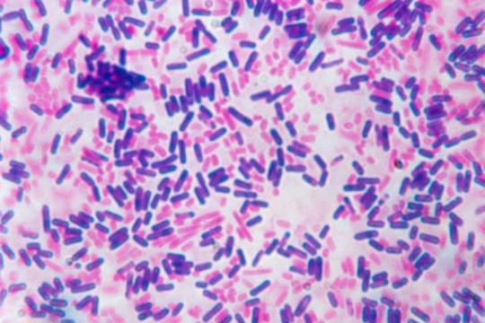
When researching your favorite species of bacteria, the first characteristic you're guaranteed to find is whether or not your bacteria is gram-positive or gram-negative. This is in reference to the Gram stain, a method of staining bacteria with dye, such that certain bacteria retain it and become purple (gram-positive), while others do not -- and after an alcohol wash, can be tinted pink with a counterstain (gram-negative). This staining procedure is almost always the first step in figuring out what kind of bacteria you're working with, and is used constantly for both clinical and research purposes.
Okay, but why? And why does the color of a dyed bacterium even matter?
The answer to these questions... lies in the structure of the cell wall. Gram-positive bacteria retain dye because their cell walls are much thicker. The typical gram-positive bacteria has one cell membrane, covered by a thick layer of peptidoglycan, which is a polysaccharide that gives structural strength to the cell. In contrast, the typical gram-negative bacteria has two cell membranes, separated by a thin layer of peptidoglycan: this layered structure is thinner and more flexible. Gram-negative bacteria can be counterstained because this outer membrane is porous, and is degraded by the alcohol wash, losing its ability to retain the original dye.
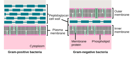
This difference in cell structure may not seem earth-shattering, but it does have some serious implications. For example, in medicine, you want to know if you're dealing with a gram-negative bacteria, because they are less susceptible to antibiotics that target the cell wall (such as penicillin): in essence, the outer membrane protects the cell. Penicillin can easily fit between the gaps in the peptidoglycan layer surrounding gram-positive cells, but not through the outer membrane of gram-negative cells, which selectively blocks the passage of toxic substances. In general, gram-negative bacteria are less vulnerable to environmental toxins.
Gram-positive bacteria have their own strengths. In fact, they have an extremely powerful tool at their disposal: unlike (almost all) gram-negative bacteria, some gram-positive bacteria are capable of forming endospores. Endospore formation is an intense survival mechanism where a bacterium creates a dormant structure inside itself (called an endospore), which is extremely durable but cannot self-replicate. When conditions have improved, the endospore transforms into a bacterium, and begins to metabolize and reproduce. Endospores can lie dormant for thousands -- and some argue, millions -- of years, and remain viable.
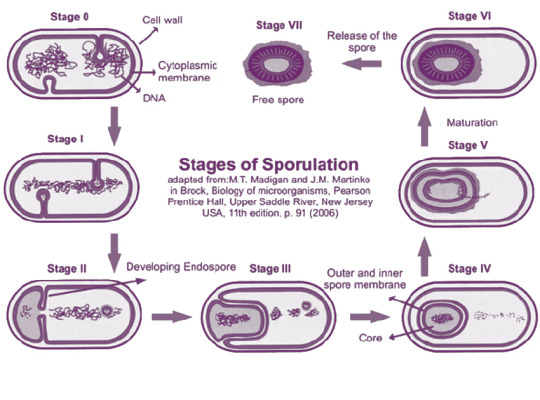
To summarize: what at first may seem like a simple question of microbial dye-jobs is actually a way to peer into the structure of a bacteria, and hopefully start to unravel the unique strategies and adaptations that the species employs for survival.
Happy Gram staining everyone!
267 notes
·
View notes
Text
Unironically eggnog can be used as a disinfectant if you've made it a certain way
Raw eggs contain disinfecting lysozyme in huge amounts, which protect the inside from bacteria during gestation. Alcohol can emulsify these and aside from its obvious antimicrobial properties it can help with the hydrolysis of the peptidoglycan wall
time's running out we need to pour eggnog on your wound before it gets infected
466 notes
·
View notes