#vascular lesions
Explore tagged Tumblr posts
Text
Many individuals with visible vascular lesions are sensitive about their marks and are increasingly seeking a clearer complexion.
Oxyderm Laser Clinic provides service for all skin types with no downtime.
#treatment#skin treatment#laser clinic#skincare#vascular lesions#laser treatment & services in edmonton#oxyderm laser clinic
0 notes
Text
Discover moments of beauty and rejuvenation at Bali Surgical Medical Spa! In Charleston, WV, our skilled professionals offer a wide range of services including Botox injections, cheek augmentation surgery, laser vascular lesion removal, and life-changing fat transfer procedures. Bid farewell to arm insecurities with our exceptional arm lift procedure. Treat yourself to a new you!
#botox charleston wv#cheek augmentation surgery#laser vascular lesion removal#fat transfer procedures#arm lift procedure
0 notes
Text
Breast cancer appears differently on ultrasound depending on various factors, including the tumor's size, shape, and location. On ultrasound images, breast cancer may appear as a hypoechoic mass, meaning it appears darker than surrounding tissues, or as an irregularly shaped lesion with indistinct borders. Other features such as macrocalcifications, increased vascularity (more blood flow), and posterior acoustic shadowing (sound blockage behind the mass) can also be indicative of breast cancer. Early detection through ultrasound screening plays a crucial role in improving breast cancer outcomes, as it allows for timely diagnosis and treatment.
#Breast cancer#Ultrasound#Hypoechoic mass#Irregular lesion#Vascularity#Acoustic shadowing#Early detection
0 notes
Text
Check Out The Superior And Best Skin Clinic Near Me In Australia
Are you tired of searching for the best skin clinic? Look no further than Medi Grace Cosmetic! Our team of expert cosmetic physicians will help you achieve beautiful, glowing skin with safe and effective solutions tailored to your unique needs. We are committed to providing our patients with the best possible care. You feel confident about your skin, whether it's a simple treatment or an utterly cosmetic procedure. Effective and safe therapies should always be available at an affordable price. Do not hesitate to contact us for more details about the best skin clinic near me.

#skin booster treatment#facial hair removal melbourne#platelet rich plasma prp injections#dr murad cosmetics#face hydration treatment#melasma treatment melbourne#best tattoo removal melbourne#silhouette soft thread lift near me#tattoo removal treatment near me#ultraformer 3#best skin clinic near me#vascular lesion treatment#thread face lift near me
0 notes
Link
#Skin Care clinic Victoria bc#Skin rejuvenation Victoria BC#Face & Neck Lift Victoria BC#Skin Tightening Victoria BC#Rosacea and vascular lesions treatment
0 notes
Text

If you are looking to get the Best Treatment for Vascular Lesion in Waterloo, then contact Bhullar’s Laser Medspa. We're a family owned business operating for seven years, providing you with results that last! We specialize in hair removal, acne scar treatment, skin rejuvenation, skin tightening. Visit:- https://goo.gl/maps/WZJstzpFaYaZaMMs8
1 note
·
View note
Text
Small health update; I was diagnosed (with 99% probability) with autoimmune thyroiditis (Hashimoto's disease), which means my own thyroid wants to eat itself, slowly degenerating, +a small tumor to biopsy, so not the best of news.
I try to focus on other things but my recent MRI also points out not only to herniated discs in my neck but also some barely noticeable vascular lesion and cortical athropy, GP said I have to wait for my neurologist to give a final statement on what or if there is something wrong. Because something is, but she said it's most likely idiopathic and is beyond her GP understanding, so I have to wait for a month now and see what gives😔
It is not as a terrible diagnosis as of now, but it is just the knowledge that a part of your body is slowly going broken (making half of your body bonkers) and can't be fixed, it's rather depressive and doesn't help my anxiety much. But at least I know with what I'm dealing with now, most of my family members have issues with thyroid gland (genetics and Chornobyl effect I say). It's just, I hoped it would develop later, but such is life I guess.

Okay, rant over, wish me luck with further tests✌️
#personal#health matters#hola thyroiditis#time for some major dietary changes :v#still MRI was a big win with my anxiety battle
35 notes
·
View notes
Text
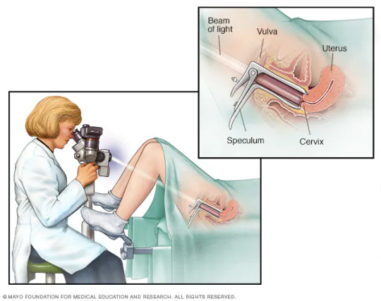
did you guys know there was a procedure where you shoot a beam of light into the vagina and look at the cervix through a microscope
A colposcope is used to identify visible clues suggestive of abnormal tissue. It functions as a lighted binocular or monocular microscope to magnify the view of the cervix, vagina, and vulvar surface. Low magnification (2× to 6×) may be used to obtain a general impression of the surface architecture. 8× to 25× magnification are utilized to evaluate the vagina and cervix. High magnification together with green filter is often used to identify certain vascular patterns that may indicate the presence of more advanced pre-cancerous or cancerous lesions.

19 notes
·
View notes
Text

Are you searching for “laser treatment near me” or “laser treatment services near me”? Look no further! New York offers a plethora of advanced laser treatment services to cater to various skin and medical needs. Whether you’re dealing with unwanted hair, skin imperfections, or medical conditions, laser treatments provide effective and non-invasive solutions.
What is Laser Treatment? Laser treatment involves using concentrated light beams to target specific areas of the body. These treatments can address a wide range of issues, from cosmetic concerns like hair removal and skin resurfacing to medical conditions such as varicose veins and skin lesions. The precision of laser technology allows for targeted treatment, minimizing damage to surrounding tissues and promoting faster recovery times.
2. Popular Laser Treatment Services in New York Laser Hair Removal:
Say goodbye to razors and waxing! Laser hair removal is a popular service for those looking to permanently reduce hair growth. Using laser technology, hair follicles are targeted and destroyed, preventing future hair growth. This service is ideal for areas like the face, legs, underarms, and bikini line.
3. Skin Resurfacing:
For individuals struggling with acne scars, wrinkles, or sun damage, laser skin resurfacing can rejuvenate the skin’s appearance. This treatment removes the outer layers of damaged skin, revealing the fresh, new skin underneath. The result is a smoother, more youthful complexion.
4. Tattoo Removal:
Regretting that old tattoo? Laser tattoo removal is an effective way to fade or completely remove unwanted tattoos. The laser breaks down the ink particles, which are then absorbed and eliminated by the body’s natural processes.
5. Treatment of Vascular Lesions:
Lasers can effectively treat vascular lesions, such as spider veins and port-wine stains. The laser targets the blood vessels, causing them to collapse and fade from view. This treatment can improve both appearance and comfort.
6. Pigmentation Treatments:
Hyperpigmentation, age spots, and melasma can be treated with laser therapy. The laser targets melanin in the skin, breaking up the pigment and evening out skin tone. This service is particularly popular among those seeking a more uniform complexion.
Why Choose Laser Treatment Services in New York? New York is home to some of the best medical and cosmetic laser treatment providers in the country. The city’s clinics are equipped with the latest technology and staffed by highly trained professionals. When you search for “laser treatment services in New York,” you’ll find a wide range of options tailored to your specific needs.
Benefits of Laser Treatments Precision: Laser treatments offer high precision, targeting specific areas without affecting the surrounding tissues. Minimal Downtime: Most laser treatments have minimal recovery times, allowing you to return to your daily activities quickly. Long-Lasting Results: Many laser treatments provide permanent or long-lasting results, reducing the need for repeated procedures. Non-Invasive: Laser treatments are typically non-invasive, meaning no surgical incisions are required.
Conclusion
If you’re looking to enhance your appearance or address a specific medical condition, consider exploring the various laser treatment services in New York. With advanced technology and expert care, you can achieve the results you desire. Don’t hesitate to search for “laser treatment near me” or “laser treatment services near me” to find a reputable clinic in your area. Whether it’s for hair removal, skin rejuvenation, or medical treatment, laser services offer a safe and effective solution for a variety of needs. Experience the benefits of laser treatments and take the first step towards a more confident you.
3 notes
·
View notes
Text
join me in pell (piss hell)
Let's talk kidneys!
Your kidneys are situated:
Inferior to the liver and the suprarenal glands
Superior to the ureters
Anterior to the posterior wall of abdomen and diaphragm
Posterior to the peritoneum (sack with yer guts in it)
Their job is to:
Regulate blood ions (like sodium) and control blood pH
Maintain blood volume (by extracting or conserving water)
Secrete hormones
Excrete toxic waste (urea, ammonia, creatinine…)
Guess what shape they are. Go on, guess.
YEAH THAT’s RIGHt – IT’S BEAN TIME, BITCHEs

[CW: beneath the cut you will find CT images of kidney trauma]
(and here is some very basic anatomy, sketched on… that same bean)
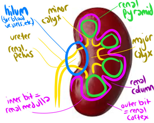
The renal cortex + renal pyramids together form the PARENCHYMA, aka the functional bit of the kidneys (aka where your peepee is made)
But HOW is that peepee made, I hear you cry?
Lemme introduce you to my good friend
The Nephron
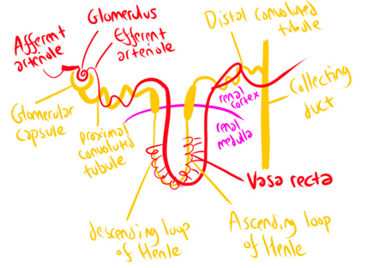
The afferent arteriole carries blood to the Glomerulus (which isn’t actually some weird DnD spell – just a knot of arteries surrounded by the Glomerular Capsule!) This arteriole then slims down considerably to form the efferent arteriole. This pressure increase forces loads of waste products and water out of the bloodstream into the glomerular capsule – but the holes in the arteriole wall are too small to release blood cells, plasma proteins, and other large molecules. This part of the nephron is called the ‘corpuscle’ (again, not a DnD spell). It’s where your blood plasma gets filtered!
The arteriole then follows the nephron around its windy path, wrapping around it at several points – notably the proximal/distal convoluted tubules, and the Vasa Recta that runs parallel to the Loop of Henle. To horrifically simplify a complex process, this provides lots of opportunities for secretion (Bad Stuff to be squeezed out of the blood – those dangerous ions and waste products we talked about earlier!) and selective reabsorption (Good Stuff (water) gets squeezed back in). It’s a careful balancing act, orchestrated in part by hormones! The end result (theoretically) is that all the stuff you DON’T want is shlorped into the nephron as urine, and all the water you need is shlorped back into the blood.
Once your kidneys have produced your peepee, it takes a fun rollercoaster ride through a series of ducts and tubes! Collecting duct -> papillary duct -> minor calyx -> major calyx -> renal pelvis -> ureter -> urinary bladder -> urethra -> you know the rest.
Your kidneys produce 180 litres of fluid a day (aka, a hell of a lot) but most of this is reabsorbed in these little nephrons, with water & useful solutes going back into the bloodstream! As a result, you only pee about 1-2 litres a day (though I swear I feel closer to the 180 litres some days)
Because kidneys are SOOOO important (your body does NOT like to be full of urea/ammonia/sodium, or acid!) they’re really, really vascular (lots of blood supply). They receive up to 25% of your resting cardiac output! So, when you’re just chilling, literally 25% of your blood is being gobbled by those hungry, hungry kidneys!
This means the kidney is VULNERABLE TO TRAUMA.
Although kidney trauma can be picked up on Ultrasound, we will take anyone who has suffered abdominal trauma through to CT, as you get better pictures there! We usually use a multiphase protocol – a longer scan, basically – to show us the extent of the injury, with a non-contrast phase (shows calculi clearly), an arterial phase (evaluates any injury to the renal arteries), a nephographic phase (shows renal lesions clearly), and a delayed phase (shows bleeding and injuries to the urinary collection system). Basically, contrast quickly moves to your kidneys from your blood stream, and filters through the collection system – so if we give a bolus of contrast and watch it flood through the renal arteries, then wait a little while, we can see how the kidneys are processing it or if it’s spilling into the surrounding space.
Kidney trauma is graded from 1 (no laceration but a haematoma (bruise) within the kidney capsule) to 5 (kidney torn away from renal vascular system and dying as a result, actively bleeding, structure of kidney shattered). Here’s a grade 5 (Left (looks like the right side of the image)) in comparison to the normal healthy kidney (Right (looks like the left side of the image)). Note the massive visible laceration + huge haematoma!
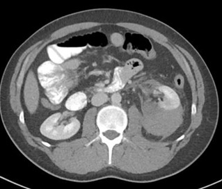

Loooooads of other stuff can go wrong with your kidneys too – but that’s a whole other post! Which I will make, one day soon, because it's super fascinating!
(Have you ever heard of a stag horn calculus? It will put you off holding onto your pee FOR LIFE. If you're sitting there kinda needing the loo but not going... GO NOW. PLEASE.)l
17 notes
·
View notes
Text
Understanding Pulsatile Tinnitus: Why Is It in One Ear Only?
Pulsatile tinnitus, often described as a rhythmic thumping or whooshing sound in the ears, can be a perplexing and distressing condition. While tinnitus itself is not uncommon, occurring in around 15% of the population, pulsatile tinnitus—where the perceived sound coincides with the heartbeat—is less frequent and often more concerning. What adds to the complexity is when this pulsating sensation is experienced in only one ear. This blog post aims to shed light on the phenomenon of pulsatile tinnitus in one ear only, exploring its potential causes, associated symptoms, diagnosis, and available treatment options.
Understanding Pulsatile Tinnitus
Before delving into the specifics of pulsatile tinnitus in one ear, it's essential to grasp the basics of tinnitus itself. Tinnitus is the perception of sound when no external sound source is present. It can manifest as ringing, buzzing, humming, or pulsating noises. While it is commonly associated with age-related hearing loss or exposure to loud noise, pulsatile tinnitus has a distinct characteristic—it synchronizes with the individual's heartbeat or pulse.
Why One Ear Only?
Pulsatile tinnitus affecting only one ear can be particularly alarming for those experiencing it. The question arises: why is it localized to just one ear? Understanding this requires an exploration of the underlying mechanisms and potential causes.
Potential Causes
Several factors can contribute to the development of pulsatile tinnitus in one ear:
Vascular Conditions: One of the most common causes is related to abnormalities in blood vessels near the ear. This could include conditions such as arteriovenous malformations (AVMs), carotid artery disease, or atherosclerosis, where blood flow becomes turbulent and produces the pulsating sound.
Middle Ear Disorders: Disorders affecting the middle ear, such as Eustachian tube dysfunction, can also lead to pulsatile tinnitus. When the Eustachian tube fails to regulate pressure in the middle ear, it can cause vascular structures to pulsate audibly.
Temporal Bone Abnormalities: Structural abnormalities or tumors within the temporal bone, where the ear is housed, can exert pressure on nearby blood vessels or nerves, resulting in pulsatile tinnitus.
Muscular Causes: In rare cases, muscular abnormalities or spasms in the neck or jaw muscles can transmit pulsations to the ear, leading to unilateral pulsatile tinnitus.

Diagnosis and Evaluation
Given the potential seriousness of some underlying causes of pulsatile tinnitus, a thorough evaluation by a healthcare professional is crucial. Diagnosis typically involves:
Medical History and Physical Examination: The healthcare provider will inquire about the nature of the symptoms, medical history, and conduct a physical examination, including a detailed examination of the ears, head, and neck.
Audiological Testing: Audiometric tests may be performed to assess hearing function and distinguish pulsatile tinnitus from other forms of tinnitus.
Imaging Studies: Imaging studies such as MRI or CT scans may be ordered to visualize the structures of the ear and surrounding areas, helping to identify any abnormalities or lesions.
Treatment Options
Treatment for pulsatile tinnitus in one ear depends on the underlying cause. Possible interventions include:
Medical Management: If the pulsatile tinnitus is associated with a treatable medical condition such as hypertension or vascular abnormalities, medication or lifestyle modifications may be recommended.
Surgical Interventions: In cases where structural abnormalities or tumors are identified, surgical intervention may be necessary to alleviate pressure on surrounding structures and restore normal blood flow.
Sound Therapy: Sound therapy techniques such as masking or habituation therapy may help manage the perception of tinnitus, providing relief from the distressing symptoms.
Cognitive Behavioral Therapy (CBT): CBT techniques can be effective in helping individuals cope with the emotional and psychological impact of tinnitus, reducing stress and improving quality of life.
Conclusion
Pulsatile tinnitus in one ear can be a challenging condition to navigate, often causing significant distress and anxiety for those affected. However, with a comprehensive evaluation and appropriate management, many individuals can find relief from their symptoms. If you or someone you know is experiencing pulsatile tinnitus, it's essential to seek medical attention promptly to determine the underlying cause and explore available treatment options. Remember, you are not alone, and there is help available to manage this condition effectively. See Also
3 notes
·
View notes
Photo
Stopping Sprouts
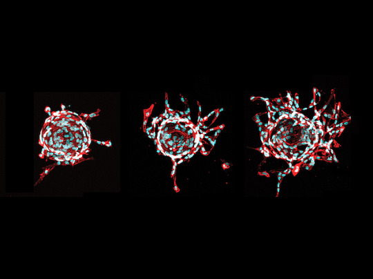
Like roads in a city, veins in your body need careful mapping. When they don’t stick to the plan, sporadic vascular malformation – malformed veins – occur, causing pain and disfigurement. These veins form lesions containing genetically faulty cells (mutants). Currently, lesions often recur after treatment. Researchers now search for new therapeutics by growing mutant endothelial cells (ECs), collected from the veins of patients, in dishes. RNA sequencing revealed mutant ECs produced the signalling molecule TGFa. Fluorescence microscopy revealed when supporting cells from lesions (stromal cells) were grown alongside normal ECs, they triggered more EC sprouting (pictured, middle, right) compared with non-lesion stromal cells (left). Next, by grafting mutant ECs into mice, they found EC-produced TGFa triggered stromal cells to release the signalling molecule VEGFA. This caused EC sprouting and malformed veins. Treating mice with afatinib, a drug that interferes with VEGF signalling, decreased lesions, suggesting it may be a useful therapeutic.
Written by Lux Fatimathas
Image from work by Suvi Jauhiainen and Henna Ilmonen, and colleagues
A.I. Virtanen Institute for Molecular Sciences, University of Eastern Finland, Kuopio, Finland
Image originally published with a Creative Commons Attribution 4.0 International (CC BY 4.0)
Published in eLife, May 2023
You can also follow BPoD on Instagram, Twitter and Facebook
#science#biomedicine#angiogenesis#blood vessels#vegf#immunofluorescence#veins#endothelial cells#capillaries#afatinib
15 notes
·
View notes
Text
Cutera Handpiece Recharge: Efficiency and Sustainability Meets
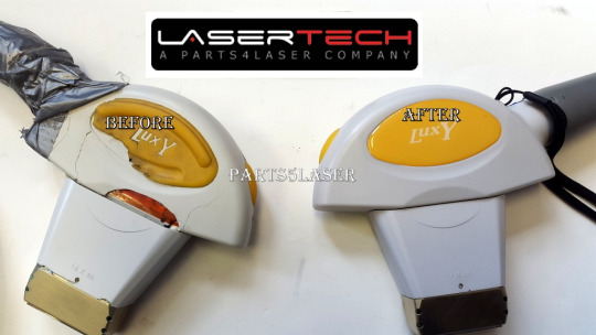
Before delving into the specifics of the handpiece recharge, let’s quickly review the Cutera mechanism and Cutera Laser Repair Services. This platform provides a selection of laser and light-based technologies intended to address vascular lesions, hair removal, fine lines, wrinkles, and other skin issues. Its interchangeable handpieces, each suited to different skin types and treatment…
View On WordPress
#Cutera Handpiece Recharge#Cutera Handpiece Refurbish#Cutera Handpiece Reset#Cutera Laser Repair Services
2 notes
·
View notes
Text
click read more to see the pathology report on my kidney they took out
Surgical Pathology Report
SPECIMEN SOURCE:
A. LEFT KIDNEY
CLINICAL HISTORY: Polycystic kidney disease
DIAGNOSIS:
A. LEFT KIDNEY:
Polycystic kidney disease with a small papillary adenoma (0.4 mm)
MD, PhD Attending Pathologist
GROSS DESCRIPTION: The specimen labeled "LEFT KIDNEY" is received fresh, now fixed in formalin and consists of 1646 gram 28.0 x 15.0 x 13.0 cm polycystic left nephrectomy specimen. The kidney alone measures 17.0 x 9.5 x 8.0 cm overall and is trabeculated and cystic. The hilum displays vessels inclusive of: ureter (8.0 x 0.2 cm); renal vein (0.6 cm in diameter) and renal artery (0.2 cm in diameter). A fixed portion of Gerota's fascia is present measuring 24.0 x 11.0 cm. The renal vein is devoid of any lesion or thrombus. The kidney is bivalved revealing brown liquid and polycystic kidney with cysts ranging in size from 0.3 x 0.3 cm to 5.0 x 4.0 cm overall. The cyst walls tan-white and smooth measuring up to 0.2 cm in thickness. The urothelium of the renal pelvis is tan-white and smooth surrounded by abundant yellow fatty tissue. The calyces, medullary pyramids, and cortico-medullary junction, and cortex are not grossly identified and totally obliterated by the cysts. An adrenal gland is not present. A lesion is not gross identified. The tissue is representatively submitted. Gross pictures are taken.
SUMMARY OF SECTIONS: A1 = Vascular margins, shaved, en face (ureter, renal vein and renal artery) inked orange A2=section of renal pelvis with cysts A3=cyst with gerota's fascia A4=cyst with hilar fat A5=cyst with perinephric fat A6=gerota's fascia and perinephric fat A7-A9= representative sections of cysts Total 9
7 notes
·
View notes
Text


The differential diagnosis of a middle mediastinal mass includes lymph nodes, foregut duplication cysts, tracheal/bronchial lesions, lipoma, esophageal lesions (benign or malignant) and vascular lesions (aortic aneurysm, varices).
This case is a 70-year-old man who presented with cough and hemoptysis. Chest radiograph reveals a right mediastinal mass that silhouettes the right hilar vessels, localizing the lesion to the middle mediastinum. There is also mass effect on the trachea and widening of the right paratracheal stripe. CT reveals a mass along the right tracheal border, which also encases the right mainstem bronchus. Biopsy revealed small cell bronchial carcinoma.
Case courtesy of Townsville radiology training, Radiopaedia.org, rID: 18285
#TeachingRounds#FOAMEd#FOAMRad#Anatomy#mediastinum#radiology#chestradiology#chestrad#cardiothoracicradiology#chestxray#CTRad
6 notes
·
View notes
Text
I hate when people use non-standard abbreviations! There's a pt with low back pain and weakness, urinary and fecal incontinence. MRI doesn't show spinal cord compression, so cauda equina ruled out. Neuro recommended w/u for autoimmune demyelinating polyneuropathies and transverse myelitis. The neurologist wrote "TM" in his note, and I didn't know what he meant, so I looked it up. Anyway, I've heard of transverse myelitis, but didn't know the workup. From UpToDate:
●Definitions – Acute transverse myelitis (TM) is a neuro-inflammatory spinal cord disorder that presents with the rapid onset of weakness, sensory alterations, and/or bowel and bladder dysfunction. Idiopathic TM is defined by its occurrence without a definitive etiology despite a thorough work-up. Secondary (disease-associated) TM is most often related to a systemic inflammatory autoimmune condition.
●Causes – Idiopathic TM usually occurs as a postinfectious complication and presumably results from an autoimmune process. Alternatively, TM can be associated with infectious, systemic inflammatory, or multifocal central nervous system disease. Acquired central nervous system demyelinating disorders that can cause TM include multiple sclerosis, myelin oligodendrocyte glycoprotein (MOG) antibody disease, neuromyelitis optica spectrum disorder (NMOSD), and acute disseminated encephalomyelitis.
●Epidemiology – TM is rare, with an annual incidence of one to eight new cases per million.
●Clinical features – The onset of TM is characterized by acute or subacute development of neurologic signs and symptoms consisting of motor, sensory, and/or autonomic dysfunction. Motor symptoms include a rapidly progressing paraparesis that can involve the upper extremities, with initial flaccidity followed by spasticity. In most patients, a sensory level can be identified. Sensory symptoms include pain, dysesthesia, and paresthesia. Autonomic symptoms involve increased urinary urgency, bladder and bowel incontinence, difficulty or inability to void, incomplete evacuation and bowel constipation, and sexual dysfunction.
●Evaluation and diagnosis – The diagnosis of TM is suspected when there are acute or subacute signs and symptoms of motor, sensory and/or autonomic dysfunction that localize to one or more contiguous spinal cord segments in patients with no evidence of a compressive cord lesion. Thus, the diagnosis of TM requires exclusion of a compressive cord lesion, usually by magnetic resonance imaging (MRI), and confirmation of inflammation by either gadolinium-enhanced MRI or lumbar puncture. When inflammation is present in the absence of cord compression, then the criteria for TM have been met, and it is necessary to evaluate for the presence of infection, systemic inflammation, and the extent and sites central nervous system inflammation.
●Differential diagnosis – The main considerations in the differential diagnosis of idiopathic TM are conditions that cause other types of myelopathy (eg, compressive or noninflammatory or vascular), the various disorders that cause secondary TM, and nonmyelopathic disorders that may mimic TM (eg, Guillain-Barré syndrome).
●Treatment – For patients with acute idiopathic TM, we suggest high-dose intravenous glucocorticoid treatment (Grade 2C). Our preferred regimens are methylprednisolone (30 mg/kg up to 1000 mg daily) or dexamethasone (up to 200 mg daily for adults) for three to five days. For patients with acute TM complicated by motor impairment, we suggest additional treatment with plasma exchange (Grade 2C). Our preferred regimen is five treatments, each with exchanges of 1.1 to 1.5 plasma volumes, every other day for 10 days; alternatively, the first two plasma exchange treatments can be given on successive days, with the remaining three treatments given every other day.
●Prognosis
•Degree of recovery – Most patients with idiopathic TM have at least a partial recovery, which usually begins within one to three months and continues with exercise and rehabilitation therapy. Some degree of persistent disability is common, occurring in approximately 40 percent. A very rapid onset with complete paraplegia and spinal shock has been associated with poorer outcomes. Recovery can proceed over years.
•Risk of recurrence – The majority of patients with TM experience monophasic disease. Recurrence has been reported in approximately 25 to 33 percent of patients with idiopathic TM, although this usually signals a systemic condition. With disease-associated (secondary) TM, the recurrence rate may be as high as 70 percent.
•Risk of multiple sclerosis – Patients presenting with acute complete TM have a generally cited risk of multiple sclerosis of only 5 to 10 percent. However, for patients with partial myelitis as an initial presentation and cranial MRI abnormalities showing lesions typical for multiple sclerosis, the transition rate to multiple sclerosis over three to five years is 60 to 90 percent. In contrast, patients with acute partial myelitis who have a normal brain MRI develop multiple sclerosis at a rate of only 10 to 30 percent over a similar time period.
2 notes
·
View notes