#membrane x computer
Explore tagged Tumblr posts
Visit Tumblr Blog
Explore Tumblr blogs with no restrictions, modern design and the best experience.
Last Seen Tumblr Blogs


Fun Fact
China blocked Tumblr because of pornography and censorship problems in 2013.
Note
It's been a while since I read your Tech Support fic and Post Florpus comics, but I just reread them and they still hit so good 🤌 but quick question bc I've been losing my mind over this but I swear to god I thought your fic had a phone call between Computer and Membrane where Membrane overhears GIR call Computer "House" and assumes his name is "Haus", was that scene from your fic??
Sorry it took me so long to answer this, I was busy and I like to hoard comments like this in my askbox for when I'm having a mental rainy day and be reminded folks still like my stuff.
Thank you! I'm glad people still like my Invader Zim art and stuff still. I hope to get back into it eventually. Because I feel it was 3 arcs away from ending the story entirely.
And yes, here's the master list of the instances that Membrane calls the Computer Haus.
This was the comic where Membrane and the Computer's friendship blossomed
This is the proper post explaining the significance of the name
This is the joke post with us coming up with the idea
But if you're talking about the content where it happens in the story.
That happened in "Jerking around the house" Which Ceph wrote and I ghostwrote and edited.
Here's the link to the Safe for work version.
(there is a nsfw version but if you want that and are of age, I trust you can find it on your own if you want it)
8 notes
·
View notes
Text
ABOUT ME!1!!1!11
I'm Meii!
I'm Chinese Malaysian, but I'm thinking of a bit Singaporean. Malaysia and Singapore were not the same!

I'm a Lesbian! So plz respect me
Fandoms I'm in: InvaderZim, CrimeWatch, Regretevator, Mouthwashing (I jst joined this fandom)
My favorite characters were: (InvaderZim: Dib Membrane, Zim, Gir) (Regretevator: Unpleasant, Jeremy, Pest, Poob, Bive, Gnarpy) (CrimeWatch: None but I don't think Julius can be counted as an character bc he's a host) (Mouthwashing: Anya, Daisuke, many ppl likes them so I like it :P)
Roblox: k0k11_k1t (Bon_UsagiAkiri)
Discord: enddiecore_88495 (I only online on computer)
Tiktok: iluvcheese_0 (UsagiMeii)
YouTube: meluvcheese_0

Btw I post arts and stuffs here. I ship ZaDr as an ragebait bc I don't think shipping alien and human are valid so I ship them as ragebait

DNI: toxic ZaDr/Bive x Split shippers, invalid ships, rudeness
I go by pronouns: She/They/Meiloid/Meiself
9 notes
·
View notes
Text
It has come to my attention
that I titled my hole here on Tumblr™ “Ashley’s dlob”, instead of what I (presumably) intended, “Ashley’s dlog”. Well how about that. The (presumably) is because I have the terriblest of memories, and I cannot recall where my mind was when I typed that out, but I’ll assume it was a typo. In any case, time for an update, as it has been a while!
I haven’t posted here as much due to the simple fact that my computer’s cooling fan decided to crap itself, and as such I spent the last couple months without one. Had I not my tablet... I might’ve gone insane, in the membrane even!! (⊙_⊙;)
But, I have it with me now, in proper working order. In that interim, I was drawing traditionally, and that really got me thinking about some stuff. Drawing really isn’t like riding a bike at all; if you don’t use it, you lose it! But, even though I am currently a NEET, it’s tough to go and draw every single day, the act is a whole body-mind-soul kind of deal, you know? If you have a poor night of sleep, you’re not gonna draw as well!
Of course, some forcing must happen, as (at least for me) I find that thinking about doing something can lead to a wretched feedback loop of thinking about X and how oh, I’m totally going to do X, I’m so ready to do X, you have NO IDEA! and I never actually do that thing. Sometimes you just have to throw yourself into the cold, open ocean at 6AM, as I did once on a trip when I was young.
All that written, I felt I was really getting somewhere with my traditional endeavors. It really goes to show that getting a tablet shouldn’t substitute drawing on paper: Though the experience of drawing is different, the skill passes over from one medium to the other, and that’s a two-way street, baby!
...
Typing this out makes me wonder, though. Was this really a “log” at all, digital or otherwise? I mean, it is my page, it is mine to do with as I see fit, but as it stands I’m just sort of rambling about random stuff, aren’t I?
Well, I started reading The Lord of the Rings in my exile. Still haven’t finished the first book, but I’m really liking what I’m reading! Not that I could gather much of the lore from what I read, given the abundance of names and places, but I’m sure there are resources that distill the info down to lesser intellects such as mine. ᵀᵒᵐ ᴮᵒᵐᵇᵃᒄᶦᴵ ᶦˢ ᵗʰᵉ ᵇᵉˢᵗ ᶜʰᵃʳᵃᶜᵗᵉʳ ᒄᵒⁿ'ᵗ ᵃᵗ ᵐᵉ
Whew, that was a lot of writing, wasn’t it. Lots of rambling I’m probably not gonna remember once I hit “Post”. But, if all goes well, I’ll be doing a lot less typing and a lot more drawing! And a lot more painting!! (๑•̀ㅂ•́)و✧

PS: I didn’t even address the “dlob” situation, did I.
1 note
·
View note
Text
Spinal Cord Tumor Treatment in Mumbai, India | Synapse Spine

Spinal cord tumors are a rare and complex type of cancer that can occur in various parts of the spine, including the spinal cord, nerve roots, and surrounding tissues. These tumors can be benign or malignant, and their treatment often requires a multidisciplinary approach involving neurosurgeons, oncologists, and other specialists. Let’s have a look into Spinal Cord Tumor Treatment in Mumbai, India at Synapse Spine.
What Are the Types of Spinal Cord Tumors?
Benign Tumors
Benign spinal cord tumors are non-cancerous growths that can cause symptoms such as numbness, weakness, or pain in the affected area. Examples of benign spinal cord tumors include:
Meningiomas: These tumors arise from the protective membranes surrounding the spinal cord and are typically slow-growing.
Schwannomas: These tumors develop from the nerve sheath and can cause numbness, weakness, or pain.
Malignant Tumors
Malignant spinal cord tumors are cancerous growths that can spread to other parts of the body. Examples of malignant spinal cord tumors include:
Metastatic tumors: These tumors originate from other parts of the body and spread to the spine, often causing significant pain and disability.
Primary spinal cord tumors: These tumors arise directly from the spinal cord or surrounding tissues and can be aggressive and difficult to treat.
Symptoms of Spinal Cord Tumors
Symptoms of spinal cord tumors can vary depending on the location and size of the tumor. Common symptoms include:
Back pain: Pain in the back, neck, or spine that worsens over time.
Numbness or tingling: Numbness, tingling, or weakness in the arms or legs.
Muscle weakness: Weakness or paralysis in the arms or legs.
Bladder or bowel dysfunction: Difficulty controlling bladder or bowel movements.
Sensory changes: Changes in sensation, such as numbness, tingling, or burning sensations.
Diagnosis of Spinal Cord Tumors
Diagnosing spinal cord tumors often involves a combination of imaging tests and physical examinations. Common diagnostic tests include:
Magnetic Resonance Imaging (MRI): This test uses strong magnetic fields and radio waves to create detailed images of the spine and surrounding tissues.
Computed Tomography (CT) scans: This test uses X-rays and computer technology to create detailed cross-sectional images of the spine.
Biopsy: This test involves removing a sample of tissue from the tumor for further examination.
Spinal Cord Tumor Treatment in Mumbai, India
Treatment options for spinal cord tumors depend on the type and location of the tumor, as well as the patient's overall health. Common treatment options include:
Surgery: Surgical removal of the tumor can be effective for both benign and malignant tumors.
Radiation therapy: This treatment uses high-energy radiation to kill cancer cells and shrink the tumor.
Chemotherapy: This treatment uses medications to kill cancer cells and slow the growth of the tumor.
Minimally Invasive Surgery Techniques for Spinal Cord Tumor Treatment in Mumbai, India
Synapse Spine offers minimally invasive surgery techniques for spinal cord tumor treatment, which can provide several benefits, including:
Reduced recovery time: Minimally invasive surgery can result in shorter hospital stays and faster recovery times.
Less tissue damage: These techniques can minimize damage to surrounding tissues, reducing the risk of complications.
Improved accuracy: Minimally invasive surgery can provide more precise removal of the tumor, reducing the risk of recurrence.
Why Choose Synapse Spine for Spinal Cord Tumor Treatment in Mumbai, India
Synapse Spine is dedicated to providing comprehensive and personalized care for patients with spinal cord tumors. Our team of experienced neurosurgeons and specialists offers a range of treatment options tailored to each patient's unique needs. With a focus on minimally invasive surgery techniques, we strive to provide effective solutions with improved recovery times.
Spinal cord tumors are a complex and challenging condition that requires a multidisciplinary approach to treatment. At Synapse Spine, we offer Spinal Cord Tumor Treatment in Mumbai, India, including minimally invasive surgery techniques, to provide effective solutions for patients with spinal cord tumors.If you or a loved one is experiencing symptoms of a spinal cord tumor, schedule an appointment with Synapse Spine today to begin your journey towards recovery by calling at 93726 71858 | 93211 24611.
#spinal fusion#degenerative disc disease#spinal stenosis#endoscopic surgery#synapse spine#spinal cord injury#spinesurgery#the spine spg#orthopedic spine surgeon#spine specialist#brain and spine tumors
0 notes
Text
Researchers Achieve Breakthrough in Silicon-Compatible Magnetic Whirls - Technology Org
New Post has been published on https://thedigitalinsider.com/researchers-achieve-breakthrough-in-silicon-compatible-magnetic-whirls-technology-org/
Researchers Achieve Breakthrough in Silicon-Compatible Magnetic Whirls - Technology Org
Researchers from Oxford University’s Department of Physics have made a breakthrough in creating and designing magnetic whirls in membranes that can be seamlessly integrated with silicon.
These hurricane-like magnetic whirls, thought to move at incredible speeds of up to kilometres per second could be used as information carriers in a new generation of green and super-fast computing platforms. The findings have been published in Nature Materials.
Artistic impression of magnetic whirls, such as merons and antimerons, generated in a free-standing and flexible membrane of hematite on a silicon wafer. Image credit: Charles Godfrey and Hariom Jani / Oxford University
Traditionally, these elusive whirls could only be produced in materials that are limitedly compatible with silicon, hindering their practical application. This obstacle was overcome by developing a new form of magnetic layers that can be detached from their original crystal hosts and transferred onto any desired platform, such as a silicon wafer.
The work was led by Dr Hariom Jani from Oxford University’s Department of Physics working in Professor Paolo Radaelli’s research group, in collaboration with the National University of Singapore and the Swiss Light Source.
Dr Jani said: ‘Silicon-based computing is much too energy-inefficient for the next generation of computing applications such as full-scale AI and autonomous devices. Overcoming these challenges will require a new computing paradigm that uses fast and efficient physical phenomena to augment current technology.’
‘We have been looking at harnessing magnetic whirls in a special class of materials called antiferromagnets, which are 100-1000 times faster than modern devices. The problem to date has been that these whirls can only be created on rigid crystal templates that are incompatible with current silicon-based technology, so our goal was to figure out a way to translate these exotic whirls to silicon.’
‘To achieve this, we fabricated ultra-thin crystalline membranes of hematite (the main component of rust and thus the most abundant antiferromagnet) that extended laterally over macroscopic dimensions,’ explains Professor Radaelli. ‘Such membranes are relatively new in the world of crystalline quantum materials, and combine advantageous characteristics of both bulk 3D ceramics and 2D materials, while also being easily transferrable.’
The hematite layer was grown on top of a crystal template that was coated with a special ‘sacrificial layer’ made from a cement component. This sacrificial layer dissolved in water, separating the hematite easily from the crystal base. Finally, the free-standing hematite membrane was transferred onto silicon and several other desirable platforms.
The group developed a novel imaging technique using linearly polarised X-rays to visualise the nanoscale magnetic patterns within these membranes. This method revealed that the free-standing layers are able to host a robust family of magnetic whirls. Potentially, this could enable ultra-fast information processing.
‘One of our most exciting discoveries was the extreme flexibility of our hematite membranes,’ continues Dr Jani. ‘Unlike their rigid, ceramic-like bulk counterparts that are prone to breaking, our flexible membranes can be twisted, bent, or curled into various shapes without fracturing. We exploited this newfound flexibility to design magnetic whirls in three dimensions, something that was previously not possible. In the future, the shape of these membranes could be tweaked to realise completely new whirls in 3D magnetic circuits.’
The group are now working on developing prototype devices that will use electrical currents to excite the rich dynamics of these super-fast whirls. Dr Jani concludes: ‘Eventually, such devices could be integrated into new types of computers that work more like the human brain – we are very excited about what’s coming next.’
Source: University of Oxford
You can offer your link to a page which is relevant to the topic of this post.
#2D materials#3d#ai#antiferromagnets#applications#Brain#cement#Collaboration#computers#computing#computing platforms#crystal#crystalline#Design#devices#dimensions#Discoveries#dynamics#energy#form#Full#Fundamental physics news#Future#green#human#human brain#Imaging#LED#Light#Link
0 notes
Text
ok, so, these things were awesome.
when i encountered one for the first time, it was in high school, as an accessibility aid. they had 12 of them or so, and they'd lend them out to students who had trouble taking notes by hand, so that they could keep up with classes by typing instead. so, perfect for people like me.
what this basically is, is a flash-memory-based text editor, no formatting tools or external storage. there are some other apps, but the main attraction is the AlphaWord program. you get 8 files, selected with those function keys up there. to get the files off, you connect it with a USB-B cable to a computer, which then treats it like a USB keyboard. when you hit Send, it just types out the entire contents of the file, really fast.
they kept making these in newer and newer models though and they're even cooler:

this is the alphasmart neo 2, it has a much nicer (albeit still rubber-membrane) keyboard than the 3000, more storage, and a way larger screen. where the 3000's screen is character-based, the neo 2 just has a pixel screen, so you can have different font sizes, and variable-width characters for much more natural reading. it has a battery life of literally days on a pair of AA's.

and this is the alphasmart dana, which has an even larger pixel screen, because in addition to having the AlphaWord program on it, it's also actually a Palm OS 4.x device, with full compatibility for its array of apps. some apps can actually use the full width of the screen (AlphaWord and a hacked version of the text editor SiEd). it also boasts dual SD card slots, which is the ideal way to load new apps on it, because i have no idea where to download whatever specific version of Palm Desktop it needs. the battery life is a bit more restrictive, at a bit less than a day under heavy use on... either 2 or 3 AA's (I forget), but it is running an entire OS there, so.
here's the great thing about these though, especially about the neo 2: these are incredibly useful tools for distraction-free writing. they need no internet connection, there are no notifications or anything else to pull your attention away, they're ultra light weight and about the size of a laptop computer so you can take them just about anywhere you want, and they have zero boot time. hit power and you're writing in seconds.
"i can write on my computer/laptop/phone/tablet," someone might say. but this thing isn't going to make noises or try to entice you into checking your email or going to research something and accidentally winding up on discord. if you have problems with focusing, one of these is ideal. best of all: they're not even that expensive second-hand; i got mine from ebay for like $50, and it seems like they're holding that value pretty well. there are always school libraries and the like trying to unload these as they move up to issuing students chromebooks or the like, so supplies are ample.
i recommend one of these if you do a lot of writing and have any kind of focus problems. it's ideal for folks on the autism spectrum or with ADHD. it saved my school career from certain doom, and damn it all, i want to spread word of that, far and wide.
the alphasmart is awesome.


Alphasmart 3000 Portable Word Processor
577 notes
·
View notes
Text
Advance Care Medical Equipment
"Advance Care Medical Equipment" could refer to a variety of medical devices or equipment designed to provide advanced care and support for patients with various medical conditions. However, since your question is quite general, I'll provide an overview of some potential categories and examples of advanced medical equipment:
Life Support Equipment:
Ventilators: Mechanical devices that assist patients in breathing or take over breathing entirely in cases of respiratory distress.
Extracorporeal Membrane Oxygenation (ECMO): A life support machine that temporarily takes over the function of the heart and lungs, used in severe cases of heart and lung failure.
Diagnostic Imaging Equipment:
Magnetic Resonance Imaging (MRI): Uses powerful magnets and radio waves to create detailed images of the internal structures of the body.
Computed Tomography (CT) Scanner: Combines X-rays with computer technology to create cross-sectional images of the body.
Cardiac Care Equipment:
Implantable Cardioverter-Defibrillator (ICD): Monitors heart rhythms and delivers an electric shock to restore normal rhythm in cases of irregular and life-threatening heart rhythms.
Pacemaker: A device that helps control abnormal heart rhythms by sending electrical impulses to the heart muscles.
Surgical Equipment:
Robotic Surgical Systems: Advanced robotic systems that assist surgeons in performing minimally invasive procedures with increased precision.
Laser Surgical Instruments: Used for various surgical procedures to cut, cauterize, or vaporize tissue.
Monitoring and Data Collection:
Continuous Glucose Monitors: Help people with diabetes monitor their blood sugar levels in real-time.
Holter Monitors: Portable devices that record a person's heart activity over an extended period, typically 24-48 hours.
Rehabilitation and Mobility Aids:
Exoskeletons: Wearable devices designed to assist individuals with mobility impairments by providing powered support to their limbs.
Functional Electrical Stimulation (FES) Devices: Use electrical currents to stimulate muscles and restore or improve function in paralyzed or weakened limbs.
Dialysis Machines:
Hemodialysis Machines: Used to remove waste and excess fluid from the blood in patients with kidney failure.
Infusion and Drug Delivery Systems:
Insulin Pumps: Deliver insulin to individuals with diabetes in a controlled manner.
Patient-Controlled Analgesia (PCA) Pumps: Allow patients to self-administer pain medication within preset limits.
Telemedicine Equipment:
Remote Monitoring Devices: Enable healthcare providers to monitor patients' vital signs and health remotely.
Telehealth Platforms: Software and hardware solutions that facilitate virtual medical consultations.
"Advance Care Medical Equipment" continues to evolve as technology advances and healthcare needs change. These are just a few examples of the many types of advanced medical equipment available to improve patient care and outcomes.
0 notes
Text
Shoulder Replacement Surgery
Your shoulder is made up of three bones: your upper arm bone (humerus), your shoulder blade (scapula), and your collarbone (clavicle). The shoulder is a ball-and-socket joint: The ball, or head, of your upper arm bone fits into a shallow socket in your shoulder blade. This socket is called the glenoid.
The surfaces of the bones where they touch are covered with articular cartilage, a smooth substance that protects the bones and enables them to move easily. A thin, smooth tissue called synovial membrane covers all remaining surfaces inside the shoulder joint. In a healthy shoulder, this membrane makes a small amount of fluid that lubricates the cartilage and eliminates almost any friction in your shoulder.
Dr. Pankaj Gunjal is one of the Shoulder Specialists in Kothrud Pune. He is an experienced Orthopedic surgeon in Karvenagar Pune.
About Shoulder Replacement Surgery
In shoulder replacement surgery, the damaged parts of the shoulder are removed and replaced with artificial components, called a prosthesis. The treatment options are either replacement of just the head of the humerus bone (ball), or replacement of both the ball and the socket (glenoid).
Is Shoulder Replacement Surgery for You?
The decision to have shoulder replacement surgery should be a cooperative one between you, your family, your family physician, and your orthopedic surgeon. There are several reasons why your doctor may recommend shoulder replacement surgery. People who benefit from surgery often have:
Severe shoulder pain that interferes with everyday activities, such as reaching into a cabinet, dressing, toileting, and washing.
Moderate to severe pain while resting. This pain may be severe enough to prevent a good night’s sleep.
Loss of motion and/or weakness in the shoulder.
Failure to substantially improve with other treatments such as anti-inflammatory medications, cortisone injections, and/or physical therapy.
At the first appointment for a shoulder replacement surgery
Your family physician may refer you to an orthopaedic surgeon for a thorough evaluation to determine if you can benefit from this surgery. An evaluation with an orthopaedic surgeon consists of several components:
A medical history – Your orthopaedic surgeon will gather information about your general health and ask you about the extent of your shoulder pain and your ability to function.
A physical examination – This will assess shoulder motion, stability, and strength.
X-rays – X-rays help to determine the extent of damage in your shoulder. They can show loss of the normal joint space between bones, flattening or irregularity in the shape of the bone, bone spurs, and loose pieces of cartilage or bone that may be floating inside the joint.
Other tests – Occasionally, your doctor may order blood tests, a computed tomography (CT) scan, a magnetic resonance imaging (MRI) scan, or a bone scan to determine the condition of the bone and soft tissues of your shoulder.
0 notes
Text
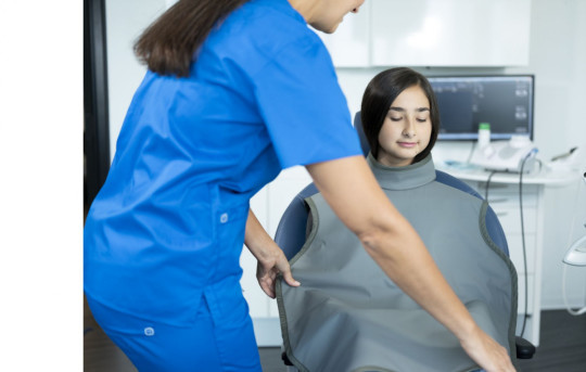
Dental Xray Machines Lowest Price
STANDARD PRECAUTIONS OF DISINFECTING YOUR DENTAL X-RAY MACHINE
Dental X Ray Machine
Many of us get question from our customers about how to properly clean and disinfect their pan-ceph or cone beam dental X-ray machine. ardent x ray machine price
STANDARD PRECAUTIONS
Standard precautions also known as universal precautions, are practices used to control infection. They are designed to protect dental professionals, their staff, and their patients from disease exposure through blood and certain bodily fluids such as saliva.
When you use standard precautions, you essentially presume that all human blood and saliva are known to be infectious. This means that everything you do to protect against cross-contamination is performed for all patients.
STEPS OF STANDARD PRECAUTIONS
FOR DENTAL X-RAY MACHINES

Wearing gloves is the best way to prevent contamination between a patient and dental staff member. All dentists and clinical team members should remove their disposable gloves and wash their hands with soap and water for at least 20 seconds between different patients. Put on new gloves in front of the patient, if possible, so they are aware of the steps you take to protect their health.

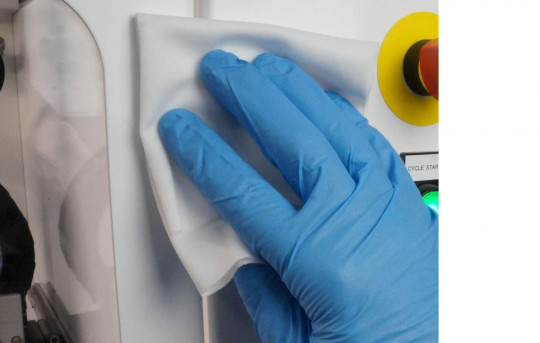
Any surface that might be touched by gloves, hands, or instruments that go into the mouth are clinical contact surfaces. For example: dental X-ray machine control panel, touch screens, exposure buttons, acquisition computer, patient positioning tools and lead apron. These are non-critical items because they are objects that might come in contact with saliva, blood, or intact skin, but not oral or mucous membranes.
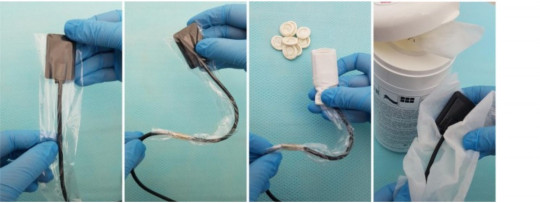
Extraoral dental X-ray machines such as panoramic, cephalometric, and cone beam systems, should use the same standard precautions for decontamination and disinfection as the other equipment in your practice. Be sure to switch the X-ray unit off before cleaning or disinfecting. Never apply sprays or liquids directly on the surfaces of the X-ray machine. Instead, apply a small amount of cleaner to a clean paper towel and use it to wipe the surface of the machine. Alternatively, you may use an alcohol-based wipe that is safe for electronics. We do not recommend any Cavicide products for dental X-ray machines because they are extremely corrosive and can cause irreparable damage to X-ray machine covers or electrical components.
Cephalometric ear post, ear post brackets, and forehead supports and/or nation pointers should all be cleaned and disinfected with an iodine-detergent disinfectant. These devices should also be covered in plastic for patient and changed after each use.
After all patient exposures are complete, the barriers should be removed and any contaminated surfaces should be re-disinfected. The lead apron should be sprayed with disinfectant and wiped as described above.
It’s important for the entire community that infectious diseases are not spread. Healthcare environments are a primary source of contamination but following standard precautions allows us to minimize the spread of bacteria and viruses and keep our staff, patients, and community safe and healthy.
#ardent x ray machine price#dr systems#fixed x ray machine#x ray india#x ray machine 300ma price in india#x-ray baggage#dental xray machine#x ray machine#v light eco#best x ray machine in india
0 notes
Text
Cidaltek E

Alcoholic Instant Instrument Spray
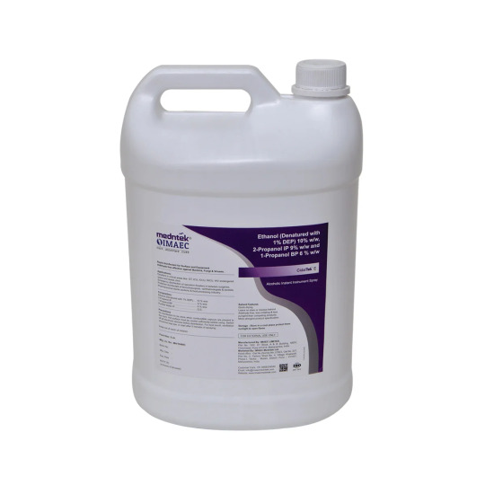
Composition: Each 100ml Contains

Pack Size: 5 Lit.
Description:
Instant Instrument/Surface Disinfectant:
CidalTek-E disinfectant having comprehensive antimicrobial activity and rapid action of ethanol & isopropanol. Alcohols are effective at eliminating vegetative bacteria and viruses from surfaces. The antimicrobial effectiveness of alcohol is through damage to bacterial cell membranes and subsequent denaturation of cellular proteins. A more effective alcohol is isopropyl alcohol (IPA), which is fast acting and possesses a broad-spectrum antimicrobial activity. When alcohols are used in combination, such as IPA and ethanol, the antimicrobial action is arguably greater. This is because IPA is slightly more efficacious against bacteria, whereas ethanol is more potent against viruses. The combination of the two makes for an effective disinfectant product.
CDC has divided noncritical surfaces in dental offices into clinical contact and housekeeping surfaces. Clinical contact surfaces are surfaces that might be touched frequently with gloved hands during patient care or that might become contaminated with blood or other potentially infectious material and subsequently contact instruments, hands, gloves, or devices (e.g., light handles, switches, dental X-ray equipment, chair-side computers). Barrier protective coverings (e.g., clear plastic wraps) can be used for these surfaces, particularly those that are difficult to clean (e.g., light handles, chair switches). Protected surfaces should be disinfected at the end of each day or if contamination is evident. If not barrier-protected, these surfaces should be disinfected between patients with an intermediate-disinfectant disinfectant with tuberculocidal claim) or low-level disinfectant (disinfectant with an HBV and HIV label claim).
Salient Features:
Ready to use alcohol-based disinfectant solution
Aldehyde-free
Instant action
Kills germs 99.99% effectively
Quick drying
Leaves no stains/ residue behind
Effective against MRSA & VRE, Drug resistance pathogens
Excellent virucidal activity.
Direction Of Use:
Rapid Dental Equipment /Instruments/Hard Surfaces Disinfection: Ready to Use. (For external use only) rapid disinfectant with comprehensive spectrum of activity for alcohol-resistance surfaces and dental devices.
CidalTek-E is suitable for the rapid disinfection of hard surfaces in the spray-wipe procedure, where a rapid effect is necessary, e.g., for dental equipment, instruments, dental chair, platform, dental tooth cast formation platform etc.
Wipe the surfaces to be disinfected, with enough ready-to use solution, ensuring complete coverage.
Rapid disinfection of hard surfaces of any dental equipment 30 seconds exposure time.
Always prefer a wipe disinfection by using lint free cloth over the spray disinfection, as it prevents the formation of aerosols and ensure best possible wetting.
When spraying, wipe afterwards, if possible, to ensure complete wetting
Contact Time: 30 seconds to 5 Minutes
Note: - Product should be used in accordance with label instruction
Area Of Application:
Rapid disinfection of dental instruments, equipment, materials, and other objects which frequently come in contact with hands.
For rapid cleaning and disinfection of sensitive dental equipment surfaces
For immediate disinfection of dental chair, equipment & accessories, waste receivers, hand pieces in kidney plates, specially used on “spitting areas” of dental chairs
Also used on inanimate surfaces in dental hospital/clinical/labs
Hard surface of frequently touch surfaces in dental hospital like dental chair handle, door knob, switches, holders, examination table, seating chairs, OPD, consulting rooms, clean touch screens, sensitive equipment, switches, dental Xray equipment, chair-side computers etc.
For cleaning and disinfection of surfaces that are most likely to become contaminated with pathogens including clinical contact surfaces in patient care areas in dental clinics such as, switches on dental chairs, computer equipment, keyboards, computer mouse, dentist chair arm, patient chair arm, dental tool handles, dental receptionist countertops, bathroom door knob, operatory cabinet handles, dentist light, clinic phones, door knobs, operatory sink faucet etc
Microbial Efficacy:
Bactericidal, Fungicidal, Yeasticidal & Virucidal.
0 notes
Text
Cidaltek E

Description:
Instant Instrument/Surface Disinfectant:
CidalTek-E disinfectant having comprehensive antimicrobial activity and rapid action of ethanol & isopropanol. Alcohols are effective at eliminating vegetative bacteria and viruses from surfaces. The antimicrobial effectiveness of alcohol is through damage to bacterial cell membranes and subsequent denaturation of cellular proteins. A more effective alcohol is isopropyl alcohol (IPA), which is fast acting and possesses a broad-spectrum antimicrobial activity. When alcohols are used in combination, such as IPA and ethanol, the antimicrobial action is arguably greater. This is because IPA is slightly more efficacious against bacteria, whereas ethanol is more potent against viruses. The combination of the two makes for an effective disinfectant product.
CDC has divided noncritical surfaces in dental offices into clinical contact and housekeeping surfaces. Clinical contact surfaces are surfaces that might be touched frequently with gloved hands during patient care or that might become contaminated with blood or other potentially infectious material and subsequently contact instruments, hands, gloves, or devices (e.g., light handles, switches, dental X-ray equipment, chair-side computers). Barrier protective coverings (e.g., clear plastic wraps) can be used for these surfaces, particularly those that are difficult to clean (e.g., light handles, chair switches). Protected surfaces should be disinfected at the end of each day or if contamination is evident. If not barrier-protected, these surfaces should be disinfected between patients with an intermediate-disinfectant disinfectant with tuberculocidal claim) or low-level disinfectant (disinfectant with an HBV and HIV label claim).
Salient Features:
Ready to use alcohol-based disinfectant solution
Aldehyde-free
Instant action
Kills germs 99.99% effectively
Quick drying
Leaves no stains/ residue behind
Effective against MRSA & VRE, Drug resistance pathogens
Excellent virucidal activity
Direction Of Use:
Rapid Dental Equipment /Instruments/Hard Surfaces Disinfection: Ready to Use. (For external use only) rapid disinfectant with comprehensive spectrum of activity for alcohol-resistance surfaces and dental devices.
CidalTek-E is suitable for the rapid disinfection of hard surfaces in the spray-wipe procedure, where a rapid effect is necessary, e.g., for dental equipment, instruments, dental chair, platform, dental tooth cast formation platform etc.
Wipe the surfaces to be disinfected, with enough ready-to use solution, ensuring complete coverage.
Rapid disinfection of hard surfaces of any dental equipment 30 seconds exposure time.
Always prefer a wipe disinfection by using lint free cloth over the spray disinfection, as it prevents the formation of aerosols and ensure best possible wetting.
When spraying, wipe afterwards, if possible, to ensure complete wetting
Contact Time: 30 seconds to 5 Minutes
Note: - Product should be used in accordance with label instruction
Area Of Application:
Rapid disinfection of dental instruments, equipment, materials, and other objects which frequently come in contact with hands.
For rapid cleaning and disinfection of sensitive dental equipment surfaces
For immediate disinfection of dental chair, equipment & accessories, waste receivers, hand pieces in kidney plates, specially used on “spitting areas” of dental chairs
Also used on inanimate surfaces in dental hospital/clinical/labs
Hard surface of frequently touch surfaces in dental hospital like dental chair handle, door knob, switches, holders, examination table, seating chairs, OPD, consulting rooms, clean touch screens, sensitive equipment, switches, dental Xray equipment, chair-side computers etc.
For cleaning and disinfection of surfaces that are most likely to become contaminated with pathogens including clinical contact surfaces in patient care areas in dental clinics such as, switches on dental chairs, computer equipment, keyboards, computer mouse, dentist chair arm, patient chair arm, dental tool handles, dental receptionist countertops, bathroom door knob, operatory cabinet handles, dentist light, clinic phones, door knobs, operatory sink faucet etc
Microbial Efficacy:
Bactericidal, Fungicidal, Yeasticidal & Virucidal.
0 notes
Text
X-Ray2EM: Uncertainty-Aware Cross-Modality Image Reconstruction from X-Ray to Electron Microscopy in Connectomics. (arXiv:2303.00882v1 [eess.IV])
Comprehensive, synapse-resolution imaging of the brain will be crucial for understanding neuronal computations and function. In connectomics, this has been the sole purview of volume electron microscopy (EM), which entails an excruciatingly difficult process because it requires cutting tissue into many thin, fragile slices that then need to be imaged, aligned, and reconstructed. Unlike EM, hard X-ray imaging is compatible with thick tissues, eliminating the need for thin sectioning, and delivering fast acquisition, intrinsic alignment, and isotropic resolution. Unfortunately, current state-of-the-art X-ray microscopy provides much lower resolution, to the extent that segmenting membranes is very challenging. We propose an uncertainty-aware 3D reconstruction model that translates X-ray images to EM-like images with enhanced membrane segmentation quality, showing its potential for developing simpler, faster, and more accurate X-ray based connectomics pipelines. http://dlvr.it/SkJFL4
0 notes
Text
Secret Santa for @crispysadisticcuddlemuffin !!

74 notes
·
View notes
Photo

No context except that this was made bc of the Moo-Ping 10 server @cephalonghost
#invader zim#professor membrane#membrane x dwicky#rapapmr#i hope that's the right ship name#membrane x miyuki#membrane x computer#Moo-Ping 10 Server
316 notes
·
View notes
Text


i really like this vibe :)
#invader zim#dib x zim#zadr#computer vibe#90s#digetalart#digital sketch#digital drawing#dib membrane#zim#fan arrt#hehe
88 notes
·
View notes
Text
owo
#my computer is so close to being finished#by so close i mean i have to buy a psu and a hdd and some fans and then im done#im so fkn excited#i also think i need to get another keyboard tbh#the one i got is green razer switches and theyre so nice and clicky but also i use my computer a lot when everyone else is sleeping#so im scared im gonna just wake them all up all the time#like im figuring out how to type quieter but i think it might be worth getting a cheap membrane board for night#idk man#im just v v excited#tazposting#feel free to interact w me abt it if ur interested at all x
0 notes