#cerebral cortex
Explore tagged Tumblr posts
Text
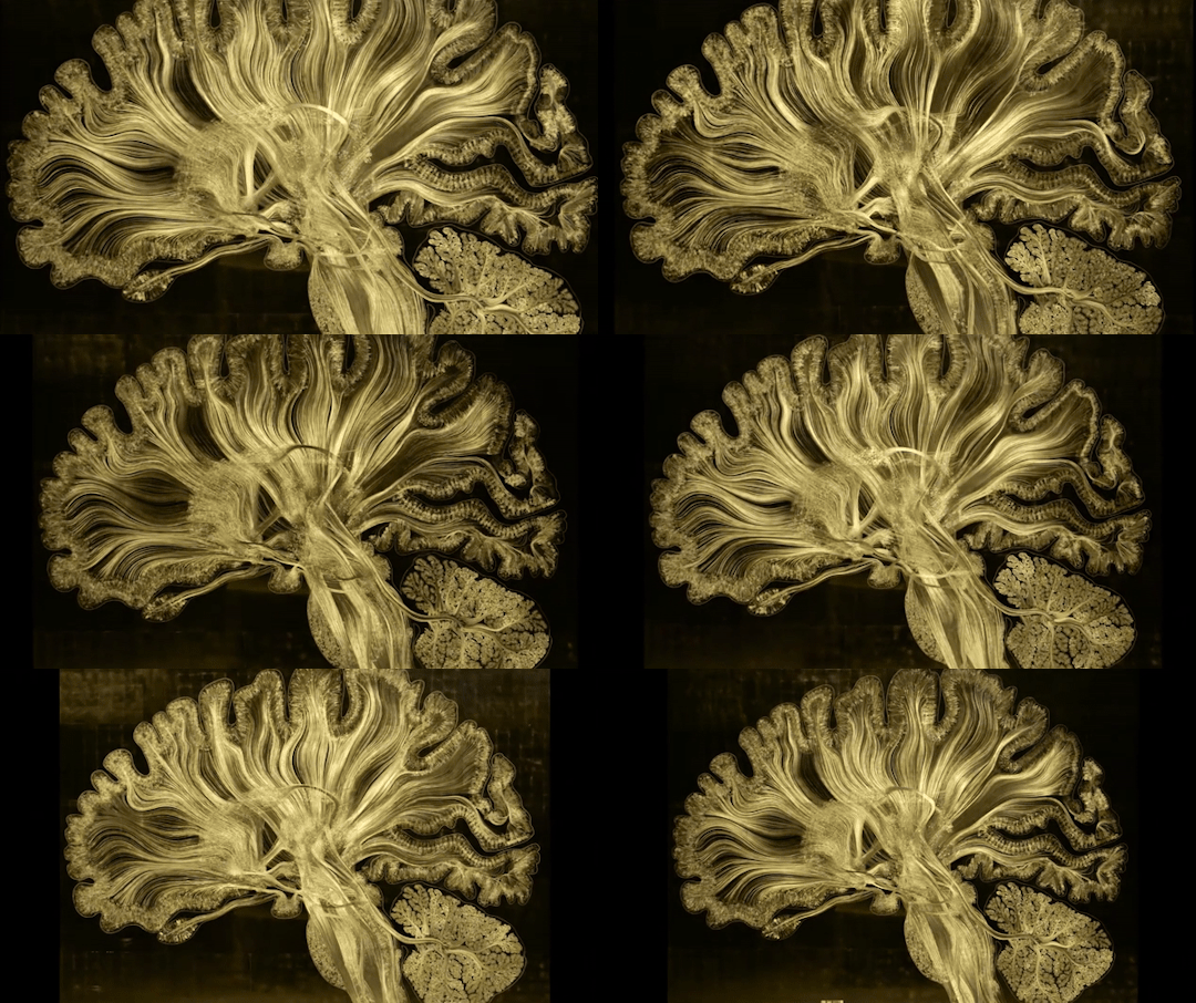
Illuminating the brain through art and science
#photography#explore#science#adorable#gifs#education#lol#human#amazing#awesome#beautiful#movement#male#female#brain#neurons#organ#connection#synapses#art#technology#self reflected#brain areas#neurology#nervous system#cerebral cortex#cerebellum#dendrites#axons#prefrontal cortex
108 notes
·
View notes
Text
One study concluded that humans have five times the information-processing capacity of cetaceans, whom they placed beneath chimps, monkeys, and some birds. But in the same study, horses—with smaller brains than chimps—were found to have five times the number of cortical neurons. Does this mean horses are smarter than chimps? A major confounding factor in these types of comparisons appears to be that every factor is itself quite confounding. Estimating numbers of neurons is a very rough science, so the raw number comparisons are crude. There are lots of different kinds of neurons, and they are arranged in different configurations and proportions in different species. We know all these variations mean something, that they will determine what brains are capable of, but we don’t know yet quite what, or how that might change from one moment to the next in different parts of the brain. There are a lot of assumptions at play, and it can be misleading to extrapolate from one brain to another.
This also applies to comparing cognitive ability. Trying to infer from brains and their structures which animals are “better” at cognition and ranking animal brains in order of “intelligence” is as treacherous as it is tempting. Stan Kuczaj, who spent his lifetime studying the cognition and behavior of different animals, put it bluntly: “We suck at being able to validly measure intelligence in humans. We’re even worse when we try to compare species.” Intelligence is a slippery concept and perhaps unmeasurable. As mentioned earlier, many biologists conceive of it as an animal’s ability to solve problems. But because different animals live in different environments with different problems, you can’t really translate scores of how well their brains perform. A brain attribute is not simply “good” or “bad” for thinking, but rather varies depending on the situation and the thinking that brain needs to undertake. Intelligence is a moving target.
What confounds this dilemma further is that individual animals within a species have varying cognitive abilities. To quote the Yosemite National Park ranger who, when asked why it was proving so hard to make a garbage can that bears couldn’t break into, said, “There is considerable overlap between the intelligence of the smartest bears and the dumbest tourists.”
— In the Mind of a Whale
#tom mustill#in the mind of a whale#science#zoology#biology#human biology#marine biology#neuroscience#ethology#brain#cerebral cortex#neurons#stan kuczaj#whales#chimpanzees#bears#intelligence
146 notes
·
View notes
Text
Brain pathways: an information superhighway
Understanding the neural network
The brain, our body's conductor, is a complex network of billions of interconnected neurons. These neurons communicate with each other via specialized pathways known as nerve tracts. These pathways are essential for transmitting sensory, motor and cognitive information throughout the body.

Main brain pathways
1. The pyramidal pathway
- Role: The pyramidal pathway is primarily responsible for the voluntary control of movement. It connects the primary motor cortex to the motor neurons in the spinal cord, enabling the initiation and control of precise skeletal muscle movements.
- Components: It comprises the corticospinal bundle and the corticobulbar bundle.
- How it works: Nerve signals from the motor cortex travel down this pathway to activate the muscles concerned.
2. Sensory pathways
- Role: These pathways transmit sensory information from the body to the brain.
- Types of sensitivity:
o Tactile sensitivity: Allows us to perceive touch, pressure and vibration.
o Thermal sensitivity: Allows us to perceive heat and cold.
o Deep sensitivity: Allows us to perceive the position of limbs in space (proprioception) and joint movements.
o Pain sensitivity: Allows us to perceive pain.
- Pathway: Sensory information is transmitted by peripheral nerves to the spinal cord, then back to the brain via various ascending pathways.
3. Specific sensory pathways
- Visual: transmits visual information from the retina to the occipital lobe.
- Auditory: Transmits auditory information from the inner ear to the temporal lobe.
- Olfactory pathway: transmits olfactory information from olfactory receptors to the olfactory bulb.
- Taste pathway: transmits taste information from the taste buds to the taste cortex.
4. Proprioception pathway
- Role: Proprioception is the sense that enables us to know our body's position in space.
- How it works: Proprioceptive receptors in muscles, tendons and joints constantly send information to the brain about the state of muscle contraction, joint angle and limb position.
- Importance: Proprioception is essential for movement coordination, balance and posture.
Nerve pathway disorders
Damage to or dysfunction of these pathways can lead to a variety of neurological disorders, such as :
- Hemiplegia: Paralysis of one side of the body.
- Paresthesia: Sensation of numbness or tingling.
- Ataxia: Loss of coordination of movements.
- Blindness: Loss of vision.
- Deafness: Loss of hearing.
In conclusion
Brain pathways are complex networks that ensure communication between the brain and the body. Understanding how they work is essential for grasping the mechanisms underlying many physiological and pathological processes.
Go further
#nerve pathways#brain#neurology#neuroscience#pyramidal pathway#sensitivity#proprioception#nervous system#neuroanatomy#brain health#neurological disorders#brain anatomy#neurons#synapses#cerebral cortex#spinal cord#peripheral nerves#senses#perception#movement#coordination
2 notes
·
View notes
Text

#gif#anime#old anime#retro#80s#abstract#variant color#art#space#neurolink#science fiction#peace#cerebral cortex#pink#pinkcore#cosmic
4 notes
·
View notes
Text
thinking about that one time i promised to do a rap battle with the gaming society president on discord not expecting him to go through with it BC ive never met him irl
then i get to the meetup and this genuinely incredibly attractive cute guy stands up and SCREAMS "WHERE IS RUBY" to demand a rap battle in front of 50 people and then they all turned to the fucking dork in a deadpool jumper should i kill myself ?
(i did not end up doing it but ive PROMISED to do it next year as long as he buys me a drink)
((i probably would have done it if he wasn't genuinely hot LMAO))
#YES I AM A NERD#bookworm#im studious#from my#cerebral cortex#to my#gluteus#back in#kindergarten#i aced#my#college#entrance exam#oh im no#rocket scientist#OH WAIT#I AM
1 note
·
View note
Text
Neuroscientists create a comprehensive map of the cerebral cortex
New Post has been published on https://thedigitalinsider.com/neuroscientists-create-a-comprehensive-map-of-the-cerebral-cortex/
Neuroscientists create a comprehensive map of the cerebral cortex
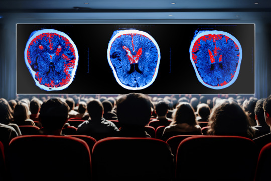

By analyzing brain scans taken as people watched movie clips, MIT researchers have created the most comprehensive map yet of the functions of the brain’s cerebral cortex.
Using functional magnetic resonance imaging (fMRI) data, the research team identified 24 networks with different functions, which include processing language, social interactions, visual features, and other types of sensory input.
Many of these networks have been seen before but haven’t been precisely characterized using naturalistic conditions. While the new study mapped networks in subjects watching engaging movies, previous works have used a small number of specific tasks or examined correlations across the brain in subjects who were simply resting.
“There’s an emerging approach in neuroscience to look at brain networks under more naturalistic conditions. This is a new approach that reveals something different from conventional approaches in neuroimaging,” says Robert Desimone, director of MIT’s McGovern Institute for Brain Research. “It’s not going to give us all the answers, but it generates a lot of interesting ideas based on what we see going on in the movies that’s related to these network maps that emerge.”
The researchers hope that their new map will serve as a starting point for further study of what each of these networks is doing in the brain.
Desimone and John Duncan, a program leader in the MRC Cognition and Brain Sciences Unit at Cambridge University, are the senior authors of the study, which appears today in Neuron. Reza Rajimehr, a research scientist in the McGovern Institute and a former graduate student at Cambridge University, is the lead author of the paper.
Precise mapping
The cerebral cortex of the brain contains regions devoted to processing different types of sensory information, including visual and auditory input. Over the past few decades, scientists have identified many networks that are involved in this kind of processing, often using fMRI to measure brain activity as subjects perform a single task such as looking at faces.
In other studies, researchers have scanned people’s brains as they do nothing, or let their minds wander. From those studies, researchers have identified networks such as the default mode network, a network of areas that is active during internally focused activities such as daydreaming.
“Up to now, most studies of networks were based on doing functional MRI in the resting-state condition. Based on those studies, we know some main networks in the cortex. Each of them is responsible for a specific cognitive function, and they have been highly influential in the neuroimaging field,” Rajimehr says.
However, during the resting state, many parts of the cortex may not be active at all. To gain a more comprehensive picture of what all these regions are doing, the MIT team analyzed data recorded while subjects performed a more natural task: watching a movie.
“By using a rich stimulus like a movie, we can drive many regions of the cortex very efficiently. For example, sensory regions will be active to process different features of the movie, and high-level areas will be active to extract semantic information and contextual information,” Rajimehr says. “By activating the brain in this way, now we can distinguish different areas or different networks based on their activation patterns.”
The data for this study was generated as part of the Human Connectome Project. Using a 7-Tesla MRI scanner, which offers higher resolution than a typical MRI scanner, brain activity was imaged in 176 people as they watched one hour of movie clips showing a variety of scenes.
The MIT team used a machine-learning algorithm to analyze the activity patterns of each brain region, allowing them to identify 24 networks with different activity patterns and functions.
Some of these networks are located in sensory areas such as the visual cortex or auditory cortex, as expected for regions with specific sensory functions. Other areas respond to features such as actions, language, or social interactions. Many of these networks have been seen before, but this technique offers more precise definition of where the networks are located, the researchers say.
“Different regions are competing with each other for processing specific features, so when you map each function in isolation, you may get a slightly larger network because it is not getting constrained by other processes,” Rajimehr says. “But here, because all the areas are considered together, we are able to define more precise boundaries between different networks.”
The researchers also identified networks that hadn’t been seen before, including one in the prefrontal cortex, which appears to be highly responsive to visual scenes. This network was most active in response to pictures of scenes within the movie frames.
Executive control networks
Three of the networks found in this study are involved in “executive control,” and were most active during transitions between different clips. The researchers also observed that these control networks appear to have a “push-pull” relationship with networks that process specific features such as faces or actions. When networks specific to a particular feature were very active, the executive control networks were mostly quiet, and vice versa.
“Whenever the activations in domain-specific areas are high, it looks like there is no need for the engagement of these high-level networks,” Rajimehr says. “But in situations where perhaps there is some ambiguity and complexity in the stimulus, and there is a need for the involvement of the executive control networks, then we see that these networks become highly active.”
Using a movie-watching paradigm, the researchers are now studying some of the networks they identified in more detail, to identify subregions involved in particular tasks. For example, within the social processing network, they have found regions that are specific to processing social information about faces and bodies. In a new network that analyzes visual scenes, they have identified regions involved in processing memory of places.
“This kind of experiment is really about generating hypotheses for how the cerebral cortex is functionally organized. Networks that emerge during movie watching now need to be followed up with more specific experiments to test the hypotheses. It’s giving us a new view into the operation of the entire cortex during a more naturalistic task than just sitting at rest,” Desimone says.
The research was funded by the McGovern Institute, the Cognitive Science and Technology Council of Iran, the MRC Cognition and Brain Sciences Unit at the University of Cambridge, and a Cambridge Trust scholarship.
#algorithm#approach#author#Brain#brain activity#Brain and cognitive sciences#brain networks#brain research#brains#cerebral cortex#cognition#cognitive function#complexity#comprehensive#connectome#data#daydreaming#Features#functions#Giving#how#human#Ideas#Imaging#Iran#it#language#learning#map#McGovern Institute
1 note
·
View note
Text
This week, on Living with Disabilities, I had the opportunity to sit down with a friend of mine Tracy Jackson. When she was younger, she earned the title of champion. She was a horse trainer back in the day.
youtube
0 notes
Text
Outer layers of the brain produce high frequency brain ways associated with sensory stimulation
Deeper regions of the brain produce low frequency alpha waves associated with control signals.
Studying six layers of the brain's Cerebral Cortex in 14 regions.
The Cerebral Cortex is responsible for higher cognitive function.
Cognitive function is a broad term that refers to mental processes involved in the acquisition of knowledge, manipulation of information, and reasoning.
Lamination is the biological process by which cells are arranged in layers within a tissue during development
1 note
·
View note
Text
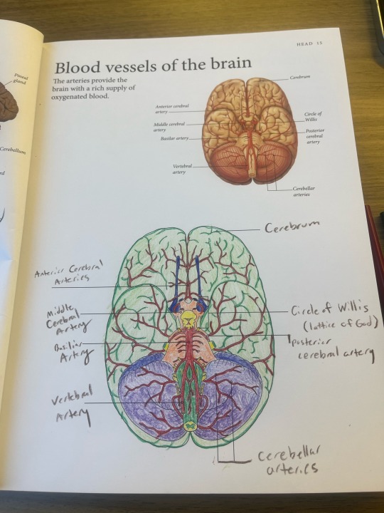
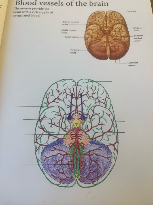
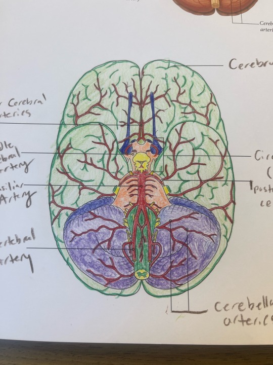
Created a God out of a human brain at the laundromat today
#cerebral cortex#art#coloring book#mildly interesting#his name is Willis as the labels imply#colored pencil
0 notes
Text
Illuminating the brain through art and science!
#photography#explore#science#adorable#gifs#education#lol#human#amazing#awesome#beautiful#movement#male#female#brain#neurons#organ#connection#synapses#art#technology#self reflected#brain areas#neurology#nervous system#cerebral cortex#cerebellum#dendrites#axons#prefrontal cortex
12 notes
·
View notes
Text
'I Can't Hear It Now' feels more like a Jinx song for Silco than for Caitlyn and Cassandra. Hear me out.
The lyrics of the song are legit more in line with Jinx and Silco from the get go.
'There is an ocean so dark down below the waves,' is literally the first line (pertaining to Silco's water motif because who else has that specific link than the one-eyed rat). And in episode 2, what to do we see?? Jinx letting him go down into the dark abyss 'below the waves'.


The next line hits even harder: 'Where you watch while these dreams gently float away'.
Everything Silco built is in ruins, his dreams for Zaun is being torn apart to the bone by power-hungry Chem-Barons, and his daughter isn't too keen on officially taking the role of revolutionary successor even when she knows it's her right or that the Undercity insisted it.
'And there is a silence so soft it's only memory
Like the way your voice always sounds when you sing to me.'

'But I can't hear it now
Just tell me how to keep breathing while pretending I'm not drowning.'
As we know, Silco never became a voice in Jinx's head (except for that one time when Isha was captured, as well as during her incarceration) and she wants him to - might have even pleaded - based on what she says to his chair in episode 4.


Also, can I just stress how Jinx probably was never scared about her dad passing because, at least, her hearing voices would mean he'd still be there?? But he didn't, and he's not able to guide her anymore or pull her out of the waters she sank herself in.
'I just watched as the door closed for good
'Cause I couldn't keep it open.'


This part is a stretch, but it might be referring to the night it all fell apart. The open door was her waiting for Vi to take Powder. Then her instincts decided for her with Silco as collateral; and when she did, Jinx took the chair.
Now, is it "disrespectful" to take Caitlyn's ballad of grief and make it about Jinx? Yep. But it leads me to a second tangent point: I didn't say all this simply to put all the attention on our favourite blue haired rebel.
The fact that the lyrics reminds me more of Jinx than Caitlyn reflects well on how the latter's perception is so warped it can't see anything past the former. Everything Cait does is centred around Jinx. When she aims her gun, it's pointing at Jinx. When she she thinks about Cassandra, there's also Jinx. Vi even says she's acting like Jinx. When I listen to this song (again, meant for Cait), I think about Jinx.
Jinx "took" this song like she did to Cait's mother, her dignity, her sanity, her identity and everything else.
#arcane#arcane analysis#jinx and caitlyn parallel each other so much in season 2 but this shifted my cerebral cortex#jinx and silco#i miss our rat father so much
187 notes
·
View notes
Text
Penfield's homonculus: a fascinating map of our brain
What is Penfield's homonculus?
Penfield's homonculus is a simplified but highly illustrative graphic representation of how our brain ‘sees’ our body. It is a neurological map that shows how the different parts of our body are represented in the cerebral cortex. Imagine a deformed little man, with disproportionately large hands and mouth: that's the homunculus!
Two homunculi, two functions
There are two main types of homunculus:
- The motor homunculus: This corresponds to the area of the cerebral cortex that controls voluntary movements. In this homunculus, the parts of the body that we use with precision (hands, lips) occupy a much larger surface area than the parts that we use less (trunk, for example). This is because these areas require finer neuronal control.
- Sensory homunculus: This is the area of the cortex that receives sensory information from our body. As with the motor homonculus, the most sensitive areas (skin, lips) are disproportionately represented.
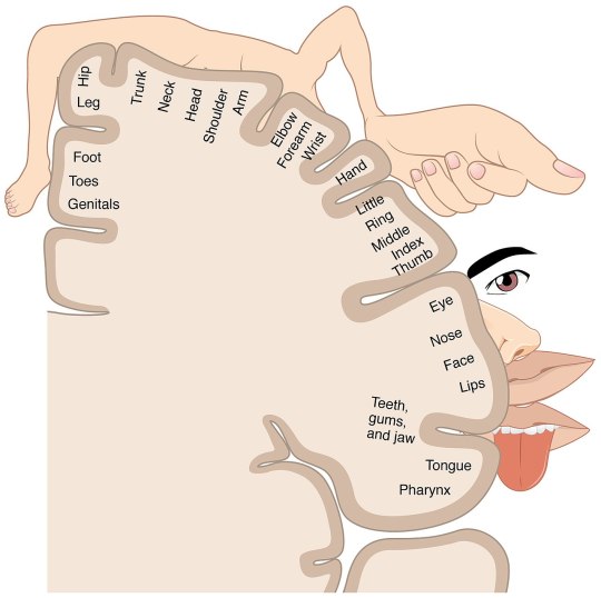
Why is this representation so distorted?
The distortion of the homonculus does not reflect the actual size of the different parts of the body, but rather their functional importance in the brain. The more sensitive an area of the body or the more precise the motor control required, the larger the surface area it occupies on the homonculus.
How was the homonculus discovered?
It was the Canadian neurosurgeon Wilder Penfield who developed this mapping in the 1930s. While operating on epileptic patients, he electrically stimulated different areas of the cerebral cortex. He realised that stimulating certain areas triggered movements or sensations in specific parts of the body. By repeating these experiments, he was able to establish a correspondence between the different regions of the brain and the different parts of the body.
What are the implications of homonculus?
The discovery of the homonculus has had a major impact in several areas:
- Neurology: It has led to a better understanding of the mechanisms behind motor or sensory disorders, such as paralysis or loss of sensitivity.
- Neurosurgery: It helps neurosurgeons to plan their operations in such a way as to avoid damaging essential areas of the brain.
- Functional rehabilitation: It guides therapists in setting up rehabilitation programmes tailored to patients who have suffered brain damage.
Beyond the homonculus: brain plasticity
It is important to note that the homonculus is a static representation of the brain, but our brain is a dynamic and plastic organ. The connections between neurons can change throughout our lives as a result of learning and experience. For example, the homonculus of someone who plays the piano will be slightly different from that of someone who does not.
In conclusion
Penfield's homonculus is a valuable tool for understanding how our brain works. It reminds us that our brain is a complex, organised structure, in which each part has a precise function. Although this representation is simplified, it offers us a fascinating vision of how our body and mind are interconnected.
Go further
#homonculus#Penfield#brain#neurology#neuroscience#brain mapping#motor skills#sensitivity#cerebral cortex#cerebral plasticity
1 note
·
View note
Text
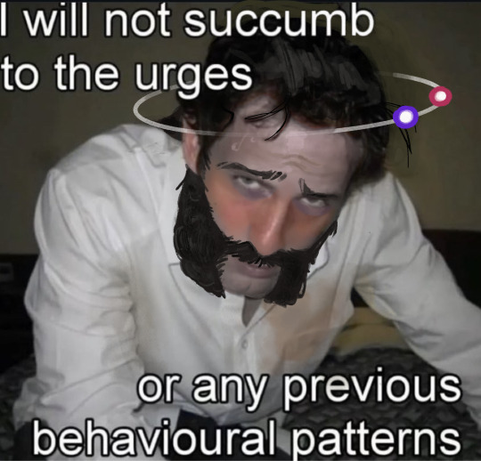
this is basically that wasteland of reality thought project right
(original pic and source below)

(source)
#i was going through my de tag for ideas and found this again and i just felt like. lightning strike my cerebral cortex.#i had to do it IMMEDIATELY#disco elysium#fanart#de fanart#disco elysium fanart#shitpost#de#my art#harry dubois#harry du bois
478 notes
·
View notes
Text


@ whoever paired them up i hope your pillow is always cold on both sides your charger works at every single angle you never stub your toe you get a raise and have an amazing year because YOU DESERVE ALL OF IT
#IF THEY KISS ON THE MOUTH IS SO JOVER FOR ME IM SO SERIOUS RN#PERCEIVE ME LIKE OFF SCREAMING HIS LUNGS OUT#I COULDN'T UNDERSTAND ANYTHING AND IT WAS STILL A SHOT OF SEROTONIN INJECTED STRAIGHT INTO MY CEREBRAL CORTEX#I LOVE EVERYONE IN THAT ROOM#papang phromphiriya#podd suphakorn#pod suphakorn#poddpapang#podpapang#IDK HOW TO TAG ANYMORE#m: txt
66 notes
·
View notes
Text
i love that if nolan’s hair isn’t styled back or it grows out enough it just falls into a natural middle part, he just like me fr


#invincible#nolan grayson#omniman#omni man#i love him#my beautiful husband who’s parasitically attached to my cerebral cortex#i headcanon’d this to my friends on discord a good while back#and I am proven yet again that I know him so well#he probably uses some kind of pomade or clay in his hair to keep it back
172 notes
·
View notes
