#biomolecular condensate
Explore tagged Tumblr posts
Text
check out this article about biomolecular condensates!
you might know that your cells contain organelles, little specialized sub-parts like nucleus, mitochondria, and ribosome, all suspended in a fluid called cytoplasm. it turns out that there are some globby bits of RNA and ugly proteins also floating around and doing important cell stuff we didn't even know about until the last couple decades!
not only are these biomolecular condensates present in eukaryotic cells (animals, plants, and fungi) where they were discovered, but also in prokaryotes (e.g. bacteria), which *don't* have organelles! this helps explain what's going on inside prokaryotic cells.
BUT ALSO this is kind of a missing link in evolutionary biology: we know how RNA and simple proteins can form from simple and abundant chemicals, but we didn't know how to get lipids, which are needed to make the membrane that comprises the outside of a cell or organelle. now we know that you can have an organelle without a membrane. that means there is a plausible and complete path from chemicals to RNA to proteins to biomolecular condensates to organelles to clusters of organelles to single cells to capturing other cells and eventually reproducing as a whole, to multicellular entities, to plants and animals and fungi!
biology was the science of least interest to me in high school, but since I've started learning more about evolutionary biology, I'm absolutely blown away by how cool everything is all the time
#biology#molecular biology#evolutionary biology#evolution#biomolecular condensate#organelle#science#cell biology#rna#protein#animal#animals#plant#plants#fungi#fungus#mitochondria#ribosome#nucleus#cell membrane#lipid#lipids#prokaryote#prokaryotes#eukaryote#eukaryotes#bacteria#cells
0 notes
Text

Beyond displays: Liquid crystals in motion mimic biological systems
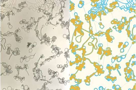
Liquid crystals are all around us, from cell phone screens and video game consoles to car dashboards and medical devices. Run an electric current through liquid crystal displays (LCDs) and they generate colors, thanks to the unique properties of these fluids: rearrange their shape, and they reflect different wavelengths of light. As the lab of Chinedum Osuji, Eduardo D. Glandt Presidential Professor and Chair of Chemical and Biomolecular Engineering, recently discovered, these fascinating molecules may be able to do even more. Under the right conditions, liquid crystals condense into astonishing structures, spontaneously generating filaments and flattened disks that can transport material from one place to another, much like complex biological systems. The insight may lead to self-assembling materials, new ways to model cellular activity and more. "It's like a network of conveyor belts," says Christopher Browne, a postdoctoral researcher in Osuji's lab and the co-first author of a recent paper in Proceedings of the National Academy of Sciences that describes the finding.
Read more.
8 notes
·
View notes
Text
Important signaling molecules called phospholipids are active throughout cells in small compartments called condensates, rather than functioning primarily in cell membranes as previously thought, according to a study from researchers at Weill Cornell Medicine. The finding helps open a new avenue of investigation in cell biology and may also be relevant to the study of neurodegenerative diseases such as amyotrophic lateral sclerosis (ALS) and Alzheimer's disease. Condensates in cells, also called biomolecular condensates, behave like oil drops within water. They are made of proteins, and often RNA molecules, that have weakly conglomerated to form distinct globules in the cell. These globules form compartments with chemical properties that differ from those of the surrounding, watery interior of the cell.
Continue Reading.
29 notes
·
View notes
Text
I have been studying nonstop for three days straight and I DESPERATELY need a break.
Please spam my inbox!
I need to talk about something besides biomolecular condensates and gene expression!
#it can be the most random thoughts#I just need some sort of conversational stimulation that isn't biology related#If I make it to see the end of Wednesday I will be a free woman
2 notes
·
View notes
Text
Postdoctoral Fellow - Cancer Research at MSKCC Memorial Sloan Kettering Cancer Center The Tulpule lab at MSKCC is seeking postdocs to study pediatric cancer, biomolecular condensates, and DNA repair. See the full job description on jobRxiv: https://jobrxiv.org/job/memorial-sloan-kettering-cancer-center-27778-postdoctoral-fellow-cancer-research-at-mskcc/?feed_id=91274 #cancer_biology #cell_signaling #pediatric_cancer #ScienceJobs #hiring #research
0 notes
Text
Cells have more mini ‘organs’ than researchers thought − unbound by membranes, these rogue organelles challenge biology’s fundamentals
0 notes
Text
Imaging-Based Quantitative Assessment of Biomolecular Condensates in vitro and in Cells
BioRxiv: http://dlvr.it/T7FM0v
0 notes
Link
0 notes
Text
#RNA-mediated ribonucleoprotein assembly controls TDP-43 nuclear retention
by Patricia M. dos Passos, Erandika H. Hemamali, Lohany D. Mamede, Lindsey R. Hayes, Yuna M. Ayala TDP-43 is an essential #RNA-binding protein strongly implicated in the pathogenesis of neurodegenerative disorders characterized by cytoplasmic aggregates and loss of nuclear TDP-43. The protein shuttles between nucleus and cytoplasm, yet maintaining predominantly nuclear TDP-43 localization is important for TDP-43 function and for inhibiting cytoplasmic aggregation. We previously demonstrated that specific #RNA binding mediates TDP-43 self-assembly and biomolecular condensation, requiring multivalent interactions via N- and C-terminal domains. Here, we show that these complexes play a key role in TDP-43 nuclear retention. TDP-43 forms macromolecular complexes with a wide range of size distribution in cells and we find that defects in #RNA binding or inter-domain interactions, including phase separation, impair the assembly of the largest species. Our findings suggest that recruitment into these macromolecular complexes prevents cytoplasmic egress of TDP-43 in a size-dependent manner. Our observations uncover fundamental mechanisms controlling TDP-43 cellular homeostasis, whereby regulation of #RNA-mediated self-assembly modulates TDP-43 nucleocytoplasmic distribution. Moreover, these findings highlight pathways that may be implicated in TDP-43 proteinopathies and identify potential therapeutic targets. https://journals.plos.org/plosbiology/article?id=10.1371%2Fjournal.pbio.3002527&utm_source=dlvr.it&utm_medium=tumblr
0 notes
Text
0 notes
Text
Hard to Drug: Protein Droplets Reveal New Ways to Inhibit Aggressive Form of Prostate Cancer - Technology Org
New Post has been published on https://thedigitalinsider.com/hard-to-drug-protein-droplets-reveal-new-ways-to-inhibit-aggressive-form-of-prostate-cancer-technology-org/
Hard to Drug: Protein Droplets Reveal New Ways to Inhibit Aggressive Form of Prostate Cancer - Technology Org
Many of the most potent human oncoproteins belong to a class of proteins called transcription factors, but designing small molecule drugs that target transcription factors is a major challenge, especially when treating aggressive forms of prostate cancer.
Surgeon during a surgery – illustrative photo. Image credit: National Cancer Institute
An international team of researchers from the Institute for Research in Biomedicine in Barcelona, the Max Planck Institute for Molecular Genetics, BC Cancer (University of British Columbia) and other institutions has now discovered a potential way to target the androgen receptor, the most prominent oncogenic transcription factor in prostate cancer, based on its propensity to form droplets also known as condensates.
The results described in this publication set the basis for the foundation of Nuage Therapeutics, a spinoff of the Institute for Research in Biomedicine and ICREA.
Super-resolution (τ-STED) imaging of the androgen receptor in human prostate adenocarcinoma cells. Image credit: MPI for Molecular Genetics
Transcription factors play essential roles in turning the genetic information encoded by genes into proteins in all cells and organisms. These regulatory proteins bind DNA, turn genes on or off, and control the rate at which DNA is transcribed into mRNA, which is needed for protein synthesis. Because of their central role in transcriptional control, many diseases can be traced back to dysregulated transcription factors.
Inhibiting their activity, especially in cancer, offers therapeutic potential, but many transcription factors have a trick up their sleeve. Their activation domains are intrinsically disordered, meaning that the chains of amino acids that make up the domain lack a clear three-dimensional structure. The lack of a stable 3D structure makes it virtually impossible to design drugs that bind to the activation domains.
A research team led by Xavier Salvatella and Antoni Riera at the Institute for Research in Biomedicine, ICREA and the University of Barcelona, Denes Hnisz at the Max Planck Institute for Molecular Genetics and Marianne D. Sadar at BC Cancer (University of British Columbia, Canada) – focused on the tendency of intrinsically disordered proteins to form so-called biomolecular condensates.
They found that the mechanisms involved in condensation could be exploited to inhibit androgen receptor activity in prostate cancer. “The rationale we have followed to optimize an inhibitor of the androgen receptor could be exploited to inhibiting other transcription factors, opening up new possibilities to address unmet medical needs,” says Xavier Salvatella.
Cellular droplets, a new approach to targeting transcription factors
Biomolecular condensates resemble proteinaceous blobs floating on water under a microscope. The condensates form in a process called liquid-liquid phase separation, similar to how oil droplets coalesce when mixed in water.
“We had previously observed that the androgen receptor forms biomolecular condensates when you add even a tiny amount of an activating molecule, such as testosterone, to cells” says Shaon Basu, now a computational biologist at the Charité and one of the study’s first authors together with Paula Martínez-Cristobal at the Institute for Research in Biomedicine.
The scientists hypothesized that there could be a link between the activation of the androgen receptor and its propensity to form droplets.
Working with biophysicist Xavier Salvatella, they used nuclear magnetic resonance techniques to identify several short pieces within the intrinsically disordered activation domain that are essential for phase separation. Moreover, the same short pieces turned out to be also necessary for the gene-activating function of the receptor.
“We discovered short sequences in the activation domain that tend to be disordered when the protein is soluble, and surprisingly, these regions seem to form more stable helices when the protein is concentrated in condensates,” explains Hnisz. The short helices create transient binding pockets that can be targeted with inhibitors when the receptor is in condensates.
Improving compounds for the treatment of prostate cancer
Working with the labs of Antoni Riera and Marianne Sadar, the team then improved an experimental small molecule inhibitor to fit almost perfectly into the transient binding pocket. They then tested in cell and mouse models whether these changes would increase the efficacy in an aggressive, late-stage form of prostate cancer.
“We modified the chemical features of the compound to match the features of androgen receptor condensation, resulting in a tenfold increase in the potency of the molecule in castration-resistant prostate cancer,” says Paula Martínez-Cristobal, also first author of the study.
“This is really important, because castration-resistant prostate cancer is an extremely aggressive cancer that is resistant to the current first-line therapeutics,” she adds.
However, more research is needed before these findings can be translated into new and safe therapeutics, the authors agree. The team hopes that the basic mechanisms they have discovered may be applicable to other transcription factors, opening the door to targeting these important molecules in many different diseases.
“We believe that the idea that there are certain sequences within intrinsically disordered protein domains that adopt a transiently stable structure in condensates is universal and likely generalizable to transcription factors,” concludes Hnisz.
Nuage Therapeutics
Founded by Xavier Salvatella, Mateusz Biesaga, Denes Hnisz and Judit Anido, the biotech company Nuage Therapeutics develops drug screening assays to target intrinsically disordered proteins that undergo biomolecular condensation, thus providing new treatments for diseases currently considered difficult to treat.
The findings now published laid the groundwork for the foundation of this company in September 2021. The potential of its science led to a €12M Seed financing round on June 2023.
Source: MPG
You can offer your link to a page which is relevant to the topic of this post.
#2023#3d#3D structure#acids#Adenocarcinoma#Aging news#amino acids#androgens#approach#biomedicine#biotech#Biotechnology news#Canada#Cancer#cell#Cells#challenge#chemical#Design#Diseases#DNA#domains#droplets#drug#drugs#experimental#factor#Featured life sciences news#Features#form
0 notes
Text
Doris Loh comments:
It feels awesome to be validated by science.
A recently published study ties together two of my peer-reviewed papers on melatonin regulation of phase separation [1, 2, 3].
How?
In my first paper on melatonin regulation of phase separation, I talked extensively about the post-translational modification of SUMOylation used by melatonin to regulate phase separation https://www.mdpi.com/2076-3921/10/9/1483/htm.
In my third paper on melatonin regulation of phase separation, I explained how melatonin can suppress viral replication by regulating phase separation of the nucleocapsid (N) protein https://www.mdpi.com/1422-0067/23/15/8122/htm
Now this peer-reviewed paper published at Nature reports that SUMOylation facilitates nucleocapsid protein phase separation, enhancing the virulence of the SARS-CoV-2 virus [1].
I published the first paper in September 2021, the third in July 2022. Some serious #lohcurve for you. Got MEL?
References:
[1] Ren, J.; Wang, S.; Zong, Z.; Pan, T.; Liu, S.; Mao, W.; Huang, H.; Yan, X.; Yang, B.; He, X.; et al. TRIM28-Mediated Nucleocapsid Protein SUMOylation Enhances SARS-CoV-2 Virulence. Nat. Commun. 2024, 15, 244.
[2] Loh, D.; Reiter, R. J. Melatonin: Regulation of Biomolecular Condensates in Neurodegenerative Disorders. Antioxidants (Basel) 2021, 10 (9). https://doi.org/10.3390/antiox10091483.
[3] Loh, D.; Reiter, R. J. Melatonin: Regulation of Viral Phase Separation and Epitranscriptomics in Post-Acute Sequelae of COVID-19. Int. J. Mol. Sci. 2022, 23 (15), 8122. https://doi.org/10.3390/ijms23158122.
0 notes
Link
Boston MA (SPX) Aug 22, 2023 Inside all living cells, loosely formed assemblies known as biomolecular condensates perform many critical functions. However, it is not well understood how proteins and other biomolecules come together to form these assemblies within cells. MIT biologists have now discovered that a single scaffolding protein is responsible for the formation of one of these condensates, which forms within
0 notes