#Transmission electron microscopy
Explore tagged Tumblr posts
Visit Tumblr Blog
Explore Tumblr blogs with no restrictions, modern design and the best experience.
Last Seen Tumblr Blogs


Fun Fact
Tumblr has 411 employees.
Text
Our ability to image the subatomic realm is limited, not just by resolution, but also by speed. The constituent particles that make up – and fly free from – atoms can, in theory, move at speeds approaching that of light. In practice, they often move much slower, but even these slower speeds are way too fast for our eyes, or technology, to see. This has made observing the behavior of electrons something of a challenge – but now the development of a new microscope imaging technique has allowed scientists to catch them in motion, in real time.
Continue Reading.
147 notes
·
View notes
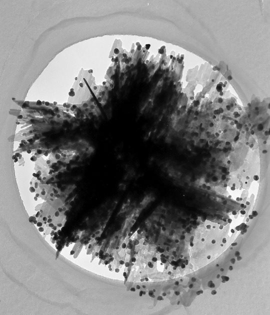
Text

Pseudomonas fluorescens
Image taken via transmission electron microscopy
Photo credit: DR TONY BRAIN / SCIENCE PHOTO LIBRARY
#pseudomonas#pseudomonas fluorescens#transmission electron microscopy#pilot hat#aviator hat#aviator hat with goggles#microbiology#microbes#biology#hats#microbes in hats#microorganisms#bacteria#protozoa#microscopy
25 notes
·
View notes
Text

Time-compression in electron microscopy: Terahertz light controls and characterizes electrons in space and time
Scientists at the University of Konstanz in Germany have advanced ultrafast electron microscopy to unprecedented time resolution. Reporting in Science Advances, the research team presents a method for the all-optical control, compression, and characterization of electron pulses within a transmission electron microscope using terahertz light. Additionally, the researchers have discovered substantial anti-correlations in the time domain for two-electron and three-electron states, providing deeper insight into the quantum physics of free electrons. Background and challenges Ultrafast electron microscopy is a cutting-edge technique that combines the spatial resolution of traditional electron microscopy with the temporal resolution of ultrafast femtosecond laser pulses. This powerful combination allows researchers to observe atoms and electrons in motion, capturing dynamic processes in materials with unparalleled clarity. By visualizing these rapid events in space and time, scientists can gain deeper insights into the fundamental mechanisms that govern material properties and transitions, helping to create advancements in research fields such as nanotechnology, optics, materials science, and quantum physics.
Read more.
#Materials Science#Science#Electron microscopy#Materials characterization#Terahertz#University of Konstanz#Transmission electron microscopy
22 notes
·
View notes
Text


#electron microscopy#transmission electron microscopy#nanoparticles#rare earth hydroxides#zinc tungstate#silver on zinc oxide#zinc titanate#original content
9 notes
·
View notes
Text
A Journey into the World of Microscopy: From Humble Beginnings to High-Tech Magnification
The science of looking into the hidden invisible Microscopy has transformed our understanding of the world around us. It can explore the universe beyond the reach of our naked eyes, with complex cellular structures, red blood cells, viruses and other viruses and microorganisms taking on amazing perspectives
The history of the microscope is a fascinating story of human curiosity, scientific genius, and relentless exploration. From the humble beginnings of simple magnifying glasses to the sophistication of modern electronic microscopes, the invention of microscopes has shaped our understanding of the microscopic world
In the 1600s, Dutch opticians such as Hans and Zachary Janssen are credited with inventing the first microscope. Known for this hybrid microscope, many lenses were used to magnify objects up to 30 times.At the end of the 17th century, Antony van Leeuwenhoek, Dutch draper some changed our perception of thumbnails. Armed with a well-made single-lens microscope, and explored the hidden reaches of nature. In 1674, Leeuwenhoek discovered microorganisms in lake water, which he aptly named “animalcules”. His discovery laid the foundations of biology and inspired generations of scientists. This incredible feat allowed him to uncover a hidden universe – the first sightings of bacteria, red blood cells, and other microorganisms.
Formation of the scientific environment (17th-19th centuries): Leeuwenhoek’s discoveries boosted scientific research. Robert Hooke, an English scientist, established these developments. In 1665, his book "Micrographia" recorded his observations with a compound microscope. Notably, the term "cell" was coined by Hooke when he examined cork tissue, laying the foundation for cell biology.Microscope systems flourished throughout the 18th and 19th centuries Joseph Lister and other scientists addressed the limitations of the early lenses, introducing improvements that reduced image distortion.
Beyond the Limits of Light: The Beginning of the New Age (19th-20th century): As the 19th century progressed, the limitations of optical microscopy became apparent and scientists yearned for a tool which can go deeper into cells. This research culminated in the development of the electron microscope in the 1930s. The 20th century was revolutionary with the invention of the electron microscope. Unlike light microscopes, which use visible light, electron microscopes use electron beams to achieve much higher magnification.Formation of the scientific environment (17th-19th centuries): Leeuwenhoek’s discoveries boosted scientific research. Robert Hooke, an English scientist, established these developments. In 1665, his book "Micrographia" recorded his observations with a compound microscope. Notably, the term "cell" was coined by Hooke when he examined cork tissue, laying the foundation for cell biology.Microscope systems flourished throughout the 18th and 19th centuries Joseph Lister and other scientists addressed the limitations of the early lenses, introducing improvements that reduced image distortion.
Beyond the Limits of Light: The Beginning of the New Age (19th-20th century): As the 19th century progressed, the limitations of optical microscopy became apparent and scientists yearned for a tool which can go deeper into cells. This research culminated in the development of the electron microscope in the 1930s. The 20th century was revolutionary with the invention of the electron microscope. Unlike light microscopes, which use visible light, electron microscopes use electron beams to achieve much higher magnification.
In the 1930s, German experts Max Knoll and Ernst Ruska made the first electron microscope. This tool let us see tiny things like cells and even atoms by using electron beams, not light, getting images many times bigger. This cool invention showed us the tiny parts inside cells, viruses, and stuff too small to see before. The 1900s brought even more cool microscopes. New kinds like phase-contrast and confocal microscopy let scientists look at live cells without using stuff that could hurt them. Now, the world of looking at tiny things is getting even better. Today, we have high-tech microscopes that use computers and lasers. These let us see and even change tiny things in ways we never could before.
Modern Microscopy's Diverse Arsenal - Today, the field of microscopy boasts a diverse range of specialized instruments, each tailored to address specific scientific needs. Here's a glimpse into some remarkable examples:
Scanning Electron Microscope (SEM): Imagine a high-tech camera that captures images using a beam of electrons instead of light. That's the essence of a SEM. By scanning the surface of a sample with a focused electron beam, SEMs generate detailed information about its topography and composition. This makes them ideal for studying the intricate structures of materials like insect wings, microchips, and even pollen grains.
Transmission Electron Microscope (TEM): While SEMs provide exceptional surface detail, TEMs take us a step further. They function by transmitting a beam of electrons through a very thin sample, allowing us to observe its internal structure. TEMs are the go-to instruments for visualizing the intricate world of viruses, organelles within cells, and macromolecules like proteins.
Confocal Microscopy: Ever wished to focus on a specific layer within a thick biological sample and blur out the rest? Confocal microscopy makes this possible. It utilizes a laser beam to precisely illuminate a chosen plane within the sample, effectively eliminating information from out-of-focus regions. This allows researchers to create sharp, three-dimensional images of cells, tissues, and even small organisms.
Atomic Force Microscopy (AFM): This technique takes a completely different approach, venturing into the realm of physical interaction. AFM employs a tiny cantilever, akin to a microscopic feeler, to physically scan the surface of a sample. By measuring the minute forces between the cantilever and the sample's surface, AFM can map its topography at an atomic level. This provides invaluable insights into the properties of materials at an unimaginable scale, making it crucial for research in fields like nanotechnology and surface science.
Fluorescence Microscopy: Imagine illuminating a sample with specific wavelengths of light and observing it glowing in response. That's the essence of fluorescence microscopy. This technique utilizes fluorescent molecules or tags that bind to specific structures within a cell or tissue. When excited by light, these tags emit their own light, highlighting the target structures with remarkable clarity. This allows researchers to visualize specific proteins, DNA, or even pathogens within biological samples.
Super-resolution Microscopy (SRM): Overcoming the limitations imposed by the wavelength of light, SRM techniques like STED (Stimulated Emission Depletion) and PALM (Photoactivated Localization Microscopy) achieve resolutions surpassing the diffraction limit. This allows researchers to visualize structures as small as 20 nanometers, enabling the observation of intricate cellular machinery and the dynamics of individual molecules within living cells.
Cryo-Electron Microscopy (Cryo-EM): This powerful technique takes a snapshot of biological samples in their near-life state. Samples are rapidly frozen at ultra-low temperatures, preserving their native structure and minimizing damage caused by traditional fixation methods. Cryo-EM has been instrumental in determining the three-dimensional structures of complex molecules like proteins and viruses, providing crucial insights into their function and potential drug targets.
Correlative Microscopy: Combining the strengths of multiple microscopy techniques, correlative microscopy offers a comprehensive view of biological samples. For instance, researchers can utilize fluorescence microscopy to identify specific structures within a cell and then switch to electron microscopy to examine those structures in high detail. This integrated approach provides a deeper understanding of cellular processes and their underlying mechanisms.
Light Sheet Microscopy (LSM): Imagine illuminating a thin slice of a sample within a living organism. LSM achieves this feat by focusing a laser beam into a thin sheet of light, minimizing photobleaching and phototoxicity – damaging effects caused by prolonged exposure to light. This allows researchers to observe dynamic processes within living organisms over extended periods, providing valuable insights into cellular behavior and development.
Expansion Microscopy (ExM): This innovative technique physically expands biological samples by several folds while preserving their structural integrity. This expansion allows for better resolution and visualization of intricate cellular structures that would otherwise be difficult to distinguish using traditional microscopy methods. ExM holds immense potential for studying the organization and function of organelles within cells.
Scanning Near-Field Optical Microscopy (SNOM): This innovative technique pushes the boundaries of resolution by utilizing a tiny probe that interacts with the sample at an extremely close range. SNOM can not only image the surface features of a sample with exceptional detail but also probe its optical properties at the nanoscale. This opens doors for research in areas like material science and photonics, allowing scientists to study the behavior of light at the interface between materials.
X-ray Microscopy: Stepping outside the realm of light and electrons, X-ray microscopy offers unique capabilities. By utilizing high-energy X-rays, this technique can penetrate deep into samples, making it ideal for studying the internal structure of dense materials like bones and minerals. Additionally, it allows for the visualization of elements within a sample, providing valuable information about their distribution and composition.
From revealing the building blocks of life to aiding in the development of new medicines, the microscope has played an undeniable role in shaping our scientific understanding. As technology continues to evolve, one can only imagine the future breakthroughs this remarkable invention holds in unveiling the secrets of our universe, both seen and unseen. These advancements hold the potential to revolutionize our understanding of biological processes, develop new materials with extraordinary properties, and ultimately pave the way for breakthroughs in medicine, nanotechnology, and countless other fields. As we continue to refine and develop novel microscopy techniques and the future holds immense promise for further groundbreaking discoveries that will undoubtedly revolutionize our perception of the world around us.
#science sculpt#life science#science#molecular biology#biology#biotechnology#artists on tumblr#microscopy#microscope#Scanning Electron Microscope#Transmission Electron Microscope#Confocal Microscopy#Atomic Force Microscopy#Fluorescence Microscopy#Expansion Microscopy#X-ray Microscopy#Super-resolution Microscopy#Light Sheet Microscopy#illustration#illustrator#illustrative art#education#educate yourself#techniques in biotechnology#scientific research#the glass scientists#scientific illustration#scientific advancements
7 notes
·
View notes
Text
The Transmission Electron Microscopy Market size was valued at USD 0.86 Billion in 2023 and the total Transmission Electron Microscopy Market revenue is expected to grow at a CAGR of 4.3% from 2024 to 2030, reaching nearly USD 1.16 Billion
1 note
·
View note
Text
I love how these kids are multilingual, they'll have a full conversation in French and just drop "TEM, motherfucker!!" in the middle of it
0 notes
Text
Estivating animals such as lungfish and amphibians form cocoon structures to reduce evaporative water loss in the dry season (17–19). African lungfish live inside their mucus cocoon for months or even years (20). Previous studies described the cocoon of anuran and urodele amphibians as shed epithelial layers, whereas the lungfish cocoon was described as a translucent brownish dried mucus structure (17, 19, 20–23). Estivating lungfish are likely more vulnerable to pathogen attack since they are immobile and continuous skin mucus secretion stops once the cocoon is formed. Thus, we hypothesized that the cocoon may have important immunological functions thus far undescribed. Our histological observations revealed that the lungfish cocoon is not a dead, dry mucus layer but, instead, is a living tissue with a well-defined cellular structure. The thickness of the lungfish cocoon was approximately 350 to 900 μm, although this may vary from body site to body site, and the distance between the epidermis and the cocoon ranged from 15 to 173 μm (Fig. 3A). The cocoon was composed of different cell types including goblet cells, epithelial cells, endothelial cells, and immune cells (Fig. 3B). Transmission electron microscopy confirmed the presence of goblet cells with many secretory mucus granules within the cocoon (fig. S3D). Moreover, abundant nests of cells were observed surrounded by long channels lined by double-layered squamous cells. A series of monolayered vessels running parallel and perpendicular to the main channel were identified. These nests of cells contained endothelium-lined vessels that could potentially serve to transport cells into and within the cocoon (Fig. 3B). Channels were not filled with mucosal secretions based on periodic acid–Schiff staining (Fig. 3C). The cocoon contained abundant granulocytic cells (fig. S2A) that were MPO+, indicating transmigration of granulocytes into the cocoon [fig. S2B; modeled in Fig. 2 (C and D)]. Scanning electron microscopy (SEM) showed that during terrestrialization, the flattened smooth epidermis of freshwater fish (Fig. 3E) became a disrupted surface covered by the cocoon where lungfish cells and groups of bacterial cells were observed

Free-swimming lungfish skin is characterized by a columnar mucosal epithelium. (A) Large numbers of multipotent stem cells with alkaline phosphatase activity (fig. S1) can be observed at the interphase between the epidermis and the dermis. Granulocyte deposits in the tissue reservoirs of free-swimming lungfish become mobilized to the skin via peripheral circulation when lungfish begin to sense lack of food and water. (B) Skin remodeling begins, with increasing numbers of granulocytes infiltrating the dermal and epidermal layers resulting in loosening of the basal membrane and inflammation. (C) The cocoon then starts to form by detachment and shedding of the inflamed epidermis. Many granulocytes are part of the cocoon, and they produce ETs in response to the high microbial load. Epithelial cells, goblet cells, and antimicrobial peptides (AMPs) are also present in the cocoon. The pool of stem cells starts to regenerate the epidermis, while granulocytes continue to arrive from reservoirs maintaining an inflammatory state. (D) In the next stages of estivation (end of the induction phase), the lungfish skin shows complete flattening of the epidermis and goblet cell exhaustion. The cocoon has several layers derived from multiple rounds of epidermal shedding and regeneration, and stem cell numbers are severely reduced. Granulocytes in the cocoon continue to undergo ETosis and are still elevated in the epidermis and dermis compared to free-swimming controls. It is unknown whether the cocoon continues to thicken beyond 2 weeks after terrestrialization
(source) when lungfish hide in the dirt in the dry season, their skin dissolves into a living cocoon full of immune cells! they have huge reservoirs of white blood cells at all times, so if they suddenly dry out they can pump them into the cocoon
6 notes
·
View notes
Note
i was going through notes for biology (woo microscopes) and there's a type of electron microscopy called transmission electron microscopy and the acronym for it is TEM which made me think of you
p.s. you cant just say "the universe wasn't mourning his loss, it was simply welcoming him home" and expect me to continue learning peacefully what the heck man
-🍁

#shouting speaks#asks#hunger au#ALDJSHDJEJDJ#OBSESSED WITH THE MICROSCOPY THING IM EVERYWHERE BABEYYYY#ppl always ask if i named myself after the undertale dog and im gonna start telling them its actually the electron microscopy#txt
14 notes
·
View notes
Text


Differences in space environment in the Moon’s near and far side
This study is led by Dr. XIAN Haiyang and Dr. ZHU Jianxi of the Guangzhou Institute of Geochemistry, Chinese Academy of Sciences. On June 25, 2024, the Chang’e-6 mission successfully achieved the world’s first sampling of the lunar farside and returned safely to Earth. In August, based on his outstanding performance in the Chang’e-5 sample research, Dr. Xian received the first batch of Chang’e-6 samples. This marks the first time in human history that samples have been directly obtained from the lunar farside, and the research team hopes to use these samples to gain insights into the space weathering characteristics on the lunar farside. The space weathering features recorded in lunar samples can sensitively capture information about the surrounding space environment, helping us better understand the conditions on the lunar farside.
Under Dr. Xian’s guidance, his student Lin Jiarui at the Electron Microscopy Center of the Guangzhou Institute of Geochemistry analyzed these precious samples one by one using a scanning electron microscope (SEM). To preserve as much of the surface information as possible, she chose to evenly distribute the fine-grained powder on conductive adhesive and then deposited a 10 nm carbon film, observing the samples at a low voltage of 3 kV. After examining nearly a thousand particles, she noted that the Chang’e-6 samples exhibited noticeably less melt drops and melt splashes on their surfaces. To further systematically study the space weathering characteristics, she selected seven mineral particles with distinct compositions using energy-dispersive spectroscopy (EDS), which together represent the main lunar mineral types.
In subsequent transmission electron microscopy (TEM) investigations, Lin and other team members prepared a feldspar particle (designated P2‑001) using focused ion beam (FIB) techniques and discovered that its surface lacked the nanophase metallic iron (npFe⁰) particles commonly found in Apollo samples. Typically, the surface of feldspar in Apollo samples exhibits a vapor‑deposited layer generated by micrometeorite impacts that contains npFe⁰. They further conducted EDS mapping on the other seven FIB sections under TEM, and the results showed no significant compositional differences between the edges and interiors of these minerals. All regions displaying observable space weathering features—including the amorphized layers, vesicles, and npFe⁰ grains—were consistent with the substrate mineral composition, indicating that these features can be attributed to the damage caused by solar wind radiation on the substrate minerals.
Lin further measured the thickness of the amorphized layers and the grain sizes of npFe⁰, and counted the solar wind tracks in pyroxene and olivine to estimate the solar wind exposure time of the particles. The study found that the solar wind exposure time of the Chang’e-6 samples was close to the minimum observed in the Apollo 11 samples, lower than that of the other Apollo samples, and slightly shorter than that of the Chang’e-5 samples. However, surprisingly, the npFe⁰ grain sizes in the Chang’e-6 samples were larger. “This might suggest that solar wind radiation in this region leads to more pronounced segregation and aggregation of iron,” she noted. These exciting new results add to the growing evidence that space weathering on the lunar farside may differ from that on the nearside, and, contrary to previous findings from Apollo and Chang’e-5 samples, solar wind radiation plays a more dominant role in the space weathering process on the lunar farside.
There are differences in the solar wind’s influence on different regions of the Moon. During each synodic month, the near side of the Moon enters Earth’s magnetotail, where the protection afforded by Earth’s magnetic field reduces its exposure to the solar wind; in contrast, the farside is continuously exposed to direct solar wind radiation. Moreover, due to orbital dynamics, different locations on the Moon experience varying impact velocities from cometary and asteroidal meteoroids. The relative velocity between the Moon’s surface and impacting meteoroids changes with the lunar phase: during a full moon, when the Moon and meteoroids move in the same orbital direction, the relative velocity increases; the opposite occurs during a new moon.
Micrometeoroid impacts and solar wind radiation are the two primary processes driving space weathering, but the effective sputtering rate from the solar wind and vapor deposit from micrometeorite impacts offset each other. Therefore, when discussing space weathering mechanisms, it is essential to consider the relative contributions of both factors under different space environments. The findings from the Chang’e-6 samples indicate that on the lunar farside, the effect of the solar wind exceeds that of micrometeorite impacts, further demonstrating that the space weathering process is regulated by variations in the space environment.
Since the first images of the lunar farside were captured in 1959, it has been evident that its topography is markedly different from that of the nearside—a phenomenon known as the Moon’s “dichotomy.” However, whether the lunar space environment also exhibits a similar “dichotomy” had until now only been inferred from remote sensing spectra. The latest analysis of the Chang’e-6 samples provides direct, sample‑based evidence for this hypothesis, highlighting the critical role of space environmental variables in the space weathering process. This discovery not only deepens our understanding of how solar wind radiation and micrometeorite impacts shape the lunar surface, but also offers important insights for studying the space weathering evolution of other airless bodies.
TOP IMAGES: Yellow rectangles indicate the positions where FIB sampling was carried out and the curved yellow lines indicate mineral boundaries. Pgt, pigeonite; Tro, troilite; Ilm, ilmenite; Aug, augite; Chr, chromite; An anorthite; Fo, forsterite. Credit ©Science China Press
LOWER IMAGES: (a) The relative influence of solar wind on different lunar locations. (b) The relative (micro)meteoroid flux at different longitudes. (c) The relative contribution of solar wind and (micro)meteoroid impact at different lunar sampling sites. Credit ©Science China Press
3 notes
·
View notes
Text
Types of Microscopes
1. Simple Microscope
2. Compound Microscope
3. Phase Contrast Microscope
4. Fluorescence Microscope
5. Electron Microscope
6. Scanning Electron Microscope (SEM)
7. Transmission Electron Microscope (TEM)
8. Dark Field Microscope
9. Dissecting Microscope (Stereo Microscope)
10. Digital Microscope
11. Scanning Probe Microscope (SPM)
12. Atomic Force Microscope (ATM)
13. Inverted Microscope
14. Acoustic Microscope
15. X-Ray Microscope
16. Polarizing Microscope
17. Metallurgical Microscope
18. Pocket Microscope
19. USB Microscope
20. Confocal Microscope
21. Laser Scanning Microscope
22. Differential Interference Contrast Microscope (DIC)
23. Near-field Scanning Optical Microscope (NSOM)
24. Raman Microscope
25. Super-resolution Microscope
26. Cryo-electron Microscope
27. Time-lapse Microscope
There is a wide range of microscopy techniques and instruments used in various fields of science and research.
#forensic#forensics#criminology#forensic science#evidence#criminalistic#forensic field#crime#forensic science notes#crime scene investigation#electron microscope#microscope
8 notes
·
View notes
Text

Yersinia pestis
Yersinia pestis is the bacterium responsible for plague, with the most common manifestations being bubonic plague, septicemic plague, and pneumonic plague.
Image taken via transmission electron microscopy. Bar = 1 μm
Photo credit: Hans R. Gelderblom, Rolf Reissbrodt/RKI
#yersinia pestis#plague#black plague#bubonic plague#propeller hat#microbiology#microbes#biology#hats#microbes in hats#microorganisms#bacteria#protozoa#microscopy
83 notes
·
View notes
Text

Tetragonal Zirconia Polycrystals (TZP)
Composition Y2O3 2-3, ZrO2 96-97 (wt%) [...] Processing Pressed powder mixtures sintered at 1400-1550ºC. Applications Where toughness, wear-resistance and refractoriness are needed e.g. extrusion dies, machinery wear parts and piston caps. Sample preparation Grind, polish, ion thin and carbon coat. Technique Bright-field TEM Length bar 600 nm Further information The comparatively low sintering temperature allows very fine grained (sub-micron), dense and so high strength as well as tough ceramics to be produced. The microstructure shows equiaxed, fine grains with little evidence of weakening grain boundary phases. Such microstructures have some of the highest values of toughness achieved in ceramics. Contributor Prof W E Lee Organisation Department of Engineering Materials, University of Sheffield
Source.
#Materials Science#Science#Microstructures#Ceramics#Magnified view#Transmission electron microscopy#Zirconium#University of Sheffield#DoITPoMS
13 notes
·
View notes
Text
MAGNUS ARCHIVES BIOLOGY ACADEMIA
requested by @adozenforks :)
- just from reading something’s scientific name, you can picture it perfectly in your mind. this is odd, as you’ve never taken any Latin before
- your DNA model twists and turns on for eternity, an endless helix of nucleotides that you can’t look away from
- you’ve computed the Punnett squares. you Knew Graham’s parents, knew their blue eyes, and it just doesn’t Make Sense that Graham’s eyes are a deep, dark brown
- you cut into the oleander leaf hoping to examine the cellular structure with transmission electron microscopy. as soon as the scalpel gets past the skin, red beads of a viscous fluid drops from the plant. if you didn’t know any better, you’d say it looked like blood
- they brought in fetal pigs to dissect during class. opening your pig, the organs are smeared with … clown makeup?
- you copy the diagrams directly from your textbook, though you think it might be an outdated copy, or maybe a misprint with incorrect proportions. you don’t remember the tibia being that Long, when you look down to check your leg it seems to match the diagram exactly. by the end of the semester, you are a perfect, textbook specimen. your Professor asks you to stay after class for some, ah, extra lab experience
#tma dark academia#tma#the magnus archives#dark academia#biology#biology academia#gothic academia#jonathan sims#martin blackwood
25 notes
·
View notes
Text
women in scanning and transmission electron microscopy
9 notes
·
View notes
Text
The data collected at Diamond contributed to a wider study of the space weathering signatures on the asteroid.
The Scientists did a chemical analysis of the astroid to see the elements and the element isotype it is made of with ZANES X-ray device. Then looked through an electron microscope ePSIC to see the grain of rock on the asteroid.
The building blocks of Ryugu are remnants of interactions between water, minerals, and organics in the early Solar System prior to the formation of Earth.
X-ray Absorption Near Edge Spectroscopy (XANES).
Diamond’s electron Physical Science Imaging Centre (ePSIC)
Without a protective atmosphere, space-exposed surfaces of airless Solar System bodies gradually experience an alteration in composition, structure and optical properties through Space Weathering
Solar wind irradiation and high-velocity micrometeoroid bombardment dominate space weathering for all airless bodies.
The mineralogy of most Ryugu grains investigated by (scanning) transmission electron microscopy is similar to that of CI chondrites, which are the most chemically primitive materials in the Solar System
#astronomy#asteroid#space weathering#science#science news#scientific research#space#outer space#solar system
4 notes
·
View notes