Text

Giardia lamblia aka Giardia duodenalis and Giardia intestinalis
Photo credit: CDC / Mahmud Tari (Public Domain)
#giardia#giardia duodenalis#giardia intestinalis#giardia lamblia#googly eyes#microbiology#microbes#biology#hat#hats#microbes in hats#microorganisms#bacteria#protozoa#microscopy#why's this guy got 3 names#who approved this
54 notes
·
View notes
Note
Hey your blog is really funny and cool keep up the good work!
P.s. I showed my science teacher your blog, now she loves your blog.
You showed it to your science teacher?!? I'm honored, truly. Thank you <3
8 notes
·
View notes
Note
Hi! Just wanted to let you know that your blog will pop up on my feed occasionally, and it always makes me happy to know that someone out there is taking the time to put microbes in hats. This blog is genius
I appreciate you! I do need to get back into being more consistent though lol. ADHD moment
9 notes
·
View notes
Text

Clostridioides difficile
Photo credit: CDC/ Lois S. Wiggs
#clostridioides difficile#clostridioides#c. difficile#birthday#birthday hat#microbiology#microbes#biology#hat#hats#microbes in hats#microorganisms#bacteria#protozoa#microscopy#this post is brought to you a day late in order for it to be on my actual birthday#last year my birthday was a thursday so it worked perfectly#but i make the rules so i can do what i want#anyway same bit 2 years in a row let's gooo!#very convenient request
67 notes
·
View notes
Note
Could you do bacillus anthracis in a burger king crown?
I know I've considered anthrax before, but haven't because all the image options kinda suck. But for you, dear follower, I will do it.
14 notes
·
View notes
Note
Diatom with those silly fakes glasses with the spring eyeballs
Ooh that's a good one. Again comes dangerously close to "does this count as a hat"...... buuut who cares I make the rules.
9 notes
·
View notes
Note
You should put parakaryon myojinensis in a hat. Maybe a detective hat and magnifying glass, since the creature is a bit of a mystery
Can do!
4 notes
·
View notes
Text

Serratia marcescens
Photo credit: Linawati Hardjito, et al.
#serratia marcescens#serratia#s. marcescens#balaclava#balaclavas#microbiology#microbes#biology#hat#hats#microbes in hats#microorganisms#bacteria#protozoa#microscopy#no I do not know why this image looks like someone printed it out (badly) and then scanned and uploaded it#or somehow used the graphics of a CRT TV to make it#but I didn't have a lot of options#but also it's kinda funny and I love committing to a bit#please appreciate the effort i put into making the balaclavas have a similar effect#also picking balaclava with such a short microbe species is definitely considered bullying#i had to severely manipulate each one to fit nicely
60 notes
·
View notes
Note
Clostridium difficile in a bday hat pls pls
Can do!
#maybe i'll do this one for my own upcoming birthday lmao#same gag two years in a row#they'll never see it coming#though this time it wont be the exact day but whatever. close enough#microbes in hats#microbe posting#ask a microbe
21 notes
·
View notes
Text
Sorry for the lack of hat-related microbial activity lately. Between work, school, and executive dysfunction purgatory, I haven't felt up to working on making posts. Might try and get back on track next week, but this week I've got a lot on my plate. No promises, though.
#microbes in hats#microbe posting#ive been procrastinating real bad on applying for college transfer and i need to work on that#and ive got calculus. and a class that i thought would be fun but is really just more work than it's worth
34 notes
·
View notes
Note
my friend wants to know if you can do/have done euplotes?
I don't think so! If you have any specific requests, I can do those, otherwise just I'll pick one that I find! (Probably whatever one I can find the best image of.)
And if you've got any hat requests, you can send that in as well.
4 notes
·
View notes
Text

Nostoc pruniforme
Photo credit: Science Photo Library
#nostoc pruniforme#nostoc#plague doctor#plague doctor hat#plague doctor mask#plague mask#microbiology#microbes#biology#hat#hats#microbes in hats#microorganisms#bacteria#protozoa#microscopy
74 notes
·
View notes
Text

Candidatus Desulforudis audaxviator
The species name of this bacterium contains the Latin phrase Candidatus (candidate) due to the fact that the species record has not been published in a taxonomically valid manner. It is not associated with any family, order, or class, but is included as a candidate under the phylum Firmicutes.
Candidatus D. audaxviator is a unique species, isolated from the Earth's surface for millions of years and a loner in its ecosystem. These bacteria do not need sunlight or chemical energy for their food or metabolic processes, instead subsisting on radioactive energy for their needs. They are able to fix their own nitrogen and cannot survive in the presence of oxygen.
The species name, audaxviator, is taken from Jules Verne’s “Journey to the Center of the Earth,” and means “descend, bold traveler, and attain the center of the Earth.” Photo credit: NASA (public domain)
#candidatus desulforudis audaxviator#desulforudis audaxviator#miner#miner's hat#microbiology#microbes#biology#hat#hats#microbes in hats#microorganisms#bacteria#protozoa#microscopy
81 notes
·
View notes
Note
Keep up with the good stuff! Hope this semester is not too hard for you. College can be a lot
Thank you! Though I'll admit my inactivity in the past couple weeks has been pure laziness, really. The semester did just start this week, and I'm finally having to take calculus and I'm scared lol. Last semester until I finally get my darn Associate's after five years. (My excuses being working part-time and not knowing what I wanted to study for the first half lmao.)
Then I get to go to a four-year and dive into more chemistry and microbiology!
#microbes in hats#ask a microbe#microbe posting#this is more than you asked about lmao#i'm a yapper at heart#it's incurable
9 notes
·
View notes
Note
it would be so cool if you did e. coli in a jason mask (my friend brought this up. doesn’t have to be e. coli tho :P)
or if you did a microbe with the markiplier pink mustache :DDDDD
I've definitely done E. coli before, but I'll absolutely take the hat suggestions! I'm always in need of those.
18 notes
·
View notes
Text

Caenorhabditis elegans
Photo credit: Unknown. All earliest source links (circa 2008) are dead.
#caenorhabditis elegans#caenorhabditis#c elegans#fez#microbiology#microbes#biology#hat#hats#microbes in hats#microorganisms#bacteria#protozoa#microscopy
159 notes
·
View notes
Text

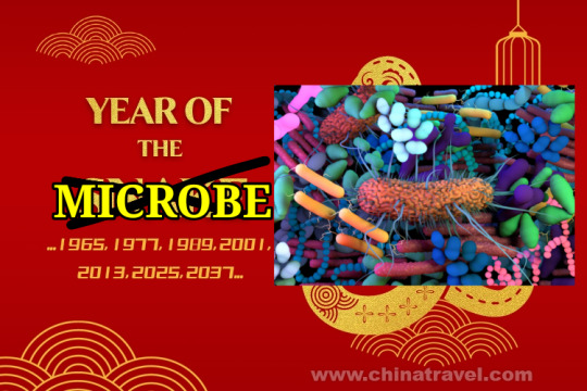
Great news everybody, it is once again the Year of the Microbe! Rejoice!
#microbes in hats#new years#happy new year#2025#microbiology#microbes#biology#hat#hats#microorganisms#bacteria#protozoa#microscopy#chinese new year#year of the microbe#technically we're a bit far off from chinese new year but if I don't post this now I WILL forget#I mean I could schedule it I suppose....#but I'm already emotionally committed to posting it now#and consistency across platforms is key! and I already posted to the discord so we're locked in
68 notes
·
View notes