#Lung Function
Explore tagged Tumblr posts
Visit Tumblr Blog
Explore Tumblr blogs with no restrictions, modern design and the best experience.
Last Seen Tumblr Blogs


Fun Fact
China blocked Tumblr because of pornography and censorship problems in 2013.
Text
#tiktok#health insurance#insurance#healthcare#health and wellness#lung transplant#lungs#lung function#disabilities#disability#disabled
6 notes
·
View notes
Text
The Interconnection Between Emotional Health and Respiratory Well-being: A Comprehensive Exploration
Emotional health is a cornerstone of overall well-being, and its influence extends far beyond mental and social aspects, deeply impacting physical health as well. Among the myriad ways in which emotional health affects the body, its influence on respiratory health is particularly significant, though often underexplored. Research increasingly points to the critical role of emotional states in…
#anxiety and breathing#emotional health#lung function#mental health benefits#mind-body connection#respiratory health#stress management
0 notes
Text
Maximizing Respiratory Health Through Proper Nutrition

Good respiratory health is crucial for maintaining overall wellness, particularly for those receiving home health care in Texas. It’s essential to recognize the role nutrition plays in supporting lung function. We understand the significance of holistic care, including dietary considerations, to enhance respiratory well-being.
Learn more:
https://www.higherstandardshomehealth.com/maximizing-respiratory-health-through-proper-nutrition
0 notes
Text
2024 Health Update
There has been a recent development in my health. As well as many others with my same cf gene. With the addition of a medication called Trikafta, It is a triple combination precision medicine of three separate drugs (ivacaftor, tezacaftor and elexacaftor). I have been taking it on and off but also having allergic reactions to. So for some time I’ve been unable to tolerate it. I have had very…

View On WordPress
0 notes
Text

Decreases stress
In a 2013 study Trusted Source pranayama reduced perceived stress levels in healthy young adults. The researchers speculated that pranayama calms the nervous system, which improves your stress response.
Improves sleep quality
The stress-relieving effects of pranayama may also help you sleep.
In clinical studies Trusted Source a technique known as Bhramari pranayama was shown to slow down breathing and heart rate when practiced for 5 minutes. This may help calm your body for sleep. Increases mindfulness
For many of us, breathing is automatic. We do it without giving it much thought at all.But during pranayama, you need to be aware of your breathing and how it feels. You also practice focusing on the present moment, instead of the past or future. This is known as mindfulness.
Reduces high blood pressure
High blood pressure, or hypertension, is when your blood pressure reaches an unhealthy level. It increases the risk for some potentially serious health conditions like heart disease and stroke.Stress is a major risk factor for high blood pressure. Pranayama can help minimize this risk by promoting relaxation.
Improves lung function
As a type of breathing exercise, the slow, forceful breathing of pranayama may strengthen your lungs.One 2019 study determined that 6 weeks of practicing pranayama for 1 hour a day could have a significant effect on lung function. The practice improved multiple parameters of lung function, according to pulmonary test results.
0 notes
Text
Navigating Spirometry and Its Clinical Applications
Spirometry, encompassing fundamental lung function assessments measuring both exhaled and inhaled air, involves the examination of three interconnected parameters: volume, time, and flow. This method stands as an objective, non-invasive, and remarkably sensitive approach, capable of detecting early deviations in lung function and offering reproducible results. The advent of portable spirometers has extended its reach to almost any setting and, with proper training, made it accessible for a broad range of individuals. The primary objectives for conducting spirometry encompass identifying the presence or absence of lung disorders, quantifying degrees of lung impairment, monitoring the impact of occupational or environmental exposures, and evaluating the effectiveness of medications.

What are the Indications for Pulmonary diseases?
Spirometry is a versatile tool used for detecting and diagnosing a range of pulmonary diseases, including:
Asthma
Chronic obstructive respiratory disease (COPD)
Cystic fibrosis
Pulmonary fibrosis
Patients with these conditions often require regular follow-up tests to monitor their lung function and disease progression. Spirometry is instrumental in assessing the effectiveness of prescribed medications and tracking any changes over time.
Furthermore, spirometry serves as a means to establish an individual's baseline lung function, offering a reference point for future comparisons and the identification of any alterations that may occur over time. This baseline measurement is especially valuable for individuals exposed to occupational hazards that increase the risk of lung disease, such as dust or toxic particles in the air.
Spirometry can also aid in investigating specific respiratory symptoms like persistent cough and dyspnoea (shortness of breath). It is a recommended diagnostic test for heavy smokers aged over 35 due to their elevated risk of pulmonary diseases.
When is Spirometry Contradicted?
If an individual has any of the following that has occurred recently, then it may be better to wait until the patient has fully recovered before carrying out spirometry.
Haemoptysis of unknown origin
Pneumothorax
Acute disorders affecting test performance, such as nausea or vomiting.
Thoracic, abdominal, or cerebral aneurysms
Recent eye surgery
Unstable cardiovascular status, recent myocardial infarction, or pulmonary embolism
Recent thoracic or abdominal surgical procedures
What is Spirometry Device?
Spirometry relies on a device known as a PFT spirometer. This medical apparatus comprises a mouthpiece and a connected tube, which is linked to a machine designed to measure the flow of air. This tool is essential for conducting spirometry tests, as it accurately records the volume, time, and flow of inhaled and exhaled air to evaluate lung function.
How is Spirometer used?
Several techniques can be employed when conducting spirometry, allowing flexibility based on the patient's comfort and cooperation:
Tidal Breathing Technique:
The patient begins with normal, tidal breaths through the mouthpiece.
A deep breath is taken while still using the mouthpiece.
This deep inhalation is followed by a rapid, full exhalation.
Immediate Exhalation Technique:
The patient starts by taking a deep breath.
They then quickly place their mouth securely around the mouthpiece.
A complete exhalation is performed promptly.
Quick Inhale and Exhale Technique:
The patient is instructed to fully empty their lungs first.
Following exhalation, they are asked to take a quick, full inhalation.
This inhalation is succeeded by a thorough exhalation.
What is a normal reading on a spirometer?
In adults, age, height, sex, and race are the main determinants of the reference values for spirometry measurement.
Spirometry measures two main components:
Forced Vital Capacity (FVC): It's the maximum amount of air exhaled after a deep breath.
Forced Expiratory Volume in One Second (FEV1): The volume of air exhaled in one second.
Results are compared to typical values for your demographic, with a normal reading being 80% or higher. Spirometry helps diagnose lung conditions like obstructive and restrictive diseases.
Spirometry results can also aid in diagnosing lung conditions, including:
Obstructive Lung Disease: Conditions that make it challenging to exhale all the air from your lungs due to lung or airway damage. Common examples include asthma, bronchiectasis, COPD, and cystic fibrosis.
Restrictive Lung Disease: These conditions prevent your lungs from fully expanding. Common causes include amyotrophic lateral sclerosis (ALS), interstitial lung disease, muscular dystrophy, sarcoidosis, and scoliosis.
Spirometry is a vital tool in the diagnosis and management of respiratory diseases. Its non-invasive nature, sensitivity to early changes, and reproducibility make it a cornerstone of modern respiratory healthcare. By navigating spirometry and understanding its clinical applications, healthcare providers can better support patients in their journey to optimal lung health.
1 note
·
View note
Text
101: Common Triggers, Treatments, and Prevention Strategies
As a respiratory disease that affects millions of people worldwide, asthma can be a debilitating condition that can significantly impact an individual’s quality of life. Asthma is a chronic illness that causes inflammation of the airways, leading to difficulty breathing and other respiratory symptoms. In this article, we will discuss the common triggers, treatments, and prevention strategies for…

View On WordPress
#allergy medication#asthma#asthma causes#asthma meaning#asthma prevention#asthma symptoms#asthma treatment#asthmatique#chronic illness#immunotherapy#inhalers#lung function#respiratory disease
0 notes
Text
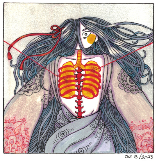
At rest, your lungs wish to deflate, and your ribcage expands outwards.
#better drawn mdzs#mdzs#wei wuxian#yiling laozu#Happy Friday the 13th!#This is scientific fact btw!#Ventilation operates through a series of active and passive forces#The active forces being muscular contraction with inhalation and exhalation having their own set of muscles.#but the interesting part is the passive forces at work:#The lungs have a certain level of elasticity to them - meaning the more they expand the more the those elastic forces are functioning-#-to try and return the system to rest (exhalation passive forces). Your diaphragm is the main force - pushing against the lungs at rest.#Your ribcage on the other hand is under a state of being pulled outwards. It *wants* to be as open as possible.#These to contradicting forces create a constant push and pull which assists in the ebb and flow of air. Most significantly with exhalation.#Now that being said - the primary action of inhalation ventilation is through control centers in your brainstem.#If you lose connection to that due to trauma you're going to need ventilation assistance.#Small note: Respiration is the cellular event of chemical exchange in the alveoli. Ventilation is airflow and pressure.#They are both important but also very different things. Sadly used interchangeably.#My anatomy nerd brain is screaming over the inaccurate ribcage...but its...recognizable. I will get it right one day.#Okay nerd rant over (I cut out a lot of stuff about pressure gradients. They are cool. To me.)#This is a redraw of an mspaint doodle I made back in april. I yearn to make the Yiling Laozu eerie as he deserves#Tear that bitch (affectionate) apart!#Been playing around with hatching for a while and its amazing how many styles there are! Not sure I'll stick with this one (but it was fun)
1K notes
·
View notes
Text
Life Update
#Sorry for the disappearance#my lungs stopped functioning LOL#However God has let me live another day and I'm about to make it everyone's problem <3#(on a brief hiatus maybe?)#(we'll see????)#littlecrittereli#(Im doing a lot better now btw!! No need to worry!! <33)
174 notes
·
View notes
Text
Whumpee doesn’t sing anymore, not since they came back…
Their friends secretly worry it’s because of profound emotional trauma. But Whumpee tries not to think anymore about why they do or don’t do things so this, too, is swept away.
It isn’t until a routine chest X-ray when they have a cough for a few weeks that whumpee really understands why they never sung after… after that place.
Their lungs were partially collapsed, had been all this time. The doc said they had been operating on about half the air the should’ve, that the lower sections deflated at some point and never filled again. They ask about past traumatic injuries.
But Whumpee only shrugged silently. They understood now, all this time- never having enough air, it wasn’t just in their head.
129 notes
·
View notes
Text

here’s what i worked on during lung function panda. that’s not what i typed but im leaving it. i dont think its finished but idk
79 notes
·
View notes
Text
Dude, I swear. This Slay the Princess game has tainted my view on literally everything.
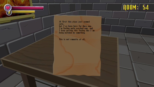
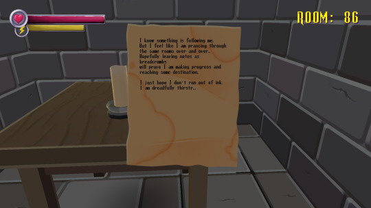
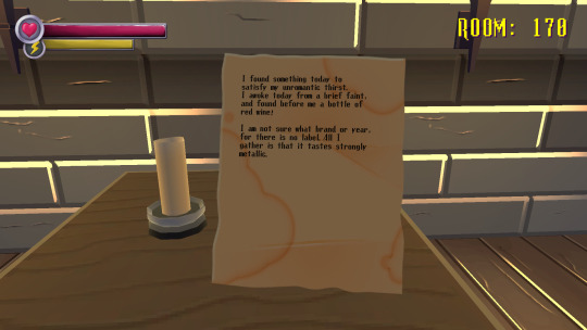
So, I recently got back into Spooky's Jumpscare Mansion (Highly recommend, btw. The HD renovation is getting a massive graphics update in the fall), and I decided to restart by playing through the story mode (Basically, the main game where you run through 1000 rooms getting chased by monsters and cardboard and whatnot).
Along the way, you can read these notes written by people who entered the mansion and didn't make it. The first collection of notes is by a guy who can only describe things as "romantic":

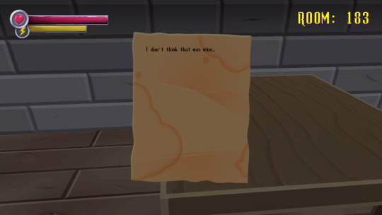
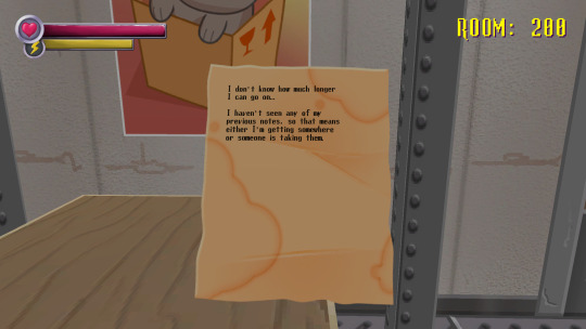

I can hear Smitten saying all of this in my head.
It must be that floaty, poetic style of writing, but still...
#ramble#slay the princess#stp voices#voice of the smitten#spooky's jumpscare mansion#it isn't just this#Smitten has also infiltrated Shakespeare and other bits of poetry#again it might just be that writing style...#other moments of my life being corrupted by Slay the Princess include:#adding “crock of shit” and “what if I throw it out the window” to my everyday vocabulary#wondering what it's like to burn and/or drown#an unhealthy want of trying to tame the crows outside of my house#seeing if I can gain function of my heart lungs liver and nerves in that order.#lol#:)
81 notes
·
View notes
Text
The Impact of Processed Foods on Respiratory Health: A Comprehensive Exploration
Processed foods have become a staple in modern diets worldwide. Their convenience, affordability, and long shelf-life make them appealing choices for busy individuals. However, the health implications of a diet heavy in processed foods extend far beyond the commonly known risks such as obesity, cardiovascular disease, and diabetes. Recent research highlights the significant impact processed foods…
#anti-inflammatory diet#asthma triggers#lung function#oxidative stress#processed foods#respiratory health
0 notes
Text
Racial Bias in Medicine Episode 3: Lung Function Tests and Race
In medicine, there is a device called the spirometer that incorrectly assumed that all Black and Asian people innately had lower lung capacity when compared to White people.
Last year, in May 2023, the American Thoracic Society recommended a race-neutral approach to interpreting spirometry. And a new study published in the New England Journal of Medicine in May 2024 showed how more Black patients would have qualified for different disease classifications, occupational eligibility, disability payments, and more had the race adjustment not existed.
@joelbervell
#black health#racial bias#medical bias#medical discrimination#myths#racial injustice#lung functioning#medical#healthcare#wellbeing#physical health#treatment#medicine#health#health and wellness#racial disparities#black people#blacklivesmatter#black lives matter#black history#health disparities#racism#racist#white supremacy#anti blackness#oppression#discrimination#instagram#pulmonaryhealth#pulmonarycare
53 notes
·
View notes
Text
2024 Health Update
There has been a recent development in my health. As well as many others with my same cf gene. With the addition of a medication named Trikafta, It is a triple combination precision medicine of three separate drugs (ivacaftor, tezacaftor and elexacaftor). I have been taking it on and off but also having allergic reactions to. So for some time I’ve been unable to tolerate it. I have had very…
View On WordPress
0 notes
Text


Free for all to enjoy. :) Thank you very much for 100k. Download here.
It's about time we had a functional round pen- this one trains agility and has custom-lockable gate so you can use it as a turn out pen! The gate is optional and has a snapping point to snap right on to the point. It also has other snapping points for decor.
#TS4#sims 4#mod#round pen#lunge#horse#equine#cc#ts4 cc#ts4 cc download#ts4 cc free#ts4 custom content#maxis match cc#ts4 cc finds#functional#objects#ts4cc
159 notes
·
View notes