#suture = fused joint between skull bones
Explore tagged Tumblr posts
Visit Tumblr Blog
Explore Tumblr blogs with no restrictions, modern design and the best experience.
Last Seen Tumblr Blogs


Fun Fact
China blocked Tumblr because of pornography and censorship problems in 2013.
Text
Thanks for the tag!!
Lots of fusion in that top fellow: singular hand and foot bones, some merging of the arms bones and leg bones, the mandible looks just about fused to the rest of the skull. Fellow's got too few ribs. Decent femurs though 5/10 Sure is made of bones!
You're right about the lower fellow's spine, there's too much of it! Also too few ribs. Their clavicles looks fused to their sternum and join it a bit low. Nice skull, can even see some decently accurate looking sutures. Generally good bone shapes. The sacrum's got no holes in it :(. Not gonna judge the carpals though, got a headache 7/10 Love this fellow, always happy to see them <3
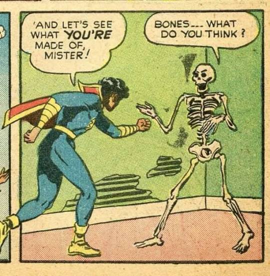
#judge your bones#tag the judge#these fellows reactions to foolishness are the best#wish i could embody their energy some day#also id already had this post judged and queued for later today when i saw your tag XD#moved the judgement to the tagged post cause i didnt wanna seem like i was ignoring you#mandible = lower jaw bone#femur = thigh/upper leg bone#clavicles = collar bones#sternum = breast bone#suture = fused joint between skull bones#sacrum = 5 fused vertebrae that form a sort of triangle that joins the spinal column to the pelvis#vertebrae = spine bones#carpals = wrist bones#the too much spine is too many lumbar vertebrae there should only be 5 here theres 8#lumbar vertebrae = 5 spine bones between the ribs and pelvis
55K notes
·
View notes
Text
Craniosynostosis Treatment: Helping Your Child’s Skull Develop Normally
Craniosynostosis is a condition where one or more of the sutures in a baby’s skull close prematurely, before the brain is fully developed. This can result in an irregular head shape and may, in some cases, lead to increased pressure on the brain. While the condition can be concerning for parents, craniosynostosis treatment options are highly effective at ensuring a child’s skull and brain develop normally. Early diagnosis and intervention are key to achieving the best outcomes.

Understanding Craniosynostosis
A baby’s skull is made up of several bones connected by sutures—flexible joints that allow the skull to expand as the brain grows. Craniosynostosis occurs when one or more of these sutures close too early. The condition is categorized into two main types:
Non-Syndromic Craniosynostosis: The most common form, occurring without associated genetic syndromes. It typically affects a single suture.
Syndromic Craniosynostosis: Associated with genetic syndromes such as Crouzon, Apert, or Pfeiffer syndromes. This type often involves multiple sutures and may come with other physical abnormalities.
Signs and Symptoms
Parents and pediatricians should watch for the following signs of craniosynostosis:
An irregular or asymmetrical head shape.
A raised, hard ridge along the suture line.
Slow or no growth of the head circumference.
In severe cases, increased intracranial pressure leading to symptoms like irritability, vomiting, or developmental delays.
Diagnosis
A pediatrician or specialist will typically diagnose craniosynostosis through a physical examination of the head shape. Imaging studies such as X-rays, CT scans, or MRIs may be used to confirm the diagnosis and determine the severity of the condition. Genetic testing might also be recommended for syndromic cases.
Read More : Role of Imaging in the Diagnosis of Craniosynostosis and Plagiocephaly in Children
Craniosynostosis Treatment Options
Treatment for craniosynostosis depends on the type and severity of the condition, as well as the child’s age. Below are the most common approaches:
1. Surgery
Surgery is the primary treatment for craniosynostosis. The goal is to correct the head shape, allow for normal brain growth, and relieve any pressure on the brain. Two main surgical options are:
Cranial Vault Remodeling (CVR): This traditional open surgery involves reshaping the skull by removing, reshaping, and repositioning affected bones. CVR is typically performed when the child is between 6 and 12 months old.
Endoscopic Surgery: A minimally invasive option, endoscopic surgery involves removing the fused suture through small incisions. This method is most effective when performed before 6 months of age and is usually followed by helmet therapy to guide skull growth.
2. Helmet Therapy
Helmet therapy is often used following endoscopic surgery to help shape the skull as it heals. The custom-fitted helmet gently redirects skull growth and ensures symmetry. Key points about helmet therapy include:
It is typically worn for 20 to 23 hours a day.
Treatment duration varies but usually lasts several months.
Regular follow-ups are required to adjust the helmet as the child grows.
3. Multidisciplinary Care
For syndromic craniosynostosis, a team of specialists may be involved in the child’s care, including:
Neurosurgeons and craniofacial surgeons for surgical planning and execution.
Geneticists to identify associated syndromes and guide future care.
Pediatricians and developmental specialists to monitor growth and developmental milestones.
Ophthalmologists if the condition affects the eye sockets.
4. Monitoring Mild Cases
In very mild cases of craniosynostosis that do not involve significant brain pressure or cosmetic concerns, close monitoring may be recommended. Regular check-ups with a specialist will help ensure that the skull’s growth is progressing normally.
The Importance of Early Treatment
Timing plays a crucial role in craniosynostosis treatment. The younger the child, the more malleable the bones of the skull are, making surgical correction easier and more effective. Most procedures are performed within the first year of life to maximize the benefits of treatment.
Recovery and Follow-Up Care
After treatment, children typically recover well and achieve normal skull and brain development. Post-operative care includes:
Hospital Stay: Most children spend 1 to 3 days in the hospital for monitoring.
Follow-Up Appointments: Regular visits to the surgical team ensure proper healing and track skull growth.
Helmet Adjustments: If helmet therapy is part of the treatment, frequent adjustments are necessary to accommodate growth.
Parents should watch for any signs of complications, such as swelling, fever, or unusual behavior, and report them to the care team immediately.
Emotional Support for Families
Receiving a diagnosis of craniosynostosis can be overwhelming for parents. Support groups, counseling, and resources from organizations specializing in craniofacial conditions can help families navigate the emotional and practical challenges of treatment.
Final Thoughts
Craniosynostosis treatment is highly effective at addressing both functional and cosmetic concerns associated with the condition. With advancements in surgical techniques and multidisciplinary care, most children go on to lead healthy, normal lives. If you suspect craniosynostosis in your child, consult a pediatric specialist promptly to discuss the best course of action. Early intervention not only corrects head shape but also ensures your child’s brain has the space it needs to grow and thrive.
0 notes
Photo

SKULL TENSEGRITY ⠀ The skull is often described as a fused bony dome, fixed in its shape and immobile. However, the skull is comprised of individual cranial bones that are connected to each other via specialized joints called cranial sutures with sutural ligament tissue within them (Pic 1-3). ⠀ As with any observation of the body when the specimen is deceased, the common conclusion is the sutures are fused and the cranial bones are “cemented together”. If one looks at the micro-anatomy of the living sutures, they can see a collagen matrix, arterioles, micro veins and nerve branches as well as mechanoreceptors. This would all indicate the sutures are a living space between the bones. ⠀ As for the links, the sutures also have direct fascial connection to the dura mater (inner-skull layering of the brain and spinal cord, Pic 4,5) via epithelial-mesenchymal transition zones.This means any changes to the cranial bones and sutures will have an effect on the dura mater (and the meninges, which are deeper and closer to the brain). Dura mater is continuous through the whole spinal cord and to the sacrum. ⠀ One can visualize this phenomenon as the driver of positive changes in “cranial manual therapies” or more violently, in negative changes after a concussion. ⠀ When the skull is compressed with normal forces, it behaves like all other tensegrity structures. ⠀ Specifically, the tension elements, including sutures and dura mater collagen, help to dissipate the forces and actually work to expand the cranial vault. Pretty amazing observations for an area many believe is fused. See G. Scarr’s article in the Journal of Osteopathic Medicine for more details (Pic 6,7). ⠀ Credit: @anatomylinks #anatomy #fascia #tensegrity #biomechanics #chiropractic #physicaltherapy #physiotherapy #yoga #yogaanatomy #osteopathy #medicine #doctor #medstudent #biotensegrity #manualtherapy #pilates #gym #tensegrity #fisioterapia #fisioterapeuta #osteopata #osteopati #osteopatia #massage #massagetherapy #massagetherapist #sportsmassage #sportsinjury #health #therapy (hier: Germany) https://www.instagram.com/p/CHfJuj4hAa-/?igshid=uyixi58z807d
#anatomy#fascia#tensegrity#biomechanics#chiropractic#physicaltherapy#physiotherapy#yoga#yogaanatomy#osteopathy#medicine#doctor#medstudent#biotensegrity#manualtherapy#pilates#gym#fisioterapia#fisioterapeuta#osteopata#osteopati#osteopatia#massage#massagetherapy#massagetherapist#sportsmassage#sportsinjury#health#therapy
3 notes
·
View notes
Photo

*Help Ibrahim Lead a Normal Life* Click here to donate: https://milaap.org/fundraisers/helpibrahim Javed and Shehnaz's happiness was at peak when they were blessed with a baby boy. Resident of 17/1, Mallipoo naga Click here to donate: https://milaap.org/fundraisers/helpibrahim r, Perambur, Chennai - 11. Javed's 1.5 year old son, Muhammed ibrahim will be two years old this November. Ibrahim showed his first symptom when he was 6 months old. His eyes started protruding and his skull was deformed. He underwent several tests and was diagnosed with Craniosynostosis, a birth defect in which one or more of the fibrous joints between the bones of the baby's skull (cranial sutures) close prematurely (fuse), before the baby's brain is fully formed. Ibrahim has already undergone three operations since he was 11 months old. The child is now facing difficulties in breathing. He cannot breath from his nose and uses his mouth to breathe. He is currently being treated at Child Trust, Nungambkam by Doctor Chidambaram. Treating Craniosynostosis involves surgery to correct the shape of the head and allow for normal brain growth. Early diagnosis and treatment will allow Ibrahim's brain adequate space to grow and develop. Your generous support can help their baby. Let not lack of resources turn their parenting experience into a bitter one. Ibrahim deserves a fair chance at life and your help will go a long way in ensuring that happens. Click here to donate: https://milaap.org/fundraisers/helpibrahim For any form of cross verification you may write to me at [email protected] *#paperplane* *#makethemostofyourgiving* Follow my writings on @YourQuote.in #yourquote #quote #stories #qotd #quoteoftheday #wordporn #quotestagram #wordswag #wordsofwisdom #inspirationalquotes #writeaway #thoughts #poetry #instawriters #writersofinstagram #writersofig #writersofindia #igwriters #igwritersclub
#quote#writersofindia#yourquote#instawriters#writeaway#igwriters#poetry#writersofinstagram#quotestagram#wordporn#stories#paperplane#quoteoftheday#makethemostofyourgiving#inspirationalquotes#wordswag#qotd#writersofig#igwritersclub#wordsofwisdom#thoughts
5 notes
·
View notes
Link
0 notes
Photo
Thanks for the tag @sentient-tent !
This fellow’s got nice hands, and the carpals are mostly decent! A bit of artistic license and the ones on the pinky finger side are a little weird but pretty good. Weird shape to the ribs though looks like that’s just the style of this fellow. The scapula seems to have an extra process at the shoulder joint and there shouldn’t be a suture all the way through the forehead (unless that’s damage?) A decent fellow in all 8/10 I want this jacket

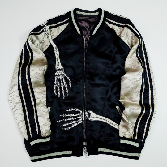
this is probably my single favorite piece of clothing ever
#judge your bones#lol this is what i look like when i hug anyone from behind (judge lore: im short)#fellow's got enough ribs enough vertebrae basically most of their bones are in the right place (little off for carpals)#carpals wise most of them are kinda the right shape and place but some are a bit off or the wrong size#also a couple are not clearly 2 separate bones instead they're partly merged but still with the shape and position they should have#tag the judge#How did i forget to put that higher up???#sentient-tent#carpals = wrist bones#scapula = shoulder blade bone#process on the scapula refers to a pokey out bit of bone (not the best description sorry)#suture = fused joint between skull bones
79K notes
·
View notes
Text
How to Get the Best Craniosynostosis Treatment New York
Bringing a new life to the world needs permanent attention from birth to fostering them young. Infants require care-taking from day one, as every organ and part of a newborn is ultra-sensitive to the outer world. As you might have heard, a newborn baby consists of more bones than an adult, and these fragmented bones have fibrous joints in between, which furthermore help them to fuse to form the final skull bone structure with the growth of your child. But sometimes, this process faces some abnormalities which result in child cranial disorders. And here, the need for craniosynostosis treatment in New York occurs.
If not treated, craniosynostosis can lead to pressure change inside the brain and outer appearance. You need to visit your doctor and get corrective treatment for the head development of your child, and you could see the results within a few weeks. But with the early-stage diagnosis, you could abort this condition.

SHORT HILLS CRANIAL CENTER is a reliable treatment center as it uses the latest technology to monitor and provides an effective cure for your child’s condition. The craniosynostosis is of different types based on the suture closure, leading to different shapes with changes in the width and orientation of the head. The infant helmet in New York helps shape and redirect the symmetrical proportions of your child’s head. The experts here provide customized helmets to support the proportionate development of the head.
So, consult the experienced and resourceful physicians here for craniosynostosis treatment now. The staff is well-trained and cordial that understands the psychological needs of your infant. For further details, please browse www.shorthillscranialcenter.com.
0 notes
Text
Pediatric Craniosynostosis Treatment Market Demand, Future Outlook And Applications
Segmentation
The global pediatric craniosynostosis treatment market is segmented on the basis of type, diagnosis, treatment, and end-user.
On the basis of type, the market is segmented into single-suture synostosis, double-suture synostosis, and complex multi-suture synostosis. The single-suture synostosis is further segmented into sagittal suture synostosis, coronal suture synostosis, metopic suture synostosis, and lambdoidal suture synostosis. The double-suture synostosis is further segmented into bicoronal, bilambdoid and sagittal plus metopic.
On the basis of diagnosis, the market is classified into the physical examination, genetic testing, and imaging studies.
On the basis of treatment, the market is classified as stereotactic image-guided endoscopic craniosynostosis repair, standard craniosynostosis surgery, baby helmet therapy, and others. The stereotactic image-guided endoscopic craniosynostosis repair is further segmented into real-time stereotactic-endoscopic craniectomy and endoscopic strip sagittal craniectomy. The standard craniosynostosis surgery is further segmented into strip sagittal craniectomy, Frontal-Orbital Advancement (FOA), and Frontal-Occipital Reversal (FOR).
On the basis of end-user, the market is segmented into hospital, clinics, diagnostic centers, and others.
Regional Analysis
The Americas dominate the pediatric craniosynostosis treatment market owing to the rising awareness about the disease and high healthcare expenditure. The total health expenditure in the United States was reported to be USD 3.2 trillion, suggested by the Centers for Disease Control and Prevention (CDC).
Europe holds the second position in the pediatric craniosynostosis treatment market. It is expected that the support provided by the government and private associations for research and development and amendments in reimbursement policies in healthcare are likely to promote the market of the European region. The rising prevalence of craniosynostosis in the European region is also expected to boost the market growth. The prevalence of craniosynostosis was found to be around 7.2 per 10.000 live-born children in the Netherlands, suggested by the Journal of Cranio-Maxillofacial Surgery in 2016.
Asia Pacific is the fastest growing region in the pediatric craniosynostosis treatment market owing to the developing healthcare technology. Healthcare expenditure is also found to be escalating in various Asian countries. As per the statistics suggested by the Australian Institute of Health and Welfare, the total health expenditure was USD 170.4 billion during the years 2015 to 2016, i.e., 3.6% higher than the expenditure of 2014 to 2015.
The Middle East and Africa hold the lowest market share in the pediatric craniosynostosis treatment market due to lack of technical knowledge and poor medical facilities.
Market Highlights
It is estimated that the pediatric craniosynostosis treatment market is expected to grow at a CAGR 6.0% during the forecast period of 2017–2023.
Craniosynostosis is a congenital disorder in which one or more of the fibrous joints between the bones of baby's skull fuse prematurely before the baby's brain is fully formed.
A number of factors such as technological advancements, escalating prevalence of craniosynostosis, increasing awareness among people, improvement in the regulatory framework, and rising funding and reimbursement are propelling the growth of the global pediatric craniosynostosis treatment market.
However, lack of skilled physicians and endoscopists, challenges in research and development, the risk of infections caused by endoscopes, and the high cost of treatment may hamper the growth of the market.
Access Report Details @ https://www.marketresearchfuture.com/reports/pediatric-craniosynostosis-treatment-market-5726
Key Players
Some of the key players in the global pediatric craniosynostosis treatment market are Arthrex Inc., B. Braun Melsungen AG, Cogentix Medical, CONMED Corporation, Cook Medical, Frontier Healthcare, Fujifilm Holdings Corporation, Hoya Corporation, Johnson & Johnson Services, Inc., KARL STORZ GmbH & Co. KG, Medtronic Plc, Minntech Corporation, Olympus Corporation, Pentax Medical, Richard Wolf GmbH, Siemens Healthcare, Smith & Nephew Plc, STERIS Corporation, Stryker Corporation, US Endoscopy Group, and others.
Browse Related Reports :
Endometrial Cancer Market Application, Growth, Trends, Size | Global Industry Analysis 2023
Palliative Care Market Research Report –Global Forecast to 2023| MRFR
Absorbable Surgical Sutures Market Outlook | Demand Overview, Share Analysis and Industry Forecast to 2023
Malaria Diagnostics Market Share, Techniques, Trends, Growth Analysis, Industry Forecast to 2023
0 notes
Text
What is a Suture Joint?

Source: OpenStax Anatomy and Physiology OpenStax Anatomy and Physiology All the bones of the skull, except for the mandible, are joined to each other by a fibrous joint called a suture. The fibrous connective tissue found at a suture (“to bind or sew”) strongly unites the adjacent skull bones and thus helps to protect the brain and form the face. In adults, the skull bones are closely opposed and fibrous connective tissue fills the narrow gap between the bones. The suture is frequently convoluted, forming a tight union that prevents most movement between the bones. Thus, skull sutures are functionally classified as a synarthrosis, although some sutures may allow for slight movements between the cranial bones. In newborns and infants, the areas of connective tissue between the bones are much wider, especially in those areas on the top and sides of the skull that will become the sagittal, coronal, squamous, and lambdoid sutures. These broad areas of connective tissue are called fontanelles. During birth, the fontanelles provide flexibility to the skull, allowing the bones to push closer together or to overlap slightly, thus aiding movement of the infant’s head through the birth canal. After birth, these expanded regions of connective tissue allow for rapid growth of the skull and enlargement of the brain. The fontanelles greatly decrease in width during the first year after birth as the skull bones enlarge. When the connective tissue between the adjacent bones is reduced to a narrow layer, these fibrous joints are now called sutures. At some sutures, the connective tissue will ossify and be converted into bone, causing the adjacent bones to fuse to each other. This fusion between bones is called a synostosis (“joined by bone”). Examples of synostosis fusions between cranial bones are found both early and late in life. At the time of birth, the frontal and maxillary bones consist of right and left halves joined together by sutures, which disappear by the eighth year as the halves fuse together to form a single bone. Late in life, the sagittal, coronal, and lambdoid sutures of the skull will begin to ossify and fuse, causing the suture line to gradually disappear. Source: Read the full article
0 notes
Text
Craniosacral Bodywork: A Balloon Filled With Hot Air Or Totally Effective Modality?
There are many topics I'm enamored of that I would like to search additionally. However, before I really do, I get that I have to know about the subject. If we go back to the times of a slower, easier life, this perfectly clearly meant going to "the stacks" of the local university library, along with libraries a little bit an excessive distance from your area. It involved using the Internet to dial up to website library IBM VM systems, and it conveyed that one was looking at the local Public Library for books, mags, and articles in newspapers concerning the subject of interest. Quite simply, a super dependence on libraries, in true to life and both virtual. Nowadays, I commence my goal with WikiPedia. I understand that the information presented is moderated, and is commonly appropriate, although on leading problems, some content articles could be skewed at times. It isn't the literal Word, routed from the Lord's Abode. Nevertheless, I've mainly been satisfied with the quality of job they do, & most content articles happen to be thoroughly sourced and they are a superb starting place for more weeks of learning. Below, I wrote an interesting article that is usually typically excerpted from WikiPedia articles and reviews. Even though I've rewritten it, I must even give credit and recognition where it is necessary. I'd be plagiarizing if I failed to, because a basic substitution of words isn't a new work, in line with the regulations. Thankfully, the Creative Commons license enables me to employ these interesting articles for my own intentions. Having said that, please delight in this quick jump into this issue. Craniosacral therapy (CST) is a kind of bodywork or some other therapy using peaceful touch to manipulate the synarthrodial joints of the cranium. A specialist of cranial-sacral remedy could also apply light details to a patient's backbone and pelvis. Practitioners think that this manipulation regulates the flow of cerebrospinal fluid and supports inches primary respiration". Craniosacral remedy originated by John Upledger, D. U. in the 1970s, because an offshoot osteopathy in the cranial subject, or cranial osteopathy, which was developed in the 1930s simply by William Produce Sutherland. In accordance with the American Cancer Culture, although CST might relieve the symptoms of stress and anxiety or strain, "available scientific facts will not support promises that craniosacral therapy helps in eliminating malignancy or any type of additional disease". CST provides been characterized as pseudoscience and its own practice has been termed quackery. Cranial osteopathy has received a similar assessment, with one 1990 paper getting there was zero scientific basis for any from the professionals ' promises the paper reviewed. The term craniosacral or cranial-sacral are based on the keywords cranium and sacrum, a bone fragments of the pelvis which links the cheapest lumbar vertebra to both hip bone fragments and the tailbone. Cranial osteopathy, a forerunner of CST, is originated simply by osteopath William Sutherland (1873-1954) in 1898-1900. While researching at a disarticulated skull, Sutherland was struck simply by the theory which the cranial sutures of the temporal bones where they meet the parietal bone fragments were "beveled, like the gills associated with a seafood, indicating articular mobility to get a respiratory mechanism. " John Upledger created Craniosacral therapy. Evaluating it to cranial osteopathy he had written: "Dr. Sutherland's finding regarding the overall flexibility of skull sutures led to the early research at the rear of CranioSacral Therapy - and both strategies impact the cranium, sacrum and coccyx - the similarities closing there. " However , present day cranial osteopaths mainly consider the two methods to be the comparable, but that cranial osteopathy provides "been educated to non-osteopaths beneath the name CranialSacro therapy. inch From 1975 to 1983, Upledger and neurophysiologist and histologist Ernest W. Retzlaff performed at Michigan State University while clinical experts and teachers. They set up a research team to research the purported pulse and additional study Sutherland's theory of cranial bone motion. Upledger and Retzlaff went on to create their particular results, which they interpreted since support pertaining to both the idea of cranial bone activity, and the idea of a cranial rhythm. Later testimonials of these studies have figured their analysis didn't meet up with enduring values to offer definitive substantiation meant for the effectiveness of craniosacral therapy and the living of cranial bone tissue movements. Professionals of both cranial osteopathy and craniosacral remedy assert that we now have little, rhythmic movements of the cranial bone fragments related to cerebrospinal liquid pressure or arterial force. The premise of CST is definitely that palpation of the cranium can be used to find this rhythmic movement from the cranial bone fragments and selective pressures may be used to change the cranial bones to accomplish a therapeutic end result. However , the amount of mobility and compliance of the cranial bones is known as controversial and can be a critically important concept in craniosacral therapy. Key respiratory system mechanism THE PRINCIPAL Respiratory Instrument (PRM), the device originally suggested by Sutherland, comes with been summarized in five ideas: The postulated intracranial fluid fluctuation can be defined by practitioners as an connections between four main components: arterial blood, capillary blood vessels (brain volume ), venous blood vessels and cerebrospinal fluid (CSF). Fluctuation from the cerebrospinal liquid There's analysis which demonstrates examiners are unable to measure craniosacral action dependably, as indicated by too little inter-rater contract among examiners. The writers of the analysis conclude this "measurement error may be sufficiently large to render many clinical options potentially erroneous". Alternate medication practitioners possess interpreted this consequence since a product of entrainment among individual and specialist, a theory which lacks technological assist. Whether craniosacral action can be dependably palpated continues to be a subject of question with studies making mixed outcomes. Mobility from the intracranial and intraspinal dural membranes In 1970, Upledger detected throughout a medical procedure around the neck what he referred to as a poor pulsating motion within the vertebral meninges. He attempted to hold the membrane still and discovered that he cannot due to potency of the action behind the mobility. Mobility of the cranial bones The extent that cranial bones can move is known as questionable and research of the lifestyle and amount of cranial motions have produced mixed results. Cranial sutures will be the areas where the eight cranial bone fragments are joined up with. During infancy, the cranial bone fragments are not rigidly fused to one another, yet are instead bound jointly by way of a membrane layer referred to as a fontanelle where two sutures join. Between your first and second 12 months of existence, the cranial bone fragments begin to go alongside one another and fuse as a normal part of development. Research examining age the closure of the cranial sutures feature reported blended findings. Closure provides been reported to occur during adolescence whilst other studies indicate better specific variability in the timing of this closure with fusion of the lambdoid stitch, sagittal stitch, and coronal sutures taking place in the 4th decade of lifestyle, but finish fusion of all sutures not occurring until advanced time (the eighth 10 years of life continues to be reported ); some studies have discovered that the sutures by no means rigidly fuse. According to Gray's Anatomy, "hen such sutures happen to be linked by sutural ligament and periosteum, nearly complete immobility benefits inches. Treatment The therapist gently palpates the patient's physique, and centers intently to the communicated movements. A practitioner's being of being in tune with an individual is described as entrainment. Sufferers often article feelings of deep rest after and during the therapy program, and may feel light-headed. While sometimes regarded as brought on by a rise in endorphins, study shows the consequences could possibly become brought about by the endocannabinoid program. You will discover couple of reports of adverse situations from CST treatment. In one analysis of craniosacral manipulation in patients with traumatic human brain syndrome, the incidence of undesireable effects from treatment was 5%. Reception According to the American Malignancy Contemporary society, although CST might ease the symptoms of anxiety or strain, "available scientific facts does not support cases that craniosacral remedy helps in curing cancer tumor or any type of various disease". Cranial osteopathy comes with received an identical evaluation, with 1 1990 newspaper finding there is no technological basis for just about any of the practitioners ' promises the paper analyzed. In Oct 2012 Edzard Ernst conducted a organized overview of randomized clinical trials of craniosacral therapy. He concluded that " the idea that CST is associated with more than non-specific effects isn't based on information from rigorous randomised clinical trials. inch Commenting especially on this bottom line Ernst commented on his blogging site that he previously picked the wording as "a courteous and technological way of expressing my thanks that CST is just phony. " Ernst also left a comment that the grade of five from the six studies he had analyzed was "deplorably poor, inches a emotion which echoed an Aug 2012 review that mentioned the "moderate methodological quality of the included studies. inch Ernst criticized a 2011 systematic critique performed simply by Jakel and von Hauenschild for addition of observational research and including research with healthy and balanced volunteers. This review figured the evidence platform surrounding craniosacral therapy as well as its efficiency was sparse and composed of research with heterogeneous designing. The authors of the review explained that currently available evidence was insufficient to draw final thoughts. The evidence bottom for CST is definitely sparse and lacks a proven biologically credible mechanism. From the absence of demanding, well-designed randomized managed studies, it's been characterized as pseudoscience, and its practice called quackery. Thank you to WikiPedia concerning the impressive information that's distributed to Internet users by dedicated staff of unpaid editors and team members. This information was gleaned from WikiPedia.org. Please sincerely reflect on DONATING BY TAPPING ON THIS LINK.
0 notes