#pericycles
Text

mlp oc redraws! i missed these silly guys and wanted to redraw their refs
#art#oc#mlp oc art#mlp oc#mlp fim#mlp fim oc#oc: misty mint#oc: zephyr comet/comet wish#oc: pericycle merrystem#oc: frosty swirl#oc: cloudsweeper#oc: hazel blossom#oc: almond hooves
10 notes
·
View notes
Text


enjoy these photos of root cells under a microscope from my bio lab
#she xylem on my cortex till i phloem#oh sorry i can’t come to the weekly hag out i’m on my pericycle
14 notes
·
View notes
Text
In most plant roots, a medial longitudinal cross section reveals long cell files that converge in the subapical region of the root (Figure 17.21A). (...) In Arabidopsis, four distinct sets of initials, all of of the which are adjacent to the QC, can be defined in terms of their position and the tissues they produce (Figure 17.21B):
Columella initials. Located directly below (distal to) the QC, these initials give rise to the central portion (columella) of the root cap.
Epidermal-lateral root cap initials. Located to the side QC, these initials first divide anticlinally to set off daughter cells, which divide periclinally to form two files of cells that mature into the lateral root cap and epidermis.
Cortical-endodermal initials. Located and adjacent to the epidermal-lateral root cap initials, the cortical-endodermal initials divide anticlinally to set off daughter cells, which then divide periclinally to form the cortical and endodermal cell layers.
Stele initials. Located directly above (proximal to) the QC, these initials give rise to the vascular system, including the pericycle.

Ablation of the QC led to another abnormal division and precocious differentiation of the adjacent initials (see Figure 17.21B), suggesting that the QC produces a mobile signal that acts on the adjacent cells to prevent their differentiation and thus maintain their ability to divide.
"Plant Physiology and Development" int'l 6e - Taiz, L., Zeiger, E., Møller, I.M., Murphy, A.
#book quotes#plant physiology and development#nonfiction#textbook#plant roots#arabidopsis#qc#quiescence center#columella#root cap#daughter cells#epidermal lateral root cap#cortical endodermal#cell division#cortex#endodermis#plant cells#stele#pericycle#cell differentiation
1 note
·
View note
Text
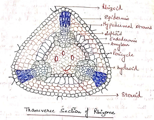
Mod 1: Bryophytes
Ngl I have no idea how to prepare for the botany paper, but I might as well try something-
Polytrichum
Classification:
Class - Bryopsida
Order - Polytrichales
Family - Polytrichaceae
Genus - Polytrichum
Gametophyte
External Structure:
The nature of gametophyte of polytrichum is differentiated into 2 parts: A horizontal underground rhizome and an erect leafy shoot.
Rhizome: The horizontally growing underground portion of the gametophore. It bears small, brown or colorless leaves in three rows and numerous rhizoids.
Leafy Shoot: The erect leafy axis arising from the rhizome. It is differentiated into a stem-like central axis which bears dark green expansions. The so called leaves are spirally arranged in 3/8 phyllotaxy.
The leaf is differentiated into a sheathing leaf base and an apical limb. The limb is narrow, lanceolate, and green. Leaf base is colorless and membranous, closely clasped around the stem.
A short transition zone is present between the rhizome and the aerial shoot. It bears small brown leaves similar to those on rhizome.

Internal Structure:
I. Rhizome: In most of the species, the rhizome is triangular in outline with rounded corners. A transverse section of rhizome shows the following regions:
a) Epidermis: The outermost layer of thick walled cells. They give rise to rhizoids.
b) Cortex: The epidermis is followed by cortex consisting of 3-4 layer of thin walled parenchymatous cells.
It is interrupted by 3 hypodermal strands that extend radially from periphery to center.
Hypodermal strands composed of prosenchymatous cells contain starch grains. Extending inwards from each hypodermal strand is a group of lignified cells containing radial strands.
Next to the cortex, there is a layer of radially elongated cells with suberized thickening on their walls. This layer can be compared to the endodermis of higher plants.
c) Pericycle: A 2-3 layered parenchymatous pericycle is present between endodermis and the central conducing strand. It is discontinuous.
d) Leptoids: There are thin walled polygonal cells present in furrows opposite to the radial strands. They are like sieve tubes of phloem hence called leptoids. The cells collectively form lepton.
e) Amylom: The innermost later of leptoids is separated from the central cylinder by a single layer of parenchymatous cells containing starch. This layer is called amylom.
f) Central Cylinder: Situated in the central region of the rhizome. Made up of 2 types of cells: thick walled living cells called stereids and empty dead cells called hydroids. Both of them together make up the hydrom.
II. Aerial Shoot: In transverse section, the aerial shoot is irregularly lobed due to the presence of leaf bases. It is differentiated into 3 regions:
a) Epidermis: The superficial uniseriate later. Discontinuous due to the persistent leaf bases.
b) Cortex: Epidermis is followed by broad cortex. Outer cortex is made of prosechymatous cells and inner is made of parenchymatous cells.
In young shoots, the cortical cells contain chloroplasts.
A large number of leaf traces are also present in the cortex.
c) Central Cylinders: The outermost later of central cylinders is discontinuous and is composed of parenchymatous cells. It represents rudimentary pericycle. The cells store starch grains.
It is followed by 3-4 layered irregular zone of elongated thin walled cells. This zone is leptom mantte ( equivalent to phloem of vascular plants)
Leptom mantte is followed by 1-2 layers of suberized cells which form hydrom sheath. The cells are rich in starch and also called amylom layers.
Next to hydrom sheath is hydrom mantte composed of think walled empty cells.
The center of shoot is occupied by hydrom cylinder made up of thick walled cells. These cells help in conduction of water and hence equivalent to the xylem of higher plants.

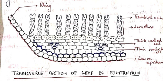
III. Leaf
A transverse section of the limb of the leaf shows a thick midrib, gradually tapering into 2 rudimentary wings.
The ventral surface is bound by a distinct epidermis. It is covered by thick cuticle. Epidermis is followed by sclerenchymatous hypodermis.
The ground tissue (cortex) is composed of parenchymatous cells.
The dorsal surface of limb is made up of vertical plates of cells called lamellae. Each lamellae has 4-8 cells; all cells contain chloroplasts except the terminal one. This terminal cell is larger than outer cells and sometimes it is (___)
The arrangement of cells in lamellae and presence of any spaces between adjacent lamellae increases the photosynthetic area.
The wing is composed of a single layer of hyaline cells. Lamellae are not present in the wing region.
The wings become dry and curved when the plants grow in dry habitats.
Reproduction: In polytrichum, reproduction takes place by vegetative and sexual methods.
I. Vegetative Reproduction: Takes place by bulbils which develop on rhizome. Besides, fragmentation of underground rhizome also helps in propagation.
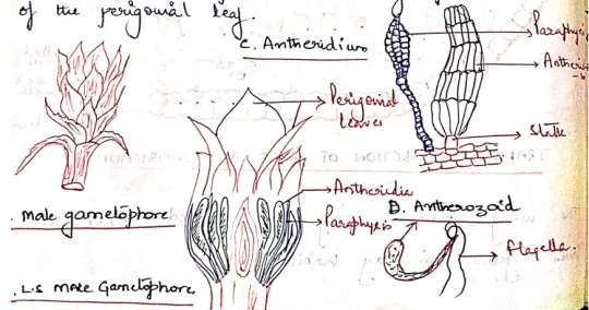
II. Sexual Reproduction: Polytrichum is dioecious, the male (antheridia) and the female (archegonia) sex organs develop at the spices of main shoots of separate plants.
Antheridia:
They are born at the apex of the male gametophore. They are surrounded by a whorl of specialized leaves, called perigonial leaves. These leaves differ from vegetative leaves in color and shape. They are red or brown in color and form a cup like structure around antheridia.
Antheridia are produced in groups at the base of the perigonial leaf.
The mature antheridium is a club shaped structure with an elongated body and a short stalk. They body has a single layered jacked of sterile cells, enclosing a mass of androcytes.
At the free distal end of the antheridium is the operculum consisting of a single or few large cells.
Intermingled the antheridia in the antheridial head are steric multicellular hair like structures called as paraphysis.
Aechegonium:
They are borne in groups at the apex of the female gametophore.
The leaves surrounding the archegonia are called the perichaetial leaves. These leaves overlap close over at the top of the archegonial (cluotes??) to form a bud-like structure called the perichaetium.
Intermingled with the archegonia in the cluotes(Still??) are paraphysis.
The mature archegonium is a flask-shaped structure consisting of a long neck, venter, and a stalk.
The venter cavity contains a single spherical egg and a ventral canal cell.
The long, narrow neck consists of 6 vertical rows of neck cells enclosing a narrow neck canal which has a row of neck canal cells (10)

Fertilization:
Water is essential for fertilization. The jacket sells of the mature antheridium swell by absorbing water. The hydrostatic pressure thus generated bursts the operculum and the antherozoids are liberated.
The neck canal cells and the venter canal cell of mature archegonium degenerate forming a mucilaginous mass.
The mucilaginous mass swells up by absorbing water and as such the cover cells present at the tip of the archegonium are pushed apart.
The antherozoids are attracted towards the archegonia and a number of antherozoids swim down the neck but only faces with the egg and this oospore is formed.
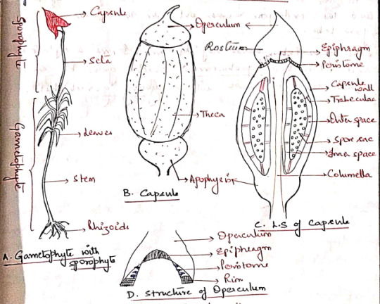
Sporophyte
The sporophyte of polytrichum is differentiated into a foot, a long seta, and an angular capsule.
Foot: A dagger-shaped structure embedded in the female gametophore. It is composed of parenchymatous cells. It acts as an anchoring and absorbing organ.
Seta: A long, slender structure between the foot and the capsule. Anatomically, seta is differentiated into epidermis, hypodermis, parenchymatous cortex and a central cylinder, 'hydroids'
The main function of seta is to raise the capsule to the height and conduction of water and nutrients.
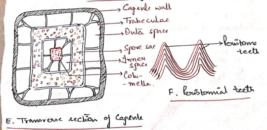
Capsule: The capsule is differentiated into 3 parts:
a) Apophysis (Basal part): The distal end of seta gradually enlarges to form a bulbous apophysis ad the base of the capsule. It has a thick walled epidermis continued with that of the seta.
Epidermis is interrupted by stomata, each stoma has 2 guard cells. It is followed by chlorenchymatous cortex and it serves as photosynthetic tissue.
The central part of apophysis is occupied by a conduction strands which is in continuation with the columella and seta (sterile).
b) Theca (Fertile): The middle fertile part of the capsule. It shows many longitudinal grooves. The wall or jacked of theca is composed of several layers of chlorophyllous cells. The outermost layer forms epidermis devoid of stomata. 'Thickwalls', 2-3 layers of cells, chlorenchyma wall layers.
An air space of lacuna is present inner to the wall layers and is divided into small chambers by transverse filaments of chlorophyll containing cells, the trabeculae.
The outer space is followed by space sac. The fertile cells present inside the spore sac constitute the archesporial tissue.
Initially the archesporium is single layered but it becomes many layered in later stages of development of the sporophyte.
The last generation of archesporial-cells differentiates into spore mother cells. Each spore mother cells will form four haploid spores.
The spore sac is also surrounded on the sinner side by an inner air space. It is also transversed by many transverse trabeculae.
The central part of the capsule is occupied by a thick column of parenchymatous cells, the columella. This is a sterile tissue which is continuous with the central axis of the apophysis.
c) Operculum: The apical part of the capsule which forms a cap-like structure at the apex of the theca. It has an extended proximal part, called rostrum, and a projects beak-like distal(?) part.
The boundaries of the operculum and theca are marked by a distinct countriction(??), the rim.
The distal end of the columella is expanded into a pale thin membranous epiphragm at the base of the operculum.
In the mature capsule the peristome is composed of 32 or 64 peristoimal teeth. The teeth are pyramidal and composed of fiber like cells several layers in thickness.
Peristome regulates spore discharge. They do not exhibit hygroscopic movements.
Dehiscence of Capsule:
At maturity, the capsule shrivels as it dries up. The columella disintegrates and the spores come to lie in the cavity thus formed.
Further drying and shriveling of the capsule wall causes the operculum to fall off and thus exposing the peristome.
After exposure of the peristome, thin walled cells of the epiphragm lying between the peristomial teeth also dry and this several minute holes are formed. The spores are dispersed through these holes.
Only a few spores are liberated every time the capsule sways in the wind. This method of spore dispersal is known as censor mechanism.
Young Gametophyte:
The spores are small sounded structures, yellow but turn green immediately after dispersal. The spore wall is differentiated into an outer exospore and an inner endospore.
At the time of germination, the exospore ruptures and the endospore comes out in the form of germ tube.
The germ tube grows rabidly and forms a septate and branched protonema, which grows by an apical cell. It grows into a young gametophyte.
Evolution of Sporophyte in Bryophytes
The sporophyte of the majority of liverworts and mosses show the same plan. It is made up of an anchoring and absorbing foot, a stalk like seta, and a capsule which contains spores and elaters.
A comparative study of the sporophytes shows that there has been a progressive reduction in the amount of the fertile tissues from Marchantials to Bryales.
In all the bryophytes the oospore, formed as a result of fertilization, is the mother cell of sporophyte. In the simplest type of sporophyte the entire oospore is utilized in the formation of spores and capsule wall.
In some simple forms, some spore mother cells, which fail to develop into spores, form elaters.
In complex forms, the oospore develops into a sporophyte which is differentiated into foot, seta, and capsule.
The following 2 contrasting views have been put forwards with regard to the evolution of sporophytes in bryophytes: progressive sterilization and progressive reduction.
Evolution of Sporophyte by Progressive Sterilization
This view, aka "theory of progressive evolution" or "theory of sterilization" was put forward by Bower and supported by Cavers and Campbell.
According to this theory, the sporophytes of the complex forms (ex: Funaria) have evolved due to progressive sterilization of the potential fertile tissue of the simpler forms (ex: Riccia)
In primitive forms the sporophyte is simple and made of the tissue of the sporophyte is fertile (???)
The progressive sterilization from Riccia to Funaria occurred through the following stages:
First Stage: The simplest known sporophyte among bryophytes is that of Ricca. It consists of only capsule, there being no trace of seta and foot.
The entire sporogenous tissue is converted into spores. Few sterile cells (nurse cells) have nutritive function. They are supposed to be ancestors of elaters.
Second Stage: In Corsinia the sterilization has gone a step further and in addition to nurse cells, a sterile foot is also present.
Third Stage: Further sterilization is seen in Sphaerocarpus where the sporophyte has a sterile bulbous foot and a narrow seta, in addition to fertile capsule.
Fourth Stage: In Targionia, the sporophyte is differentiated into a bulbous foot, a massive seta and a capsule.
Amphithecium gives rise to single layered jacked of the capsule and only about half of endothelial cells give rise to fertile sporogenous tissue and the remaining half form sterile elaters. Thus there is further sterilization of sporogenous tissue.
Fifth Stage: In Marchantia, the sporophyte shows foot, seta, and a capsule.
Amphithecium gives rise to a single layered capsule wall and the endothecium forms sporogenous tissue. Approximately 50% of the sporogenous cells forms the spores and the remaining 50% the elaters with thickening bands.
Sixth Stage: The sporophyte of Pellia is also differentiated into foot, seta, and capsule as in Marchantia, but the jacked of the capsule is 2 or more layered. Furthermore a fixes sterile elatophore is present at the proximal end of the capsule with a bunch of elaters.
Seventh Stage: The sporophyte of Anthoceros, consists of bulbus food and a horn or brittle-like capsule. The capsule wall is made up of 4-6 layers of parenchymatous cells.
The outer wall later is epidermis which possesses stomata. The cells of the inner layers of capsule wall have chloroplasts. Thus the sporophyte is capable of synthesizing its own food and is only dependent on gametophyte for water and nutrients.
Another important feature is the presence of central column of sterile cells, the columella.
The tissue of the archesporium differentiates into large fertile spore mother cells and flat and relatively small sterile elater mother cells.
Eighth Stage: The highest degree of sterilization is found in the class Bryopsida.
Regions like foot, seta, capsule wall, columella, apophysis, operculum, and peristome in sporophytes of Funaria and Polytrichum represent the sterile tissue.
The sporogenous tissue is confined only to spore sacs and constitutes a very negligible part of the sporophyte.
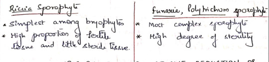
Evolution of Sporophyte by Progressive Reduction
Aka Simplification
Kashyap, Church, Goebel, and Evans are the supporters of the retrogressive theory of evolution.
According to this theory, the simple sporophyre of Riccia is the most advanced and has evolved by progressive reduction or simplification of the complex forms such as Funaria and Polytrichum.
The significant steps in the reduction series are:
1. Simplification of the dehiscence apparatus.
2. Reduction of the green photosynthetic tissue in the capsule wall.
3. Disappearance of stomata and intercellular spaces in capsule wall.
4. Decrease in the thickness of the capsule wall.
5. Gradual elimination of seta and foot.
6. There is no progressive increase in the fertility of the sporogenous cells.
Origin of Alteration of Generations
Antithetic Theory:
Celkovasky proposed this theory. On the basis of this theory, the gametophyte or sexual plant represents the original generation.
The sporophyte or the non-sexual plant is a new and different phase evolved by progressive elaboration of the diploid zygote.
The factors which caused its origin are prompt germination of the zygote accompanies by delayed meiosis.
The result is the production of a small sporophyte of Riccia consisting simply of a spore case.
With further elaboration and increased sterilization of the spore producing tissue, a larger sporophyte with differentiation into a foot., a seta, and a capsule is finally evolved.
Homologous Theory:
Pringsheim proposed this theory. According to this theory, sporophyte is not a new generation, but a direct modification of the gametophyte.
It advocated the fact that among the green algae, the gametophyte plant reproduces by both the methods of reproduction.
It bears spores and also the gametes.
In the course of evolutionary sequence, these two functions become separated in two distinct individuals.
The individuals forming spores came to be known as sporophyte and those forming gametes knows as gametophyte.
These two individuals occur regularly alternating in the life cycle.
Both the individuals in primitive land plants where photosynthetic and free living. Gradually the sporophyte became attached to and partly parasitic on the gametophyte. Consequently it became reduced.
Marchantia Thallus
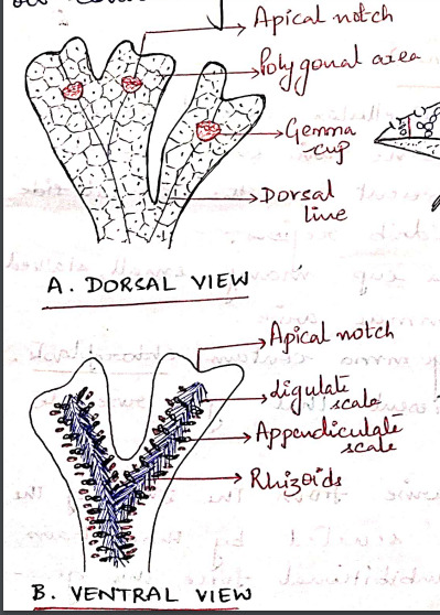
The gametophyte of Marchantia is prostrate, dorsiventral and dichotomously branched. A distinct midrib is present in each branch of thallus.
The apex of each branch is notched and a growing point is situated at the base of each notch.
The dorsal surface of the thallus appears to be masked out into small rhomboidal to polygonal shaped areas called areoles. Which represent the outline of underlying air chambers.
Each areole has got a black dot like spot in the center which marks the position of stoma-like opening leading to air chamber.
Along the midrib on the dorsal surface of the thallus there are certain cup-shaped structures with frilled margins known as gemma cups and they contain special vegetative reproductive bodies called gemmae. (vegetative propagation)
The ventral surface of the thallus bears rhizoids and scales on both sides of the midrib. The rhizoids are usually colorless and unicellular.
Rhizoids are of 2 types: smooth walled and tuberculate. Their function is to:
i) Fix the thallus to substratum.
ii) Absorption of water and nutrients.
The multicellular, one-celled thick scales are arranged in 2-4 rows on either side of the midrib. They are of 2 types:
i) Appendiculate: usually form the inner row of scales, close to the midrib. Have apical appendage.
ii) Ligulate: relatively small and do not have any appendage.
The function of scales is to:
i) Protect the growing point.
ii) Retain some water by capillary action.
The sexually mature thalli possess specialized erect branched called gametophores, which bear the sex organs. These branches arise from the growing apex situated in the apical notch.
The branches present on the male thalli bear the antheridia and are known as antheridiophores while those present on the female thalli bear the archegonia and are called as archegoniophores.
#exam season#send help#biology#notes#plants#botany#bio#bryophytes#bryophyta#polytrichum#plant science#science
9 notes
·
View notes
Text
Organic reactions are chemical reactions involving organic compounds. The basic organic chemistry reaction types are addition reactions, elimination reactions, substitution reactions, pericyclic reactions, rearrangement reactions, photochemical reactions and redox reactions. In organic synthesis, organic reactions are used in the construction of new organic molecules. The production of many man-made chemicals such as drugs, plastics, food additives, fabrics depend on organic reactions.
WIKIPEDIA
0 notes
Text
What are the topics in Organic Chemistry 2?
Following are the topics for Organic Chemistry 2:

Alcohols
Epoxides
Oxidation & Reduction
Grignard Reagents
Dienes & MO Theory
Diels Alder & Pericyclic Reactions
Aromaticity
Reactions of Aromatic Molecules
Synthesis of Aromatic Molecules
Aldehydes and Ketones
Carboxylic Acid and its Derivatives
Enolates (Alpha Carbon)
Carbohydrates
Amines
Amino Acids
Read More Here
https://onlinecollegehomeworkhelp.com/organic-chemistry-tutors/
0 notes
Text


Some Notes on Pericyclic Reactions
2 notes
·
View notes
Text

*cartoon crashing and banging noises*
4 notes
·
View notes
Text
Although it is impossible to define their boundaries with absolute precision, the division of the root into the following zones provides a useful spatial framework that is relevant to our discussion of the underlying mechanisms (Figure 17.20). (...) Even with an understanding of how a graded distribution of auxin across the root can be achieved, some explanation is still required for how these concentration differences evoke a variety of downstream responses, including in the localized zones of cell division, elongation, and differentiation (see Figure 17.20).


"Plant Physiology and Development" int'l 6e - Taiz, L., Zeiger, E., Møller, I.M., Murphy, A.
#book quotes#plant physiology and development#nonfiction#textbook#auxin#cell division#cell elongation#cell differentiation#root#plant cells#maturation zone#elongation zone#meristematic zone#root cap#lateral root#root hair#pericycle
0 notes
Link
Chemical Science Pericyclic Reaction Class Notes with Assignment Career Endeavor Classes UGC NET Handwritten Notes by CSIR is one of the best study materials Handwritten notes are carefully selected from real classroom coaching students.
And now you can also join our telegram channel for various official exam notes and latest study material Click on this link to join our telegram channel (https://t.me/educom_iq)
We at EducomIQ are committed to providing quality notes at affordable prices.
For Queries:-
Call or What's app @7827422314
For ordering visit our site EducomIQ.com
0 notes
Text
Mod 3: Gymnosperms
Pinus Needle T.S.
It is circular in outline in P. monophylla, semicircular in P. sylvestris and triangular in P. longifolia, P. roxburghii, etc.
Outermost layer is epidermis, which consists of thick-walled cells. It is covered by a very strong cuticle.
Many sunken stomata are present on the epidermis.
Each stomata opens internally into a substomatal cavity and externally into a respiratory cavity or vestibule.
Below the epidermis are present a few layers of thick-walled sclerenchymatous hypodermis. It is well developed at ridges
In between the hypodermis and endodermis is present the mesophyll tissue.
Cells of the mesophyll are polygonal and filled with chloroplasts. Many peg-like infoldings of cellulose also arise from the inner side of the wall of mesophyll cells.
Few resin canals are present in the mesophyll, adjoining the hypodermis. Their number is variable but generally they are two in number.
Endodermis is single-layered with barrel-shaped cells and clear casparian strips.
Pericycle is multilayered and consists of mainly parenchymatous cells and some sclerenchymatous cells forming T-shaped girder, which separates two vascular bundles. Transfusion tissue consists of tracheidial cells.
Two conjoint and collateral vascular bundles are present in the center. These are closed but cambium may also be present in the sections passing through the base of the needle.
Xylem lies towards the angular side and the phloem towards the convex side of the needle.
Idk where to find xeric nature info
Williamsoniaceae
Occurrence of Williamsonia
Williamsonia belongs to family Williamsoniaceae of Bennettiales.
It has been reported from Upper Triassic period but was more abundant in Jurassic.
This was earlier discovered under the name Zamia gigas by Willamson in 1870 but has now been named as Williamsonia.
Professor Birbal Sanhi (1932) described W. sewardiana from Rajmahal Hills of Bihar (India).
External Features of Williamsonia
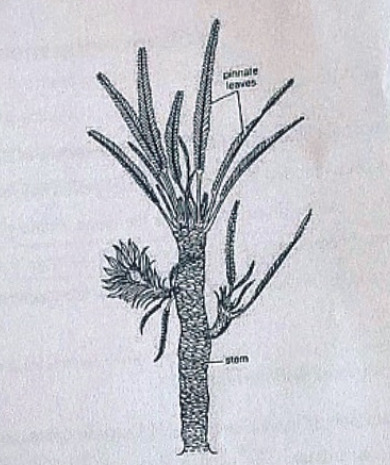
Williamsonia resembled Cycas in appearance, but its best-knows species is W. sewardiana. The plant had an upright, branched, and stout stem covered by persistent leaf bases.
A terminal crown of pinnately compound leaves was present. For the stem genus Bucklandia, Sharma (1991) opined that features of leaf bases such as their shape, size and arrangement pattern are of taxonomic significance.
He observed that leaves in Williamsoniaceae show syndetocheilic stomata with rachis possessing collateral endarch vascular bundles.
Reproduction in Williamsonia
The fructifications of Williamsonia were large and attained a diameter of about 12 cm.
They were borne on a peduncle.
Many spirally arranged bracts were present around the base of the floral axis.
In W. gigas the cones were present among the crown of leaf bases while in W. sewardinia they were present on the short lateral branches.
Williamsonia plants were unisexual.
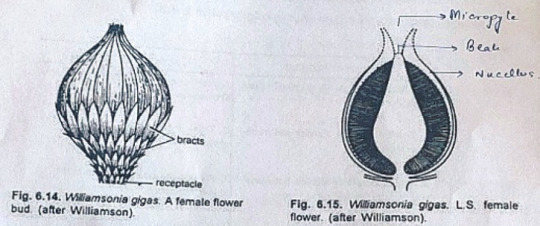
Female Flower
The female 'cones' of W. gigas and W. sewardiana have been investigated in detail. Instead of 'strobili' or 'cones', Sporne (1965) proposed to use the term 'flower'.
The conical receptacle was surrounded by many perianth-like bracts. The ovules were stalked.
The apex of the receptacle was naked and sterile. The nucellus was surrounded by a single vascularize integument, which was fused with the nucellus. The nucellus had a well-marked beak and a pollen chamber. In young ovules the micropylar canal was long and narrow.
In mature ovules, the canal widened because of the formation of nucellar plug and disappearance of interlocking cells. In the apical part of the endosperm, Sharma (1979) observed 2 or more archegonia.
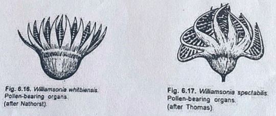
Male Flower
Male flowers consisted of a whorl of microsporophyll's which were united to form a more or less cuplike structure. In majority of the investigates species the sporophylls were un-branched but in some species they were also pinnately branched.
Sitholey and Bose discovered W. santalensis from Upper Gondwana, and observed that microsporophyll's in the species were bifid.
One of the branches of microsporophyll was fertile while the other was sterile. The fertile part has finger-like structures called synangia. Each synangium had two rows of chambers enclosing microsporangia.
The fertile branch of the bifid sporophyll possessed many purse-like capsules, in each of which there were present many monocolpate pollen grains.
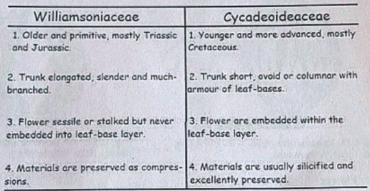
Cycadeoideaceae
Classification:
Division - Cycadeoidophyta
Order - Cycadeoideales
Family - Cycadeoideaceae
Genus - Cycadeoidea
Introduction:
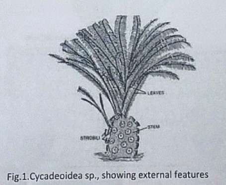
Cycadeoidea is the only genus of family Cycadeoidaceae, represented by thirty species. They are entirely extinct and resemble cycads in the outward stumpy appearance of trunk and an apical crown of pinnate compound leaves. This fossil group of plants flourished during the Triassic to Cretaceous periods of the Mesozoic era. They are reported from various places in the world, in India the Cycadeoidales are found in Rajmahal Hills in Bihar. The petrified trunks of C. entrusca are the oldest fossil ever collected by man.
External Features:
The genus Cycadeoidea had a short, branched, or unbranched spherical, conical, or irregular trunk. The diameter of the trunk is 50cm and the highest rarely reached a meter except in C. jenneyana, it attended the height of several meters. These trunks are covered by rhomboidal leaf bases having multicellular hairs in between. Crown of 10ft long pinnate compound leaves are present at the top.
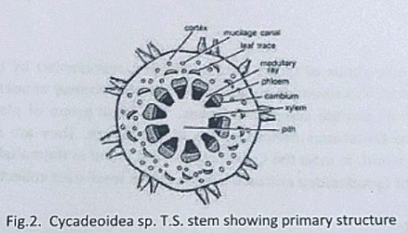
Anatomy of Stem:
The transverse section of the stem shows roughly a circular outline. The epidermis is not very distinct due to the presence of heavy armor of leaf bases. The cortex is parenchymatous and traversed by mucilage canals and numerous leaf traces. The primary vascular structure consists of a ring of endarch, collateral, conjoint, and open vascular bundles encircling the pith. Pith is wide and parenchymatous. A ray-like extension passes between the vascular bundles that make their appearance discrete.
There is a cambium ring with a thin zone of secondary wood. The secondary wood encircles the primary xylem and consists of tracheids with scalariform and bordered pits. The secondary medullary rays traverse the secondary xylem and secondary phloem.
The C-shaped leaf traces arise singly from the primary vascular strand and entering the cortex divided into several masarch strands and enters straight into the leaf.
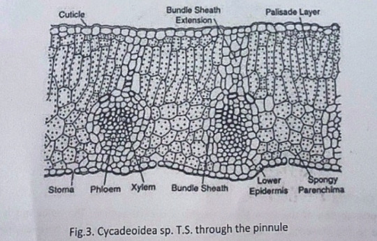
Anatomy of Leaf:
The pinnules show xerophilous structure. The upper and lower epidermis is heavily cutinized and thick walled. The mesophyll cells are distinguished into palisade and spongy parenchyma. The vascular bundles are mesarch and surrounded by bundle sheath.
Reproduction:
The reproductive structure is represented by flowers. In most of the species, the flowers are bisexual and arise in the axil of each leaf.
Structure of Flower:
The flowers are bisporangiate, stalked, and partially sunken in the leaf base armor. Rach such mature flower is 5-10cm in diameter and 10cm long. From the base of such flowers about 100 to 150 hairy bracts arise in close spiral little below the apex. These bracts formed a perianth like structure and protect the megasporangiate and microsporangiate parts of a flower. The microsporophyll or androecium forms a whorl united at the base into a sheath. The megasporophyll or gynaecium consists of numerous stalked ovules born around a central receptacle. Between the ovules, interseminal scales with expanded tips are present. These expanded tips fused to form a continuous surface with pores, through which the micropyle of ovules extended. The vascular supply of flowers consists of many branches from leaf traces.
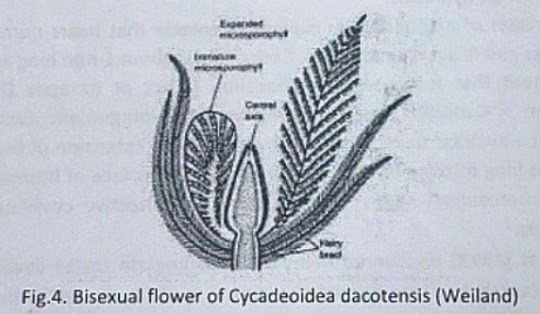
Microsporophyll or Androecium:
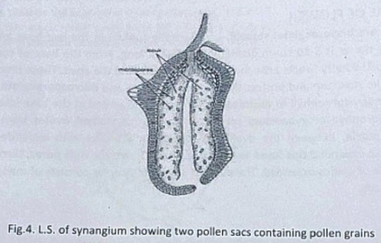
The microsporophyll is 10-12cm long, consists of a central rachis bearing numerous pinnae. The pinnae bear two rows of bean-shaped shortly stalked pollen capsules or synangia. These pollen capsules are born on the trabeculae within the fertile region of microsporophyll. A line of dehiscence is also visible at the base of each microsporophyll. This suggest that the entire microsporophyll might have been shed as a unit. The pollen capsule or synangia measures about 3.5x2.5mm and its wall is several layers thick, the outer layer made up of palisade like cells, and the inner layer is made up of thin-walled cells followed by a tapetum. The tapetum was not demarcated. A ring of microsporangia arranged around the periphery of each synangium. The microsporangia dehisce longitudinally and release the microspores into the synangial cavity. At maturity, the synangia liberate these microspores outside by an apical opening that splits into two valves. The liberated microspores or pollens are oval, measures up to 68µ that represents the male gametophytes. Pollen grains of Cycadeoidea are multicellular.
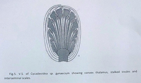
Megasporophyll or Gynoecium
The gynoecium consists of a spherical or conical receptacle that bears numerous stalked orthotropous ovules and interseminal scales. Each ovule is about 1mm long and consists of the single integument that fused with the nucellus except at its apex.
According to Lignier, in C. morieri, nucellus is free from the integument. Each ovule has a pollen chamber and a nucellar beak. This nucellar beak is the extension of the integument. The ovules also have long micropyle, extended from the flat surface of interseminal scales. The fused tips of interseminal scales form an external protective covering or pericarp surrounding the seeds.
Crepet and Delevoryas discovered many of bisporangiate cones from the Cretaceous of black hills. They studied the structure of these ovules in detail. These ovules are urn-shaped and resemble with the ovules of C. wellsii. According to them the micropyle of these ovules are funnel-shaped due to the constriction below the flaring. The inner wall of the micropyle is lined with large cells, considered to be epidermal cells. The integument has three distinct layers. The outer fleshy layer of radially elongated cells, the middle stony layer made up of thick-walled cells, and the inner layer is fleshy.
The young nucellus is made up of thin-walled cells. The cells at the micropylar end are much elongated (80µ long) in comparison to the cells of the chalazal end. The cell at the nucellar tip is pointed up tp whereas cells on either side are bend outward to give the nucellus a distinct shape.
Crepet and Delevoryas reported a linear tetrad or row of three cells in the center of the nucellus.
The seeds are somewhat elongated or oval and possessed two cotyledons.
#exam season#send help#biology#notes#science#botany#gymnosperms#pines#pine trees#pine needles#plants#plant science#plant biology#nature#long post
8 notes
·
View notes
Link
The handwritten notes of DIAS Coaching Pericyclic Response for IAS Admission are spiral-bound notes taken from the student. and our printing is the best
We at EducomiQ are committed to providing quality notes at affordable prices.
For Queries:-
Call or What's app @7827422314
For ordering visit our site EducomIQ.com
#handwritten notes of DIAS Coaching Pericyclic#Pericyclic Response handwritten notes#Pericyclic Response class notes#Pericyclic Response for IAS Mains#EducomIQ
0 notes
Text
this might be the ultimate dumb bitch moment but it didn’t even occur to me that Diels and Alder were two people who discovered the diels alder reaction and not one guy until my prof said “diels and alder won the Nobel prize in the 50s for it”
0 notes
Text
Looking at your study materials like ´What am I ready to put up with today? NOT FUCKING THIS´
#stereochemistry will kill me#and if stereochemistry doesn't manage pericyclic reactions WILL#guys I know that I'm taking a lot of time to answer and write and THIS is the reason#I BLAME IT ALL ON CHEM
6 notes
·
View notes
Last Seen Blogs


architect-chicago
Bez tytułu

lilacsimps
SID
uberstip
iSteve
auto-responder-ai-blog
Alpha robot, Come and get me.