#hydrocephalus medicine
Explore tagged Tumblr posts
Text

Throwback to the best ad I've ever gotten on tumblr
#it's for draining cerebrospinal fluid in patients with hydrocephalus#seems like a good product but I don't think tumblr is the right place to advertise it you know?#doesn't seem like the ad is targeted at patients#and I'm not exactly qualified for craniofacial surgery#invisishunt#tumblr ad#advertising#medicine#skull#shunt
2 notes
·
View notes
Text
बेशर्म I Ipomoea_carnea I Meningitis_Hydrocephalus_Arthritis_Leucoderma_HIV_CVD_Convolvulaceae_diy
Safer-Effective-Better-Remedy
#बेशर्म #Ipomoea_carnea #Meningitis #Hydrocephalus #Arthritis #Leucoderma #HIV #CVD #Convolvulaceae #diy #remediate_pitta #चिंता #Blood_Pressure #रक्तचाप #Diabetes #मधुमेह #convolvulaceae #Neurodegenerative_problems #brain #free_radical_scavenger #wound_healer #inflammation #lipid_peroxidation #life_longivity #besharam #antifungal #alopecia_inducer #anxiety #healthylifestyle #healthylifestyle…
youtube
View On WordPress
#Ayurveda#Besharam#Cultivation#Ethnomedicines#Herbal#HIV#Homoeopathy#hydrocephalus#Ipomoea_carnea#Medicinal plants#Meningitis#One health concept#Remedies#Youtube
0 notes
Text
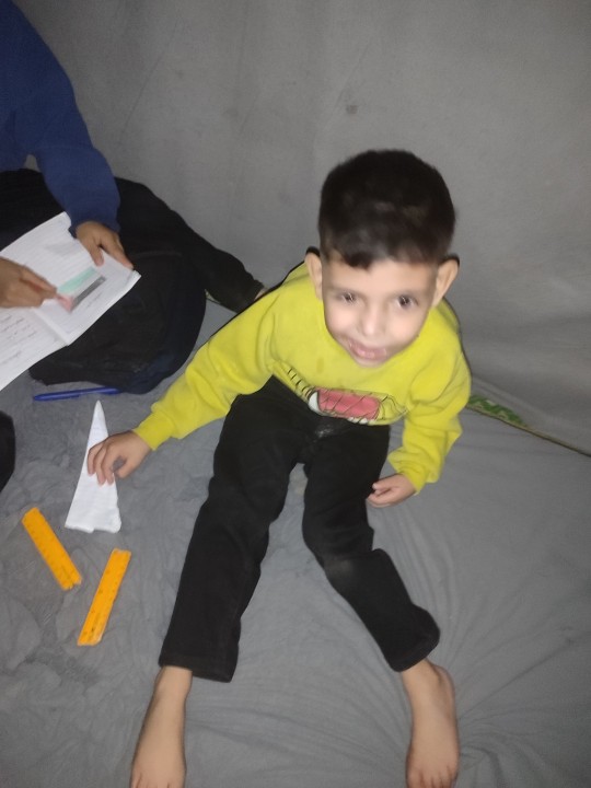
"Urgent Appeal for Mohammed's Family in Gaza🙏🏻💔🙏🏻


I am the father of the child Mohammed, who suffers from partial paralysis on the left side since birth, loss of vision in the left eye, and brain atrophy. A shunt was installed in his brain due to abnormal fluid accumulation (hydrocephalus). Mohammed suffers from frequent neurological seizures and is in urgent need of healthcare and social support, special medications, and follow-up at the eye and neurology clinics.


The situation in Gaza makes it extremely difficult to provide the necessary care for Mohammed, my three children, and my family. We are currently living in a tent, and the weather is very cold. After our home was completely destroyed, we do not have access to medicines, food, or drink, and we urgently need suitable clothing and food for Mohammed and my three children.
We urgently need your support to donate for:
- Providing necessary medicines and medical care for Mohammed
- Ensuring healthy food and water for our family
- Providing warm clothing to face the severe cold
- Attempting to leave Gaza for treatment and providing a better and safer life for our children
Every donation, no matter how small, can make a big difference in our lives. We kindly ask you to share this post and help in any way possible.
Thank you for your support and care
I am Ross from the United States, and I have created this fundraiser to help the Alostaz family in Gaza through the father of Mohammed, Mahmoud. All raised funds will be sent through me to the family Alostaz.
@90-ghost @heritageposts @gazavetters @neechees @butchniqabi abi @fluoresensitivearchived @khangerinedreams @autisticmudkip @beserkerjewel @officialspec2 @palhelp @batekush @appsappsapps @nerdyqueerandjewish r @butchsunsetshimmer @biconicfinn @stopmotionguy @willgrahamscock @strangeauthor @bryoria-annafaye-hall-blog @shesnake @legallybrunettedotcom @lautakwah @sovietunion @evillesbianvillainarchive @antibioware @akajustmerry @neptunerings @dlxxv-vetted-donations @vague-humanoid @buttercupart @sayruq @sar-soor @northgazaupdates2 @feluka @dirhwangdaseul-archived @jdon @ibtisams-blog @sayruq @memingursa @schoolhatergirl @ot3 @lapithae @ryo-yamada @opencommunion @anneemay @killy @schooloutfitideas @bisexualr2d2
@chronicschmonic @feluka-blog-blog @halalchampagnesocialist @ihavenoideashelp @irhabiya @jezior0 @kordeliiius
#free gaza#gaza aid#gaza genocide#gaza fundraiser#free palestine#gaza solidarity encampment#help gaza#gazaunderattack#gaza strip#rafah
571 notes
·
View notes
Text


Hi, all.
First, I just want to thank everyone who became a part of Angelo (Tutoy's) journey. You are a true blessing to his family.
I need to create a new post for this fundraising as the first one was already too long and was not gaining any traction anymore. I need to make a noise for this 12-year-old's life.
To cut the story short, Tutoy has hydrocephalus. Everyday, he and his family battle for his life -- food, medicines, checkups, surgeries.
This March 28-31, a good samaritan from a charitable organization here in the Philippines offered assistance for Tutoy's check-up. This batch of medical doctors are different from his "usual" doctors so somehow, they would start from scratch and see if his condition would get better while saving up for the major surgery.
If all turns out good, then all we have to do is gather funds for his head/brain surgery -- a minimum of $1000. This is an urgent call for help since every day is not an assurance for his life.
If you reached this point, hopefully you can take part in saving his life -- reblog this post or donate if you have an extra amount with you. Any amount is of BIG value to them.
Again, thank you very much!
P@yp#! - @camillefadriquelan
$20/1000
#donations#help#please donate#send help#donate if you can#fundraiser#signal boost#mutual aid#crowdfunding#important#surgery#save children#save family#help families#family#hospital#hydrocephalus#donation drive#donate#donation post#very urgent#urgent#emergency#paypal#fund raising#funding#please boost#signal boooooost#boost#billsponsor
82 notes
·
View notes
Text
Fake Life, Real Love


Original story by Lovely-Sj92 on wattpad, go support them :)
Kim Seokjin x bottom male reader
Where Jin kidnaps y/n and makes him live a fake life with him
Warnings: mentions of sex, kidnapping, angst?
★★★★★★★★★★★★★★★★★★★★
Jin remembers it as if it were yesterday, it was a Sunday, December 15th, with a horrible weather, you entered the emergency room on a stretcher
Seokjin thought he saw an angel.
Hydrocephalus caused by a brain tumor was fucking killing you, and as family doctor it was up to Seokjin to save you and he was fucking going to do it
The surgery lasted between six and seven hours and it was no coincidence that the entire medical team involved in the surgery believed that you had died
Seokjin had never been attracted to anyone, he'd never had any friends, and his family was shit. Seokjin didn't know anything other than medicine, he lived only for it
So, when he felt his heart beating for something more than just need, he told himself that for no apparent reason could he let that beautiful boy get out of his hands
It would be his, by fair means or foul
And the crying family, nor the devastated man that was the your boyfriend and was going to change his mind
That was the end of his life
That was the beginning
★★★★★★★★★★★★★★★★★★★★
Seokjin abandoned everything weeks later, his management position at the hospital, his fucking expensive apartment in the city and hid in a cabin in the middle of the forest
Next to you
You didn't remember anything about yourself, not your name, not your family, not even the broad-shouldered man who called himself your husband
But he was the only person you had at the time. Seokjin told you a lot about yourself, your family, your wedding and why you didn't remember anything about your life
He told you about your friends, your likes, dislikes, fears and and things that made you happy
And even though you still saw him as a stranger, you couldn't help but start to love him, because after all, he was your husband
Right?
-I'm home babe - You looked at Seokjin and smiled lightly
-Welcome - You murmured, helping him with the bags of groceries. Seokjin went to the village every Sunday to buy groceries and other things for the house. You and him lived in the middle of the forest, in a beautiful two-story cabin overlooking a lake and a small stable
It was a beautiful, it was four hours from the town, ten from the city and about two hours from the neighbors. Seokjin was a doctor before moving there as far as you knew, he had left the profession after the accident
That supposed accident that caused that now you won't even remember who your parents are. Seokjin didn't tell you more, as it wouldn't be good for you if your first memory was a bad experience
You believed him obviously
-Uh, you got some hot chocolate - You murmured happily
-You asked me, obviously I was going to get it - he kissed your cheek, and although you weren't entirely comfortable with those displays of affection, you let him. You didn't want to make Seokjin feel bad, besides he was your husband, those things were normal between husbands
Right?
-Do you want to watch a movie?
-Sounds good to me - You murmured- I'll make some hot chocolate, okay?
-Yes - He kissed your cheek and left you alone in the kitchen
Why did this felt so weird? You erased those thoughts from your mind, because even though you felt Seokjin like a stranger, you knew it was impossible for him to be lying to you
Right?
★★★★★★★★★★★★★★★★★★★★
You began to get used to it as the days went by, You no longer felt uncomfortable waking up next to Seokjin, you began to feel comfortable next to the older man, your laughter and smiles went from being fake to real and without realizing it, kissing Seokjin was as natural as sleeping at night and waking up in the morning
You found yourself eagerly awaiting his arrival, being the first to kiss him and being the first to hold him tight every night and yet, there was always something at the end of the day, before you closed your eyes, before your last minute of consciousness, an unknown feeling in your chest that, despite everything, would not leave you alone
But even with that inside of you, you couldn't help but feel more and more in love with Seokjin
Your husband
★★★★★★★★★★★★★★★★★★★★
It was early one morning after your twenty-fourth birthday that it happened. You and him had gone to bed after eating cake and watching a movie, you and him were facing each other, Seokjin naturally had his hand on your waist, giving gentle caresses from top to bottom
Both of you were silent, but your eyes did not separate from each other. It was an oddly intimate moment
Seokjin kissed you, gave you another, another, and another. It was natural to end up naked under the sheets of the bed
Jin treated you sweetly and gently, delicately caressing your skin and kissing you as if you were the most beautiful guy in the world and that's how he made you feel. He found himself whispering an "I love you" against your lips as they reached the height of pleasure while having sex
You slept in Seokjin's arms, feeling protected
And not even the discomfort in his chest managed to make you feel less loved
★★★★★★★★★★★★★★★★★★★★
It was three years later when the real problems began. You loved his house, it was beautiful, you adored the peace and the nature that surrounded it. But you also wanted to see the outside world, the nearby towns and, why not, the city
But Seokjin always showed rejection at the mere mention of it, and you were getting tired of it. You were not a fucking kid, and wanting to see other places didn't mean wanting to leave or change the life you had
Or at least, of which you were aware
You didn't want to distrust Seokjin, because you fucking loved him and you fucking didn't want to believe that he was lying to you
But he hid things from you, he hid many things from you. So that Wednesday, after Seokjin left for work, you began your search. What were you looking for? You had no idea, but you would find something
You searched through Seokjin's closet, his office, then went to the basement and finally to the attic. "Shit," You whispered, dropping to the dusty floor
You were not going to find anything at all?
After a few seconds of rest, you stood up, feeling like an idiot for distrusting your husband and launching a pathetic search for evidence in your own fucking house
But before going down you saw a box on top of an old closet, you analyzed it for a few seconds and after a sigh you went to get it
You had nothing to lose, right?
After almost falling by climbing onto an old chair to reach the box, you managed to get it down safely
You sat down on the floor once more, no longer caring about getting covered in dust, you opened the box and began rummaging through it without any interest
Pictures?
You began to see them one by one, in the pictures you were alongside people you didn't recognize at all. You felt his heart beat faster when you came across a picture of a handsome man hugging you from behind
He was handsome
You put all the photos on the floor and looked at them.
-But...
You picked them up and stumbled down to the living room, quickly picking up a small album from one of the pieces of furniture
They were the same fucking picture, but with different people next to you.
You compared those of your parents
Those of your brothers
That of your friends
You gulped, and after looking at them carefully you noticed that the ones of your supposed family, the ones Jin showed you, were fake
He took the photo again where the handsome guy was
And the more you looked at him, the more things came to your mind and the more you looked at him, you discovered that you were living a lie. If you were not suffering from hallucinations and your memories weren't false
So who the hell was Kim Seokjin?
★★★★★★★★★★★★★★★★★★★★
You denied yourself what you had remembered, denied that it was your memories and tried to continue with your life as you knew it. But you couldn't, and how could you if Seokjin didn't exist in your memories? And how could you if in your memories you loved another man and not Seokjin?
Everything exploded one Sunday afternoon.
When you inadvertently shed a tear when you saw yourself being hugged from behind by the other man
-Honey? What's wrong? - he asked worriedly.
But when he placed a hand on your face, you suddenly pulled away, as if his simple touch burned you
-What did you do? -the brown-haired boy whispered- What the hell did you do!?
Seokjin was startled, but still tried to approach you once more, receiving rejection from you again
-What's going on?
-What's wrong? -you laughed sarcastically- Who are you?
Jin looked at you confused.
-I'm your husband, Kim Seokjin.
-Y-You're not, I-I have a boyfriend - you whispered, Seokjin turned pale- and I love h-him and I don't even know who you really are...
-Honey, you're hallucinating...
-Don't treat me like I'm crazy! - you shouted, pushing him- You better start talking right now or I swear I'll walk out that door and you'll never see me again in your life
Jin looked at you, silently begging you to stop, to forget all this and continue loving him blindly but he could no longer continue lying like that to the person he loved most in the world, you
-I fell in love with you, -he whispered- I saw you and I knew I would love you forever. -He approached you, feeling relieved when you didn't run away from him- You came into the emergency room, you were dying from a brain tumor that had caused hydrocephalus.
-And then...? -Seokjin looked away- Seokjin...
-I made everyone believe you were dead.
You abruptly pulled away from him, staring at him in disbelief.
-W-What?
-I-It was the only way, I knew you would probably suffer from memory loss, then you would forget about him and you could fall in love with me...
-God... You're crazy - You whispered agitatedly, feeling yourself drowning in your own tears.
-I love you.
-Do you love me? - You laughed- You were selfish, Seokjin, you took my family, my memories, my life, is that love for you?
-What else could I do? You were with him and I had no chance.
-And this was your best idea?
-Y/N...
-Give me the car keys.
-My love...
-Give me the fucking car keys!
Seokjin handed them to you and you walked towards the doo
-I love you, please...
-I'm sorry Jin, but I can't keep up with your charade. - Yougulp, looking at him- Someone is going to love you, but that someone isn't me, not anymore...
You walked out of there, without looking at the man who lied to you
To the man you still loved
★★★★★★★★★★★★★★★★★★★★
You drove in a state of shock to what you once remembered as your home, it was a long journey of hours to get there. You stopped in front of the house and got out of the car. You felt weird to absolutely everything.
You walked until you were on the sidewalk in front of it, looking through a window at the living room, very different from the one you remembered
Maybe your boyfriend no longer lived there
But there you saw him, as handsome as ever, with short hair and an aura of maturity that made him look simply wonderful.
But a guy comes and hugs him like you used to and when they are snuggled up on the couch, a little girl soon arrives to keep them company
There he is, the man you imagine a life with, next to someone else.
There is his daughter, the one that you dreamed of adopting with him
And there was your family, to which you no longer belonged.
And how could you go, knock on the door and ruin that family's life?
After all, you were dead
You had nothing
You didn't have your family
You didn't have Seokjin
You didn't even have a place to belong
You were just a dead guy wandering around that night
Just like that, you left everything behind and walked away alone
40 notes
·
View notes
Text
Very aware that this is a joke, a gag, a bit...
However, as a person who has a hole drilled into her skull, I can recommend it, if you actually need one. Just get a neurosurgeon to do it, don't buy a drill yourself. 😂
(Medical explanation under the cut for those who may be curious.)
It's called a burr hole.
They don't drill into the brain itself, just the skull.
Burr holes can be used for temporary drains to treat emergencies such as subdural or epidural haematoma (blood collecting in the skull that puts pressure on the brain).
It's also how they get the proximal catheter (short slim tube) of a shunt to the ventricles of the brain, to treat Hydrocephalus (a build up of cerebrospinal fluid in the skull, which puts pressure on the brain).
A shunt is usually permanent rather than temporary, and involves the proximal catheter, a valve to control flow rate, and a distal catheter (longer slim tube) which is fed through to either the peritoneal cavity of the abdomen, or sometimes the atria of the heart. It allows the excess fluid to be drained out of the skull and reabsorbed by the body.
This is why I have a burr hole! I'm an outlier, diagnosed with Hydrocephalus in my 30s rather than infancy or 60+, and without significant brain trauma to cause it. A Spiders Georg of the condition, if you will.
So while I certainly wouldn't recommend trepanning yourself, burr holes are still used by neurosurgeons in modern medicine.
The more you know! 🌈🌟
you're nor tired sad or unmotivated, your brain is simply begging to have a hole drilled into it. go to my tiktok shop buy one trepanning drill this will change.your.life.
42K notes
·
View notes
Text
Discovering the Best Neurologist Hospital in Patna: Your Guide to Quality Neuro Care
Neurological disorders can greatly impact a person’s quality of life. From persistent headaches and seizures to more complex conditions like stroke, Parkinson’s disease, or brain tumors, neurological issues demand timely diagnosis and expert treatment. Patna, the capital of Bihar, has made significant strides in healthcare, especially in the field of neurology. Whether you are searching for the best neurologist hospital in Patna or looking for a trusted neuro doctor in Patna, this blog will help guide you through your options and what to look for when choosing a neuro hospital in Patna.
Understanding Neurology and Its Importance
Neurology is the branch of medicine that deals with disorders of the nervous system, which includes the brain, spinal cord, and peripheral nerves. A neurologist doctor in Patna is trained to diagnose and treat various neurological conditions such as:
Epilepsy
Migraine and chronic headaches
Stroke
Multiple sclerosis
Alzheimer’s and other dementias
Parkinson’s disease
Neuromuscular disorders
In more severe cases that require surgical intervention, a neurosurgeon hospital in Patna can offer advanced treatments through surgical procedures. These are often necessary in cases involving tumors, brain injuries, or severe spinal conditions.
What Makes a Neuro Hospital in Patna Stand Out?
A top-quality neuro hospital in Patna should offer:
Experienced Medical Team: The presence of certified and experienced neuro physicians in Patna, along with specialized brain specialist doctors in Patna, ensures high-quality diagnosis and care.
Advanced Technology: Tools like MRI, CT scans, and EEGs are vital for accurately diagnosing neurological conditions.
Surgical Expertise: Access to trained neurosurgeons who can perform critical operations when required.
Emergency Care: Neurological emergencies like strokes or head trauma need prompt treatment, making 24/7 emergency care essential.
Multidisciplinary Approach: Involving physiotherapists, psychologists, and rehabilitation experts to offer holistic care.
Top Services Offered by a Neurologist Hospital in Patna
Choosing the right neurologist hospital in Patna means getting access to a wide range of services, including:
Neurological diagnostics
Treatment of chronic neurological diseases
Emergency neurology care
Stroke management and rehabilitation
Pediatric neurology
Neuro-oncology
Epilepsy management
Spinal disorders and injuries
Hospitals that offer these services under one roof become a go-to destination for anyone seeking a neuro physician in Patna.
When Should You Visit a Neuro Doctor in Patna?
Many people delay consulting a neuro doctor in Patna because they are unaware of the symptoms that require neurological attention. Some common signs include:
Frequent and severe headaches
Sudden loss of coordination or balance
Numbness or tingling in limbs
Persistent dizziness
Difficulty speaking or understanding speech
Memory problems
Muscle weakness
If you experience any of the above symptoms, it is crucial to consult a brain specialist doctor in Patna immediately.
Neurosurgeon Hospital in Patna: When Surgery Becomes Necessary
Some neurological conditions are not treatable through medication alone. This is where a neurosurgeon hospital in Patna comes into play. Neurosurgeons handle cases such as:
Brain and spinal tumors
Traumatic brain injuries
Herniated discs and spinal stenosis
Hydrocephalus
Congenital defects like spina bifida
Brain hemorrhages
Modern neurosurgeon hospitals in Patna are equipped with state-of-the-art operation theaters, neuro-navigation systems, and postoperative care units that ensure better outcomes.
Role of a Brain Specialist Doctor in Patna
A brain specialist doctor in Patna is primarily focused on the diagnosis and medical treatment of disorders related to the brain. While neurologists do not perform surgeries, they play a crucial role in:
Prescribing medications
Developing rehabilitation plans
Conducting neuropsychological assessments
Coordinating with neurosurgeons when surgical treatment is required
Whether you have epilepsy, migraine, or a neurodegenerative disease, a brain specialist doctor in Patna can provide the guidance you need.
Choosing the Right Neuro Physician in Patna
While many doctors may be labeled as neurologists, choosing the right neuro physician in Patna can make all the difference in your treatment journey. Here’s what to consider:
Qualifications and Certifications: Ensure your doctor is an MD or DM in Neurology.
Experience: Opt for someone with at least 5–10 years of experience in treating neurological disorders.
Patient Reviews: Check online reviews and testimonials from other patients.
Hospital Affiliation: Doctors affiliated with a reputable neurologist hospital in Patna are generally more trusted.
Approachability: Choose a physician who takes time to explain the condition and treatment plan.
Advantages of Getting Treatment from a Neuro Hospital in Patna
Choosing a specialized neuro hospital in Patna offers several advantages over general hospitals:
Access to the latest neurological research and clinical trials
Dedicated neuro ICU and emergency services
Specialized staff trained to handle neurological emergencies
Post-surgical care and long-term rehabilitation programs
Counseling and support groups for neurological patients
This focused approach ensures that patients receive the most up-to-date and efficient care available.
Importance of Early Diagnosis and Regular Checkups
Early detection of neurological issues can prevent complications and improve treatment outcomes. A neurologist doctor in Patna can help diagnose conditions in their early stages through advanced testing and clinical evaluations. Regular checkups also help in managing chronic conditions like Parkinson’s disease, epilepsy, and multiple sclerosis more effectively.
Affordable and Quality Care in Patna
One of the standout features of a neurologist hospital in Patna is that it provides high-quality care at relatively affordable rates. Compared to metro cities, Patna offers comprehensive neurological treatments without burning a hole in your pocket. Many hospitals also offer health insurance coverage and cashless treatments, making it easier for families to access necessary care.
Conclusion
Whether you’re dealing with persistent headaches or more serious conditions like seizures or spinal disorders, finding the right medical help is crucial. With advanced technology, skilled doctors, and comprehensive treatment plans, a good neuro hospital in Patna can be your partner in recovery.
If you or your loved one is suffering from a neurological issue, don’t delay. Reach out to a reputed neurologist doctor in Patna or consult a brain specialist doctor in Patna for a complete evaluation. Timely diagnosis and treatment at a professional neurosurgeon hospital in Patna could change your life for the better.
0 notes
Text
Advancements in Treatment and Care for Spina Bifida: What You Need to Know
Spina Bifida, a congenital condition that affects the spine, has long posed significant challenges for those living with it. However, advancements in medical technology and care have transformed the landscape for individuals with Spina Bifida, providing them with better opportunities to lead active, fulfilling lives. Let’s take a look at the key advancements in treatment and care that are shaping the future for people with this condition.
Fetal Surgery: A Groundbreaking Option One of the most significant advancements in recent years is fetal surgery, a procedure performed on the fetus while still in the womb. This surgery involves closing the opening in the spine before birth to prevent further damage to the spinal cord and nerves.
Improved Mobility Outcomes Studies have shown that children who undergo fetal surgery for Spina Bifida have better mobility outcomes compared to those who undergo surgery after birth.
Reduced Risk of Hydrocephalus The surgery has also been linked to a decreased need for shunt placement to manage hydrocephalus (fluid buildup in the brain), a common complication of Spina Bifida.
While fetal surgery is a complex procedure, it offers hope for better quality of life and long-term health outcomes for children born with Spina Bifida.
Advances in Neurosurgery and Spine Care Traditional neurosurgical procedures continue to evolve, offering better outcomes for patients with Spina Bifida. Modern neurosurgery techniques have become less invasive and more effective in managing symptoms and complications associated with the condition.
Shunt Systems for Hydrocephalus Innovations in shunt technology have improved the management of hydrocephalus, reducing the risks of infection and malfunction.
Tethered Cord Surgery Many individuals with Spina Bifida develop tethered cord syndrome, where the spinal cord is abnormally attached to surrounding tissues. Advances in tethered cord release surgery now provide safer and more effective solutions to alleviate pain and prevent further neurological damage.
These advances ensure that individuals with Spina Bifida can maintain greater mobility and experience fewer complications as they grow older.
Enhanced Mobility Solutions and Assistive Devices Modern mobility equipment has greatly improved the independence and quality of life for people with Spina Bifida.
Custom Wheelchairs Advanced wheelchair designs, such as lightweight models with better maneuverability, offer greater comfort and freedom for people with limited mobility.
Exoskeletons Although still in early stages of development, wearable robotic exoskeletons hold promise for providing mobility assistance, allowing people with Spina Bifida to walk with mechanical support.
These technological advancements make daily tasks easier and give individuals greater autonomy, both at home and in public spaces.
Multidisciplinary Care Approaches The complexity of Spina Bifida requires a multidisciplinary approach to care, involving a team of specialists such as neurologists, orthopedic surgeons, urologists, and physical therapists. Advances in this model of care have led to better coordination between healthcare providers, ensuring comprehensive treatment for the physical and emotional needs of individuals with Spina Bifida.
Coordinated Care Patients now benefit from integrated care plans, where all aspects of their health are addressed, from mobility and bladder management to mental health support.
Telemedicine With the rise of telemedicine, individuals with Spina Bifida have greater access to expert care, even if they live far from specialized healthcare facilities. Telehealth services allow for regular check-ups and consultations without the need for frequent travel.
This holistic approach not only improves medical outcomes but also enhances overall well-being.
Stem Cell Research and Regenerative Medicine One of the most exciting areas of advancement is stem cell research. Scientists are exploring the potential of stem cells to repair spinal damage caused by Spina Bifida.
Spinal Cord Regeneration Early studies suggest that stem cell therapy could help regenerate damaged spinal tissues, offering a potential breakthrough in treating Spina Bifida at the cellular level.
Long-Term Potential While still in experimental stages, stem cell therapies could one day become a routine part of Spina Bifida treatment, improving mobility and reducing complications.
These developments represent a significant step forward in understanding how to repair the nervous system and offer hope for more effective treatments in the future.
Improved Urological and Bowel Management Many individuals with Spina Bifida face challenges related to bladder and bowel function. Recent advancements in medical devices and treatments are providing better control and comfort.
Bladder Augmentation Surgery New techniques in bladder augmentation help improve urinary continence and reduce the risk of infection.
Intermittent Catheterization Innovative catheters and better training have made bladder management easier and more hygienic for individuals with Spina Bifida.
These advancements allow for better management of one of the most common complications of Spina Bifida, improving quality of life and reducing health risks.
Rehabilitation and Physical Therapy Rehabilitation plays a key role in helping individuals with Spina Bifida maximize their physical abilities. Modern approaches to physical therapy focus on improving mobility, strength, and overall fitness.
Adaptive Physical Therapy Tailored exercises and therapies help individuals develop strength, balance, and flexibility, even with limited mobility.
Assistive Technology in Therapy Devices like robotic-assisted gait trainers and virtual reality systems are being integrated into rehabilitation programs to enhance therapeutic outcomes.
By continuing physical therapy, people with Spina Bifida can maintain their independence and improve their functional abilities over time.
Advancements in treatment and care for Spina Bifida have opened new doors for those living with the condition. From fetal surgery and mobility aids to stem cell research and coordinated care, these developments are helping individuals with Spina Bifida lead healthier, more independent lives. With ongoing research and innovation, the future looks bright for improving the quality of life for people with Spina Bifida.
Source:
0 notes
Text
I'd like to give a few examples.
🧪The man known as the father of chemistry (or alchemy, our teacher said both are used for him), Jabir ibn Hayyan. He wrote a book named Kitab al-Kimya, "kimya" means chemistry, and the word chemistry originated from that as well. He invented aqua regia, he had the first chemistry lab, discovered the methods of refining and crystallizing nitric acid, hydrogen chloride and sulfuric acid, and discovered diethyl ether, citric acid, acetic acid and tartaric acid. He developed the "retort" and literally introduced the concept of "base" to chemistry.
📐The father/ founder of algebra, Al-Khwarizmi. He wrote a book called Al-Jabr and the word "algebra" comes from "jabr". He presented the first systematic solution of linear and quadratic equations. One of his achievements in algebra was his demonstration of how to solve quadratic equations by completing the square, for which he provided geometric justifications. He introduced the methods of "reduction" and "balancing". The word "algorithm" literally comes from his name. He also produced the first table of tangents.
📐Biruni, who proposed that the radius be accepted as a unit in trigonometric functions and added secant, cosecant and cotangent functions to it. He made many contributions to astronomy that are too detailed for me to write here because this is long enough already, but for medicine, he managed to make a woman give birth by C section. He wrote Kitabu's Saydane which describes the benefits of around 3000 plants and how they are used.
🩺The father of early polymeric medicine, Ibn Sina. His books, The Law of Medicine and The Book of Healing were taught as the basic works in medical science in various European universities until the mid-17th century. He discovered that the eye was made up of six sections and that the retina was important for vision, performed cataract surgery. He performed kidney surgery, diagnosed diabetes by analyzing urine, identified tumors, and worked on diseases such as facial paralysis, ulcers, and jaundice. He used "anesthesia" in surgeries, invented instruments such as forceps and scalpels to remove catheters and tumors. He was the first physician in history to mention the existence of microbes, at a time when there was no microscope. He made contributions to so many fields: astronomy, physics, chemistry, psychology (he suggested treating patients with music).
🩺Al-Zahrawi wrote Kitab al-Tasrif, a thirty-volume encyclopedia of medical practices. The surgery chapter of this work became the standard textbook in Europe for the next five hundred years. He pioneered the use of catgut for internal stitches, and his surgical instruments are still used today to treat people. He did so much work in surgery that I can't write them all here. The first clinical description of an operative procedure for hydrocephalus was given by him, he clearly described the evacuation of superficial intracranial fluid in hydrocephalic children. He was also the first physician to identify the hereditary nature of haemophilia and describe an abdominal pregnancy, a subtype of ectopic pregnancy that in those days was a fatal affliction, and was first to discover the root cause of paralysis.
✈️Abbas ibn Firnas devised a means of manufacturing colorless glass, invented various planispheres, made corrective lenses, devised an apparatus consisting of a chain of objects that could be used to simulate the motions of the planets and stars, designed a water clock, and a prototype for a kind of metronome. He also attempted to FLY, and he did fly a respectable distance but forgot to add a tail to his wings and didn't stick the landing.
Women also became scholars in the Islamic society. An example would be Maryam al-Ijliyya, who was an astronomer and an astrolabe maker, who measured the altitude of celestial bodies with the astrolabes she made. Another example would be Fatima al-Fihri, who founded the oldest university in the world, the University of Qarawiyyin.
Baghdad was the dream place anyone in academia now would want to go, it was a peaceful place of inclusivity and research. So many scholars advanced so many fields of study. Ibn al-Haytham invented camera obscura (and pinhole camera), Ibn al-Nafis was the first to describe the pulmonary circulation of blood, father of robotics Ismail al-Jazari invented the elephant clock and his list of contributions to engineering are so long that I can't write them here...
These are just a few examples, of course. I hope this encourages people to do research on this topic more. I even added some emojis to make this more fun to read.💁🏻♀️
was talking to my mom about how white people ignore the contributions of poc to academia and I found myself saying the words "I bet those idiots think Louis Pasteur was the first to discover germ theory"
which admittedly sounded pretentious as fuck but I'm just so angry that so few people know about the academic advancements during the golden age of Islam.
Islamic doctors were washing their hands and equipment when Europeans were still shoving dirty ass hands into bullet wounds. ancient Indians were describing tiny organisms worsening illness that could travel from person to person before Greece and Rome even started theorizing that some illnesses could be transmitted
also, not related to germ theory, but during the golden age of Islam, they developed an early version of surgery on the cornea. as in the fucking eye. and they were successful
and what have white people contributed exactly?
please go research the golden age of Islamic academia. so many of us wouldn't be alive today if not for their discoveries
people ask sometimes how I can be proud to be Muslim. this is just one of many reasons
some sources to get you started:
but keep in mind, it wasn't just science and medicine! we contributed to literature and philosophy and mathematics and political theory and more!
maybe show us some damn respect
#islam#golden age of islam#history#medicine#science#engineering#mathematics#astronomy#academia#chemistry#biology
10K notes
·
View notes
Text
Make the Horoscope of your Child by the NT Scan

A good radiologist or a foetal medicine specialist can be an expert horoscope maker most of time better than an astrologer because he or she has a better proved scientific approach.
HOW it is done you may wonder?
NT scan
NT scan is the key for him or her to make the horoscope. NT means Nuchael Translucency. It is the thickness of fluid behind the neck in the subcutaneous plane. NT scan is done between 11 weeks and 13 + 6 weeks (not 10 weeks 6 days or at 14 weeks) when the CRI- (total length from crown to rump) is between 45mm and 84mm.
The astrologer needs the correct time of birth and the child’s gender. Here the Doctor needs 5 things to make the horoscope chart.
The CRL
NT measurement
Nasal bone present or absent — all these obtained from scanning the fetus
From the maternal blood, he or she collects the levels of PAPP-A and
Free Beta HCG
He or she feeds all these to a computer which is loaded with special software which gives the result as follows
(a)Low risk (b) Intermediate risk (c) High risk
If the result is low risk, we will give a pass mark and allow the pregnancy to continue. No need to worry. This baby will have a more or less a normal life and will live up to death like any of us with all the pangs and happiness of life.
If the result is intermediate risk, we will give one more chance to the pregnancy by advising NIPT – a blood test for mother. If NIPT is negative, they are like the group mentioned above but if it is positive they fall into high risk category.
The high risk category��� It is in this group, unfortunately the horoscope predictions will be bad for these children.
What to do with the high risk group?
To draw a “Thalakury” and predict the future instead of “Star Positions” we make it with four main factors.
Adenini
Quaninei
Cystinom and
Thyminym which the scientist call nucleotides
For more accurate reading of the Grahanila position we add three components of the nucleotides
(a) Phosphate group (b) A sugar molecule and (c) A nitrogen base
How is this data collected for the horoscope?
We do CVS (chorionic villus Sampling) before 14 weeks of pregnancy or amniocentesis after 16 weeks of pregnancy. These samples contain fetal cells. We culture it outside and multiply the cells. Then we analyse the chromosome and DNA by which we get all the data to draw the Grahanila.
Predictions
Will the parent get a full term baby or will the pregnancy end up in intra uterine fetal death?
If the DNA test comes as Edwards syndrome — trisomy 18 (Extra chromosome 18); PATAVU Syndrome (Trisomy 13); TURNER Syndrome (Monosomy X) ; TRIPLOIDY Hydatid mole ; Osteogenesis imperfect type Il, severe case of congenital diaphragmatic hernia; SMITH LEMLI — OPITS Syndrome
In the above conditions one can predict that most probably the pregnancy will end up in IUD.
By scanning you can decide whether it is a male or female child (We do not reveal this to the patient, as this is against Indian law)
If it is a male child will it have IUD?
X linked recessive disorders (DUCHENNE MUSCULAR DYSTOPHY; Haemophilia A and B ; X linked Hydrocephalus ; X linked severe combined immino deficiency ( SCID); X linked ICHTHYOSIS (severe form of a skin disease), X linked lympho proliferative syndrome ; Menkes disease; X linked MYOTUBULAR MYOPATHY; HUNTER SYNDROME; ORNITHINE TRANSCARBAMYLASE Deficiency – All usually will lead to IUD.
If it is a female fetus will it live up to term?
TURNER SYNDROME (MONOSOMY X); REIT Syndrome; Congenital adrenal hyperplasia; LEIGH syndrome; GOLTS Syndrome, severe form of cystic fibrosis etc. may end up in IUD
In the horoscope we can predict this “BOY” may not reach adulthood — like the story of MARKENDAYAN in Hindu mythology or we can predict the “GIRL” may not reach adulthood.
Also like “SARPADOSHAM” we can indicate this female child will potentially pass the disease to their offsprings.
0 notes
Text
Case of an Hydrocephalus Internus, successfully treated by Mercury
First I want to post the following several statements from here concerning a mercury formulation called calomel ─ the author is explaining how such a toxic compound came to practically be deemed a miracle medicine well into the 19th Century: ❝How is it then that calomel, along with other metal salts, was commonly used by physicians for some five hundred years as standard therapy for almost every…
0 notes
Text
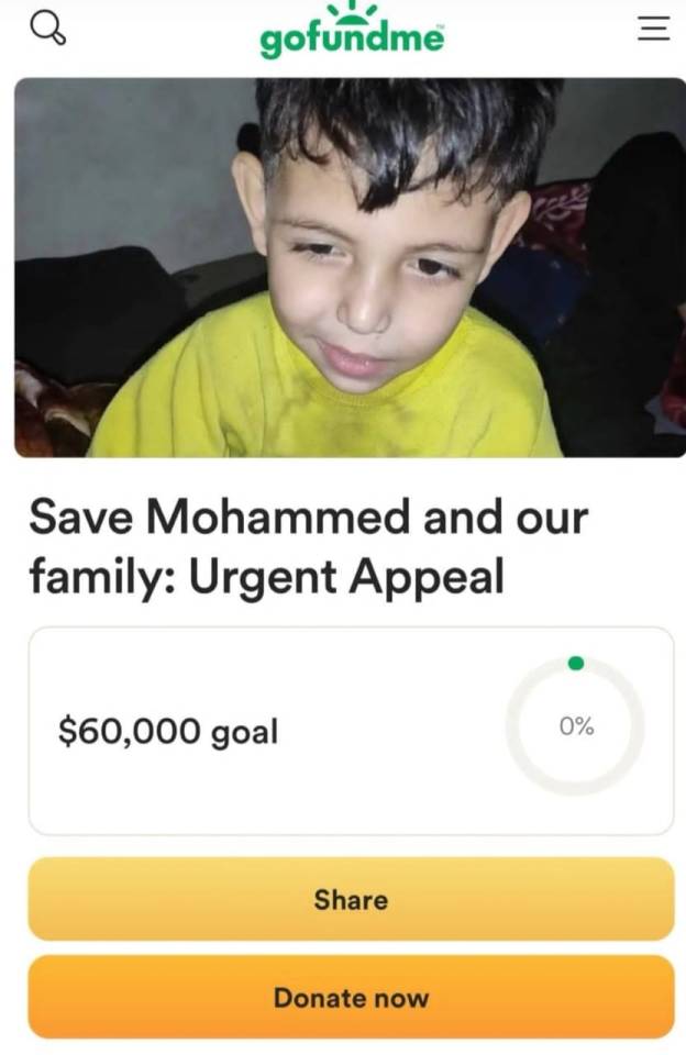
Save Mohammed and Our Family🙏🏻🙏🏻🙏🏻

"Urgent Appeal for Mohammed's Family in Gaza
I am the father of the child Mohammed, who suffers from partial paralysis on the left side since birth, loss of vision in the left eye, and brain atrophy. A shunt was installed in his brain due to abnormal fluid accumulation (hydrocephalus). Mohammed suffers from frequent neurological seizures and is in urgent need of healthcare and social support, special medications, and follow-up at the eye and neurology clinics.


The situation in Gaza makes it extremely difficult to provide the necessary care for Mohammed, my three children, and my family. We are currently living in a tent, and the weather is very cold. After our home was completely destroyed, we do not have access to medicines, food, or drink, and we urgently need suitable clothing and food for Mohammed and my three children.
We urgently need your support to donate for:
- Providing necessary medicines and medical care for Mohammed
- Ensuring healthy food and water for our family
- Providing warm clothing to face the severe cold
- Attempting to leave Gaza for treatment and providing a better and safer life for our children
Every donation, no matter how small, can make a big difference in our lives. We kindly ask you to share this post and help in any way possible.
Thank you for your support and care."
I am Ross from the United States, and I have created this fundraiser to help the Alostaz family in Gaza through the father of Mohammed, Mahmoud. All raised funds will be sent through me to the family Alostaz.
Please help me and donate to treat my son Muhammad and get him abroad and get us safely out of this fierce war
@halalchampagnesocialist @spooksier@jonahmagnus @artemis-pendragon @lesbian-hannibal
@turtletoria @bulkhummus @smOkebreaks @doubleca5t @wuntrum
@mysharona1987 @fairycosmos @watermotif @vague-humanoid @mavigator
@legallybrunettedotcom@brucespringsteendotcom
@pjharvey @doublism@cigaretteuncle
@odinsblog@chainmail-butch @bestlesbiancave @t4tails @electricpurrs
@mordhiobhail @bOnkcreat @lemon-wedges @holedyke @jerseyclown
@butchfeygela @danijaci @pinayelf @dogesterone @professorllayton
@bakwaas @eastgaysian @tf2yuri@bongjoonheaux
@romanceyourdemons
@valtsv@cryptotheism @coughloop
@cottoncandylesbo @jame7t
@amygdalae @pointnclick @psygull @wolvierinez
@0047 silver
#free palestine#@tamarrud#gaza aid#gaza fundraiser#gaza solidarity encampment#help gaza#gaza strip#gazaunderattack#free gaza#gaza genocide
586 notes
·
View notes
Text
Case of an Hydrocephalus Internus, successfully treated by Mercury
First I want to post the following several statements from here concerning a mercury formulation called calomel ─ the author is explaining how such a toxic compound came to practically be deemed a miracle medicine well into the 19th Century: ❝How is it then that calomel, along with other metal salts, was commonly used by physicians for some five hundred years as standard therapy for almost every…
0 notes
Text
Ultrasound Scans in Pediatrics: Ensuring Healthy Growth and Development
At Ovum Hospital, we prioritize the health and well-being of children, understanding that early detection and monitoring of medical conditions are crucial for ensuring healthy growth and development. One of the most valuable diagnostic tools in pediatric medicine is the ultrasound scan. This non-invasive imaging technique is essential for diagnosing a range of conditions and monitoring various aspects of a child's health. Here’s an in-depth look at the importance of ultrasound scans in pediatrics.
What is an Ultrasound Scan?
An ultrasound scan, also known as sonography, uses high-frequency sound waves to create images of structures within the body. Unlike X-rays or CT scans, ultrasounds do not use ionizing radiation, making them a safer option for imaging, especially in children. Ultrasound scans are performed using a device called a transducer, which emits sound waves and captures the echoes as they bounce back from internal tissues and organs. These echoes are then converted into real-time images.
Common Pediatric Conditions Diagnosed with Ultrasound
Ultrasound scans are incredibly versatile and can be used to diagnose a wide range of conditions in children, including:
Abdominal Issues: Ultrasounds can detect problems in the abdominal organs such as the liver, kidneys, spleen, and intestines. Conditions like appendicitis, gallstones, kidney stones, and liver disease can be accurately diagnosed with an ultrasound scan.
Congenital Anomalies: Prenatal ultrasounds are crucial for detecting congenital anomalies before birth. Postnatal ultrasounds continue to monitor these conditions, such as congenital heart defects or kidney malformations, ensuring early intervention.
Head and Brain Issues: In infants, ultrasounds can evaluate the brain's structure and check for conditions like hydrocephalus, where there is an accumulation of cerebrospinal fluid, or any other brain abnormalities.
Musculoskeletal Disorders: Ultrasounds can assess soft tissue injuries, joint problems, and developmental dysplasia of the hip in infants, helping to ensure proper growth and mobility.
Thyroid Problems: Ultrasound scans are used to examine the thyroid gland, checking for abnormalities such as goiters, nodules, or other thyroid-related issues.
The Benefits of Ultrasound Scans in Pediatrics
Safety: One of the most significant advantages of ultrasound scans is their safety profile. Since they do not use ionizing radiation, they are safe for repeated use, making them ideal for monitoring chronic conditions or performing follow-up exams.
Non-Invasive: Ultrasounds are non-invasive, which means they do not require any incisions or injections. This aspect is particularly beneficial for children, who may be more sensitive to invasive procedures.
Real-Time Imaging: Ultrasound scans provide real-time images, allowing for immediate assessment and diagnosis. This real-time capability is especially useful in emergency situations where quick decision-making is required.
Versatility: Ultrasounds can be used to examine almost any part of the body. From the abdomen to the brain, and from muscles to joints, the versatility of ultrasound scans makes them indispensable in pediatric care.
Cost-Effective: Compared to other imaging modalities like MRI or CT scans, ultrasound scans are generally more affordable. This cost-effectiveness makes them accessible and a preferred first-line diagnostic tool in many cases.
How Ultrasound Scans Contribute to Healthy Growth and Development
Early Detection and Intervention: Early detection of abnormalities or conditions through ultrasound scans can lead to timely intervention and treatment. For example, identifying a congenital heart defect early on can allow for surgical correction before it causes significant health issues.
Monitoring Growth and Development: Regular ultrasound scans can monitor a child’s growth and development, ensuring that organs are developing correctly and identifying any potential issues early.
Guiding Treatment Plans: Ultrasound scans provide detailed information that can guide treatment plans. Whether it's determining the exact location of a kidney stone or assessing the extent of an abdominal infection, the information gathered from an ultrasound scan is invaluable for creating effective treatment strategies.
Reassurance for Parents: For parents, having access to a safe and reliable diagnostic tool like ultrasound provides reassurance. Knowing that any potential health issue can be detected and monitored without exposing their child to harmful radiation offers peace of mind.
Choosing the Right Facility for Pediatric Ultrasound Scans
When it comes to ensuring the best care for your child, choosing the right medical facility is crucial. At Ovum Hospital, we pride ourselves on being a leading provider of pediatric healthcare services, including ultrasound scans. Our state-of-the-art equipment and experienced team of pediatric specialists ensure that every ultrasound scan is performed with the highest level of precision and care.
If you are looking for a reliable place for an ultrasound scan Bangalore, Ovum Hospital is your best choice. Our commitment to providing comprehensive and compassionate care makes us a trusted name in pediatric healthcare.
Conclusion
Ultrasound scans are a cornerstone of pediatric diagnostics, offering a safe, non-invasive, and effective way to monitor and ensure healthy growth and development in children. At Ovum Hospital, we utilize advanced ultrasound technology to provide accurate diagnoses and guide treatment plans, reinforcing our reputation as a top gynecologist hospital Bangalore residents trust. If you need an ultrasound scan Bangalore for your child, trust Ovum Hospital to deliver the highest standard of care.
0 notes
Text
What is the Cost of Brain Tumor Surgery in India?

What is the Cost of Brain Tumor Surgery in India?
A mass of cancerous cells in the brain is called a brain tumor. It has the potential to harm the brain. A brain tumor surgery is the surgical excision and removal of the abnormal outgrowth in the brain. A brain tumor leads to several symptoms like headache, nausea, vomiting, vision abnormalities, memory problems, speech disturbances, etc.
In India, every year, 40,000 to 50,000 people are diagnosed with brain tumors, out of which 20 % are children. Treatment of brain tumors in India will depend on symptoms, size, and location of the brain tumor: treatment options include chemotherapy, radiotherapy, immunotherapy, brain tumor surgery, and radiosurgery.
The majority of the cost of brain tumor surgery in India will depend on the type of tumor, its grade, size, and location. On average, brain tumor surgery will cost you around 6000 USD to 10000 USD.
What is the purpose of brain tumor surgery in India?
To remove the tumor completely
To remove a part of the tumor to delay the progression and to relieve the symptoms
To drain hydrocephalus, which is a fluid built in your brain
To improve the success rate of chemo and radiation therapy
To relieve seizures caused by brain tumors.
Brain Aneurysm Surgery Cost in delhi | Deep Brain Stimulation Surgery Cost in India | Brain Tumor Surgery Cost | Robotic Knee Replacement Surgery Cost in India | Robotic Prostatectomy Surgery Cost in Delhi, India
Types of Brain Tumor Surgery Performed in India and its cost
Type of Surgery
Explanation
Cost
Craniotomy
Surgeon will make a small incision on your scalp and cut an area of bone from your skull to get its access and remove tumor cells.
6000$
Neuroendoscopy (TNTS)
Your surgeon in India will make a hole in your skull through which he will insert an endoscope in the brain and remove tumor cells.
6500$
MRI Guided Laser Ablation
A minimally invasive technique that uses laser ablations to destroy tumor cells.
6000$
Radiosurgery
It is used to treat benign brain tumors and brain cancers.
5000$
Zap - X Radiosurgery
Indraprastha Apollo hospital has started Asia’s first Zap-x radiosurgery. It is a painless , non-invasive single sitting OPD based procedure.
8000$
Gamma Knife
It is also a form of radiosurgery for precise removal of brain tumors. Radiation is given in high doses for a shorter duration which is highly precise in nature.
7500$
Cost of Brain Tumor Surgery in India: Advanced Neurosurgery, Affordable Costs
Minimum Cost: $6000
Average Cost: $7000
Maximum Cost: $10000
The cost of brain tumor surgery in Delhi will depend on the following factors:
Admission fee
Oncologist fee
Age of patient
Severity of tumor
The medical condition of the patient
Type of brain tumor surgery performed
Post-procedure complications
Type of hospital
The admission room you choose
Diagnostic tests
When the above factors are considered, the Cost of Brain Tumor Surgery in India varies from USD 1300 to USD 6500, whereas the same surgery in the US costs around USD 73,000.
Cost of Brain Tumor Surgery in Top Hospitals of India
Hospital
Cost
Indraprastha Apollo Hospital
10000$
Max Healthcare Saket
7500$
Venkateshwar Hospital
5600$
Aakash Healthcare
6000$
Fortis Hospital
7500$
Cost Breakdown
Pre-procedure tests cost: $500
Cost of surgery: $5500 to $9500
Follow-up consultation fee: $15 per session (first consultation is free)
Medicines & Post-Op Treatment: $30
Cost of tests to check progress: $50
ICU stay for 1 to 2 days: Part of the surgical package.
Hospital room stay - Part of the surgical package.
Hotel - 20$ per day
Food - 10$ per day
Taxi - Complimentary from hospital
Sim Card - Complimentary
Language Translator - Complimentary
Dedicated Patient Care Executive - Complimentary
Connect with the best medical consultants in India today!!
Cross Border Care is the best medical consultant in India. We will help you avail yourself of all the latest treatments for brain tumor surgery in India at the most affordable rates. You can get in touch with us for robotic thyroidectomy, robotic prostatectomy, pituitary tumor surgery, and spine surgery.
To connect with the top doctors and top hospitals in India and to know more about our facilities, please get in touch with us today at [email protected]
Read Also These blogs where can get best and useful information:-
Best Three Hospitals for Bone Marrow Transplant Surgery in India The Best Three Hospitals for Osteosarcoma Surgery in India The Best Three Hospitals for TAVI Surgery in India Top Three Doctors for Kyphoplasty Surgery in India Top Three Hospitals for Kyphoplasty Surgery in India The Best Three Doctors for EPS and RF Ablation in India
0 notes
Text
Postdoctoral Research Associate in Functional Genomics Washington University School of Medicine Jin Lab @WashUGenetics is looking for a functional genomics #postdoc. Please DM me or apply here #Brain #Heart #Hydrocephalus #Genomics See the full job description on jobRxiv: https://jobrxiv.org/job/washington-university-school-of-medicine-27778-postdoctoral-research-associate-in-functional-genomics-4/?feed_id=76905 #cell_culture #functional_genomics #in_vivo #mitochondria #zebrafish #ScienceJobs #hiring #research
0 notes