#fungal microscopy
Explore tagged Tumblr posts
Visit Tumblr Blog
Explore Tumblr blogs with no restrictions, modern design and the best experience.
Last Seen Tumblr Blogs


Fun Fact
69% of Tumblr users are millennials.
Text


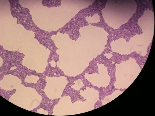
Teleomorphs of an Ascomycete (this was a cup fungi but the term refers to any fungi producing spores on what is referred to as the ascus). These are shown under the microscope with Lugol's solution at a recent fungal microscopy workshop. My last fungal microscopy workshop was on Basidiomycetes and it was cool to see the difference in cutting pieces of specimens to make slides, and the difference in spore-bearing structures under magnification. Lugol's solutions and other reagents can loosen tissues and dissolve pigments and mucilage, making spores easier to observe. Reagents can also produce color changing reactions that can inform species identification
#teleomorph#fungal microscopy#microscopy#fungus#fungi#mycology#mushrooms#mushroom#ascomycete#ascomycota#cup fungi#science
45 notes
·
View notes
Text
That post that called the relationship btwn plants and mycorrhizal fungi “yuri stuff” lives rent free in my head
62 notes
·
View notes
Text
Rhodotorula
“Rhodotorula mucilaginosa cells, methylene blue stain, magnification 400 x, notice the oval yeast cells as well as filamentous structures (pseudohyphae).” - via Wikimedia Commons
#fungi#fungus#microbiology#wikipedia#wikipedia pictures#wikimedia commons#nature#microbes#mycology#medical mycology#medical#medical microbiology#infectious diseases#medicine#methylene blue#methylene blue stain#specimen staining#fungal infection#fungicore#mycologycore#microscope#microscopy#yeast
12 notes
·
View notes
Text
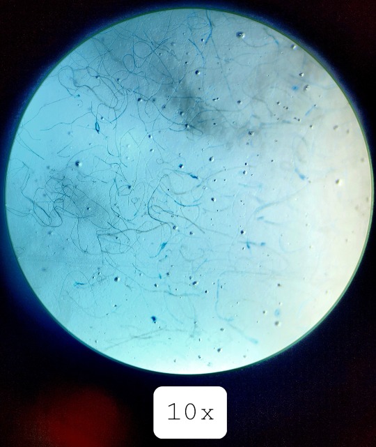

Fungal filaments under 10x
#studyblr#microbiology#microbio#microbio lab#microbiology lab#microbio laboratory#microbiology laboratory#fungi#fungal microbiology#fungi microbiology#microscopy#microscope#bio#biology
29 notes
·
View notes
Note
Hello ! I have read -per your tags- that you are a fungi biologist !!! First this is very cool, second i have a question about MOLD if its ok :)
Can you eat jam where the mold has been removed ? What is the general rule if there is one behind NO eating bc mold or ok eating if removed ? Im really curious about it +is it true that mold can grow in your body ? (I had a pack of unfortunately moldy tobbaco and a friend insisted on smoking it and i had to use the argument that they would become Moldy From The Lungs to stop them.... but is this true ?)
Anyhow, thank you for your time and keep on fungin' <3
Hey pal! First of all, thank you for the question (and for thinking fungal biology is cool haha), it was absolutely delightful for me to get this. I wish people talked to me about fungi outside of work more often! 🍄
As a disclaimer: my work is mostly molecular biology work on Saccharomyces cerevisiae aka Brewer's Yeast, and occasionally Candida albicans, and neither of these is closely connected to the mold question. I'm by no means a food safety expert either, but with that out of the way:
The short answer is that I would not eat jam with mold on it, even if you remove the mold. I would say that the rule to go by is that if it's a soft food with mold on it, discard the whole thing. If it’s something "hard" like a hard cheese, then it's probably safe to cut away the mold and eat the cheese. I found this little guide that seems quite reasonable and helpful to me, and it’ll tell you more about what types of food grow mold and how to handle them if you scroll to the end!
The long answer: Molds, like a lot of fungi, grow multicellular filaments called hyphae (Brand & Gow, 2009; Brand, 2012) which are only micrometres long. Meaning that you would need a microscope to be able to see them. Think of them as invisible roots or arms that can spread through your food. The fuzzy, green/grey/white manifestation that you see of the mold is a “colony” or in other words a network of cells or hyphae (Miguélez et al., 1999) depending on the organism. If you can see it, it is because it has been growing for a while, and that most likely means that your food is already contaminated with the “roots”. And these aren’t harmless; because depending on the type of mold, they can produce mycotoxins. Mycotoxins are biochemicals the cell makes which can cause serious harm to your health (Gonkowski et al., 2020). They have been known to cause allergic reactions, gastrointestinal issues, and poisoning. This is all dependent on the type of mycotoxin and the concentration of it, and the individual’s health, but I reckon this is the case where you want to be safe not sorry. From Gonkowski et al: “It is known that mycotoxins may act on many internal organs and systems, including, among others, nervous, reproductive and immunological systems, metabolic processes and endocrine glands” (Rykaczewska et al., 2019). In some cases, bacteria can also grow with the mold and cause illness. So, the long answer also boils down to: no, don’t eat it, it’s really not worth the risk.
And, yes, mold can grow in human lungs if the spores are inhaled. Granted that is most likely to happen with immunocompromised individuals as far as I know, but! I would advise against sniffing it at any rate haha.
I'm including the references in the text and at the end for fun, in case you’re interested to read more about this or see the microscopy images in the papers!!
Happy and safe eating 🍄😊
Brand, A. (2012) ‘Hyphal growth in human fungal pathogens and its role in virulence’, International Journal of Microbiology, 2012, pp. 1–11. doi:10.1155/2012/517529.
Brand, A. and Gow, N.A. (2009) ‘Mechanisms of hypha orientation of fungi’, Current Opinion in Microbiology, 12(4), pp. 350–357. doi:10.1016/j.mib.2009.05.007.
Gonkowski, S., Gajęcka, M. and Makowska, K. (2020) ‘Mycotoxins and the enteric nervous system’, Toxins, 12(7), p. 461. doi:10.3390/toxins12070461.
Miguélez, E.M., Hardisson, C. and Manzanal, M.B. (1999) ‘Hyphal death during colony development in streptomyces antibioticus: Morphological evidence for the existence of a process of cell deletion in a multicellular prokaryote’, The Journal of Cell Biology, 145(3), pp. 515–525. doi:10.1083/jcb.145.3.515.
Rykaczewska, A. et al. (2019) ‘Imbalance in the blood concentrations of selected steroids in pre-pubertal gilts depending on the time of exposure to low doses of Zearalenone’, Toxins, 11(10), p. 561. doi:10.3390/toxins11100561.
#this is sooo many words I'm sorry#absolutely loved it though thanks for letting me be a nerd for a bit <3#asks#mara talks for ts
9 notes
·
View notes
Text
Why You Shouldn't Ignore Ear Discomfort: Seek Expert Help for Timely Diagnosis and Treatment
A broad and varied category of illnesses affecting different components of the human auditory system is known as ear disorders. Ear ailments are typically inflammatory and infectious in nature; they are less frequently brought on by traumas or endocrine problems.
Since ear disorders are typically accompanied by severe discomfort, hearing loss, and other unpleasant symptoms, they cannot be ignored. However, self-medication using "folk remedies" is not only risky but also useless. The lack of proper diagnostics and qualified medical care for ear pain can lead to serious complications - irreversible deafness, acute mastoiditis, meningitis, and sepsis. That is why, if any alarming ENT symptoms from the ears appear, it is important to see a doctor as soon as possible.

Be sure to visit an ear specialist in Chandigarh if you have:
pain in the ear, which may radiate to the eyes, temples, or lower jaw (sometimes when chewing, swallowing, and walking);
a headache that is widespread on the side of the affected ear
inflammation and redness of the outer ear;
itching in the auricle;
noise or ringing in the ears;
discharge of serous fluid, pus, blood, or ichor from the ear (otorrhea);
sensation of fluid flowing in the ear;
cervical lymph node enlargement;
decreased or complete loss of hearing;
giving one's voice into a blocked ear (autophony);
respiratory symptoms: chills, loss of appetite, overall weakness, and fever (38 °C and higher);
nausea, vomiting, dizziness, and poor motor coordination;
sleep disorders.
Please note! Many ear diseases develop rapidly, quickly become chronic, and cause serious complications. Therefore, if even minor discomfort in the ear appears (not to mention acute pain), do not delay a visit to the otolaryngologist!
Classification of ear diseases
Among ear pathologies, inflammatory, non-inflammatory (including hereditary) and oncological diseases are distinguished.
Ear Inflammatory Disorders:
Otitis: Ear infection from bacteria or fungi.
Otomycosis: Fungal ear infection in the external canal.
Labyrinth - A serious ear infection that requires immediate attention.
Mastoiditis: Infection causing swelling behind the ear.
Auricular perichondritis: Condition involving infection and swelling in ear cartilage.
Tympanosclerosis: Chronic otitis inflicting tissue growth inside the middle ear.
Non-inflammatory Ear Disorders:
Earwax: Blockage brought on by too much wax.
Deafness: Hearing loss, either whole or partial.
Sudden deafness: Caused by infections or underlying issues.
Sensorineural Hearing Loss: Inner ear or nerve damage.
Otosclerosis: Bone adjustments affecting listening.
Neuritis of VIII Nerve: Permanent hearing loss due to nerve damage.
Ear Injuries: Damage from physical trauma or pressure changes.
Benign Tumors: Non-cancerous ear growths.
Outer Ear Cancer: Rare Skin cancer occurring on the outer ear.
Due to the similarity of signs across diverse ear problems, only an ear specialist chandigarh can accurately diagnose and choose the best course of treatment for different ear pathologies.
Ear illness diagnosis and treatment
Advanced diagnostics and thorough treatment of ear disorders are provided by the best ear specialist.
During the first visit, a skilled otolaryngologist performs an examination and orders several more tests utilizing state-of-the-art diagnostic tools. The following may be necessary to make a diagnosis:
otoscopy,
audiometry,
tympanometry,
hearing test using a tuning fork,
otoneurological tests,
biochemical and serological blood tests for infections,
bacterial culture of ear discharge for microflora with determination of sensitivity to antibiotics,
microscopy of ear discharge,
immunological research and other tests.
Assisting patients with ear diseases, an ear specialist in chandigarh uses all the possibilities of conservative and surgical methods. Each patient receives a personalized treatment plan based solely on their clinical image and the type of sickness they have. Immunomodulators, antiseptics, antifungals, antihistamines, and local and systemic antibiotics may be administered to the affected individual. If necessary, the doctor performs a dry or wet toilet of the ear, opens furuncles and perforates the eardrum by pumping out pus, and removes foreign bodies. These and other surgical manipulations are performed by Ear doctors using modern radio waves, endoscopic, and laser equipment of high precision and advanced technology.
In Conclusion
You must seek expert medical attention if you feel any ear discomfort, including pain, hearing loss, or discharge. To avoid more issues, an ear expert will do a comprehensive evaluation with cutting-edge diagnostic equipment and customized treatment alternatives. Early intervention can ensure the best outcomes and protect your hearing and overall health.
For more information please visit our web page :- Dr. Ram ENT Hospital
#ear doctor chandigarh#ear specialist#ear specialist chandigarh#ear specialist in chandigarh#ear specialist in mohali#ear hospital#best ear doctor#best ear hospital#best ear specialist#Digital Hearing Aids#Bone-Anchored Hearing Aids#cochlear implant surgery#cochlear implant
0 notes
Text
Effective Solutions for Vaginal Infection Treatment in Thane

Vaginal infections are a prevalent health issue affecting women of all ages. These infections can cause discomfort, embarrassment, and serious complications if untreated. With advanced facilities and specialists like Dr. Aditi Godbole, women in Thane can now access exceptional vaginal infection treatment in thane, ensuring tailored care and effective solutions.
Understanding Vaginal Infections
Vaginal infections, or vaginitis, occur due to an imbalance in the vaginal ecosystem caused by bacterial overgrowth, fungal infections, or parasitic organisms. Common symptoms include itching, burning sensations, abnormal discharge, and unpleasant odors. Contributing factors include poor hygiene, hormonal changes, and certain products like scented soaps or douches.
Types of Vaginal Infections
Bacterial Vaginosis (BV): An overgrowth of harmful bacteria causes BV, leading to thin, grayish discharge with a fishy odor. While not sexually transmitted, it can be triggered by sexual activity.
Yeast Infections: Caused by Candida albicans, these result in thick, white discharge, intense itching, and swelling. Women with weakened immune systems or on antibiotics are more prone.
Trichomoniasis: A sexually transmitted infection caused by Trichomonas vaginalis, presenting yellow-green frothy discharge and discomfort during intercourse.
Non-Infectious Vaginitis: Allergic reactions to products like spermicides or detergents can cause irritation and inflammation.
Diagnosis of Vaginal Infections
Accurate diagnosis is essential for effective treatment. Dr. Aditi Godbole and other specialists in Thane employ comprehensive diagnostic methods, including:
Medical History: Assessing symptoms, lifestyle, and medical background.
Physical Examination: Conducting pelvic exams to detect inflammation, discharge, and abnormalities.
Laboratory Tests: Analyzing vaginal discharge samples to identify causative agents through microscopy or culture tests.
Treatment Options Available in Thane
Thane offers cutting-edge gynecological facilities with personalized treatment plans for vaginal infections. Key treatment options include:
Medications:
Antibiotics: Oral or topical antibiotics like metronidazole or clindamycin for bacterial infections.
Antifungal Agents: Creams, suppositories, or oral medications like fluconazole for yeast infections.
Antiparasitic Drugs: Metronidazole or tinidazole for trichomoniasis.
Lifestyle Modifications:
Avoiding scented products and harsh chemicals.
Maintaining hygiene and wearing breathable cotton underwear.
Practicing safe sex with protection.
Probiotic Supplements: Enhancing diet with probiotics to restore healthy vaginal flora and reduce recurrent infections.
Hormonal Therapies: Using estrogen creams or vaginal tablets for postmenopausal atrophic vaginitis.
Preventive Measures
Preventing vaginal infections requires healthy habits and proactive care. Experts in Thane recommend:
Balanced Diet: Including nutrients, probiotics, and water to support overall health.
Regular Check-Ups: Routine gynecological visits for early detection and management.
Proper Hygiene Practices: Washing with mild, unscented soap and avoiding over-washing or douching.
Why Choose Thane for Vaginal Infection Treatment?
Thane stands out for quality medical care, with professionals like Dr. Aditi Godbole offering holistic solutions. Benefits include:
Personalized Care: Tailored treatment plans addressing individual concerns.
Advanced Diagnostic Techniques: Modern facilities for precise diagnoses.
Expertise and Experience: Skilled specialists with extensive knowledge in gynecological conditions.
For women in Thane, seeking timely medical attention and expert guidance ensures effective management of vaginal infections.
How to Reach Dr. Aditi Godbole’s Clinic in Thane?
Dr. Aditi Godbole’s clinic is conveniently located to serve patients from across Mumbai and beyond.
Clinic Address: Ground Floor, Gaala No. 15, Sai Tirth Towers, Station Rd, Sidharth Nagar, Kopri, Thane East, Thane, Maharashtra 400603
Directions to the Clinic:
Western Line: Take a train to Thane Station. From there, the clinic is just a short 10-minute walk or a 5-minute auto-rickshaw ride.
Central Line: Reach Thane Station via the Central Line, and follow the same directions as above.
Out-of-Mumbai Patients:
By Train: Arrive at Thane Station, well-connected by long-distance trains.
By Road: Use the Eastern Express Highway for a hassle-free journey to Thane East.
By Air: From Mumbai Airport, take a cab or local transport to Thane East, approximately 60-90 minutes away.
The clinic’s accessible location and proximity to Thane Station ensure convenience for all patients.
0 notes
Text
Best ENT specialist in bangalore
Why is my ear discharging?
What causes Ear Discharge ? Most cases , the ear discharges the earwax that might be seen as discharge. However some cases , with a ruptured eardrum or a perforated ear drum, it may cause discharge of blood or fluids from the middle ear. The underlying cause of this is an infection in the middle ear or mastoid bone.
What is Otitis Media ( Middle ear infection) ? The ear can be divided briefly into 3 parts , the external middle and inner EAR. Otitis media is the inflammation of the lining of the middle ear that causes Recurrent otorrhoea ( ear discharge) through a perforation in the ear. This could be Acute Otitis media: Acute inflammation , less than 3 weeks Duration, usually caused by bacterial or viral infections with pus in the middle ear ,may or may not be associated with perforated eardrum Otitis Media With Effusion : without active inflammation, proceeded by partially treated Otitis media , causing glue like discharge from the ear. Chronic Otitis Media : Chronic inflammation more than 4 weeks with a Persistent eardrum perforation , on and off ear discharge not completely resolving Mastoiditis : Acute inflammation of the mastoid bone (bone located behind the ear ) causing ear pain and swelling behind the ear
What are the symptoms ?The symptoms include Ear discharge ( Profuse or scanty; mucoid /mucopurulent or watery ;greenish /whitish /blood tinged ; foul smelling/ not foul smelling ). The ear discharge might not be obvious , sometimes only when u puta cotton in ear ,it wets the ear Ear Blocked sensation Hearing impairment Red , painful tissue in the ear canal Ear pain / pain behind the ear Fever / giddiness / vomiting Headache Facial weakness or facial palsy
When to approach a doctor if I have ear discharge ? It is advisable to consult an Ent surgeon immediately when you have an ear discharge ,since it is can cause complications if left untreated . At home adjuvant measures include avoid taking headbath , avoid swimming, avoid using ear buds . Symptoms of complications include Unresolving headache , pain behind the ear Neck pain /neck stiffness Projectile vomiting Impaired consciousness Giddiness (sensation of spinning ) High Grade fever What are the investigations that I have to do ? Once you approach you clinician ,they might take one or few of the investigations to confirm the diagnosis , chronicity, treatment plan and prognosis Ear swab : The discharge from the ear may be sent for culture and gram stain under microscopy to identify the organism and specific antibiotic of choice Otoscope/ Endoscopic Examination: to assess the status of external ear and ear drum Audiogram / hearing test : to assess the level & impairement of hearing , status of ossicles ( bones in the ear ) CT scan of the temporal bone : to identify the extent of infection ,the nidus of infection, & complications if any MRI scan : in select cases only when suspected intracranial complications How is it treated ? Depending on your underlying cause and disease extent ,your clinician may advice Oral / intravenous antibiotics Topical ear drops Analgesics and Antipyretics Steroid nasal sprays When do I need surgery? This is subjective to patients disease progression. Any ear discharge that is chronic more than 4 weeks ,usually needs surgery which could be either Tympanoplasty ( ear drum perforation repair) Grommet insertion ( for glue ear , placing small ventilation tube in the ear ) Mastoidectomy ( surgery to completely extirpate the infected mastoid air cells and ear discharge
How can I prevent / avoid ? Ear discharge can occur due to many bacterial / viral infections of middle ear or even sometimes Fungal infection of external ear. If you get ear discharge it is always advisable to immediately approach an ENT doctor and start treatment immediately to avoid the complications such as permanent hearing impairement/ spread of infection . Also if you have risk factors such as
Uncontrolled Diabetes
Smoking
Immunocompromised status due to any chronic illness , Avoid self medications without prescription and seek immediate care.
0 notes
Text
Pathogenic Microorganism Detection in Invasive Pulmonary Aspergillosis(IPA) Patients Using Bronchoalveolar Lavage Fluid

Pathogenic Microorganism Detection in Invasive Pulmonary Aspergillosis(IPA) Patients Using Bronchoalveolar Lavage Fluid in Biomedical Journal of Scientific & Technical Research
Background: To evaluate the diagnostic values of different forms of pathogenic microorganism detection from the bronchoalveolar lavage fluid (BALF) for Invasive Pulmonary Aspergillosis (IPA) diagnosis.
Methods: Bronchoalveolar lavage fluid (BALF) was collected from 100 patients with suspected clinical pulmonary Aspergillus infections by means of bronchoscopy. The smear microscopy, fungal culture and PCR were used to detect the Aspergillus from the BALF.
Results: 15 cases were diagnosed by pathological data(the proven group), while 12 confirmed cases were supported by clinical examinations including radiology and etiology (the probable group). The 73 cases left were not IPA, among which 21 cases of common pneumonia were taken as the control group. For the practise of smear microscopy method, the sensitivity, specificity, and accuracy of the diagnoses were 40.7%, 100.0%, and 66.7% respectively. As for the fungal culture, the sensitivity, specificity, and accuracy were 37.0%,100.0%, and 64.6% respectively. For PCR, the sensitivity, specificity, and accuracy were 55.6%, 100.0%, and 77.1% respectively. With the combination of the three methods mentioned, the final results were 92.6%, 100.0% and 95.8%.
Conclusion: The pathogenic detection of bronchoalveolar lavage fluid (BALF) is an ideal diagnostic method for Aspergillus infections. The smear microscopy method, the fungal culture and PCR has their own limitations in diagnosis. The detection of combining the three methods is proved to improve the efficiency of IPA diagnosis significantly.
For more articles in Journals on Biomedical Sciences Click here Bjstr
Follow on Twitter : https://twitter.com/Biomedres01 Follow on Blogger : https://biomedres01.blogspot.com/ Like Our Pins On : https://www.pinterest.com/biomedres/
#journals on biomedical imaging#journals on medical microbiology#biomedical open access journals#open access clinical and medical journal#medical and medicinal journal
0 notes
Link
1 note
·
View note
Text
Pathology Lab Services in Dhanori Pune - CT Nursing Home
At CT Nursing Home, we understand the importance of accurate and timely diagnostic results in ensuring the best possible healthcare outcomes. Our pathology lab services in Dhanori, Pune, are designed to provide you with comprehensive and reliable diagnostic solutions.
Pathology Lab Services in Dhanori, Pune:
Our pathology lab is equipped with state-of-the-art technology and staffed by experienced pathologists and technicians. We offer a wide range of pathology lab services, including:
Blood Tests: Complete Blood Count (CBC), Blood Chemistry, and other specialized tests.
Urine Tests: Urine Routine, Urine Microscopy, and Urine Culture.
Tissue and Cell Analysis: Histopathology, Cytopathology, and Molecular Pathology.
Microbiology Tests: Bacterial Cultures, Fungal Cultures, and Viral Tests.
Immunology Tests: Autoimmune Disorder Tests and Allergy Tests.
CT Nursing Home in Dhanori offers special discounts and packages for pathology tests. They have a dedicated pathology lab that follows a stringent quality control system and provides internationally accepted quality diagnostic services. The lab offers a range of tests, including blood tests, urine tests, and tests on stools and bodily tissues.
Our Clinic provides discounts and packages for various pathology tests, ensuring that patients receive accurate and reliable diagnostic results at affordable prices. We are committed to providing you with the best possible healthcare outcomes through our comprehensive pathology lab services in Dhanori, Pune. CT Nursing Home Best Pathology Lab in Pune Under the supervision of a qualified pathologist, the Laboratory follows a stringent quality control system.

#BestPaediatricHospitalNearMe#General Surgery Hospital in Dhanori#Sonography Services In Dhanori#BestInfertilitySpecialistInDhanori#Color Doppler Services In Dhanori#Top Maternity Hospital in Dhanori#Maternity Hospital in Dhanori#Pathology Lab Services in Dhanori
0 notes
Text
Two +ssRNA mycoviruses cohabiting the fungal cultivar of leafcutter ants | Virology Journal | Full Text
See on Scoop.it - EntomoNews
Leafcutter ants are dominant herbivores in the Neotropics and rely on a fungus (Leucoagaricus gongylophorus) to transform freshly gathered leaves into a source of nourishment rather than consuming the vegetation directly. Here we report two virus-like particles that were isolated from L. gongylophorus and observed using transmission electron microscopy. RNA sequencing identified two +ssRNA mycovirus strains, Leucoagaricus gongylophorus tymo-like virus 1 (LgTlV1) and Leucoagaricus gongylophorus magoulivirus 1 (LgMV1). Genome annotation of LgTlV1 (7401 nt) showed conserved domains for methyltransferase, endopeptidase, viral RNA helicase, and RNA-dependent RNA polymerase (RdRp). The smaller genome of LgMV1 (2636 nt) contains one open reading frame encoding an RdRp. While we hypothesize these mycoviruses function as symbionts in leafcutter farming systems, further study will be needed to test whether they are mutualists, commensals, or parasites.
Two +ssRNA mycoviruses cohabiting the fungal cultivar of leafcutter ants | Virology Journal | Full Text
Published: 04 September 2024
------
NDÉ
Traduction
Deux mycovirus +ssRNA cohabitant avec le cultivar fongique des fourmis coupe-feuille
Les fourmis coupe-feuille sont des herbivores dominants dans les régions néotropicales et dépendent d'un champignon (Leucoagaricus gongylophorus) pour transformer les feuilles fraîchement cueillies en source de nourriture plutôt que de consommer la végétation directement.
Nous rapportons ici deux particules semblables à des virus qui ont été isolées de L. gongylophorus et observées à l'aide de la microscopie électronique à transmission. Le séquençage de l'ARN a permis d'identifier deux souches de mycovirus +ssRNA, Leucoagaricus gongylophorus tymo-like virus 1 (LgTlV1) et Leucoagaricus gongylophorus magoulivirus 1 (LgMV1).
[...]
Bien que nous ayons émis l'hypothèse que ces mycovirus fonctionnent comme des symbiotes dans les cultures des fourmis coupe-feuille, des études supplémentaires seront nécessaires pour vérifier s'ils sont des mutualistes, des commensaux ou des parasites.
[Atta sp / Acromyrmex sp]
0 notes
Text
Unlocking High-Definition Microscopy in Life Sciences with 13MP USB Camera
Microscopy is a vital tool in the life sciences that allows researchers to examine biological processes, microbes, and cellular structures at a detailed level. The possibilities of microscopes increase with technology, especially when high-resolution cameras are used. The adoption of a 13MP USB camera is one such development that is transforming high-definition microscopy. This blog examines the ways in which a 13MP USB camera, with its unmatched clarity and detail, improves life sciences research.
The Development of Camera Integration and Microscopy
Microscopy has come a long way from its rudimentary beginnings. The integration of digital cameras into microscopes marked a significant shift from analog to digital imaging. Traditionally, high-resolution imaging required expensive and complex systems. However, the advent of the 13MP USB camera has democratized access to high-definition microscopy by combining superior resolution with the simplicity of USB connectivity.
A 13MP USB camera, with its high-resolution capabilities, offers researchers an edge in capturing detailed images of microscopic specimens. This resolution translates to greater clarity, allowing for more accurate analysis and documentation of biological samples.
Why Choose a 13MP USB Camera for Microscopy?
1. Superior Image Quality
The 13MP USB camera stands out due to its impressive resolution, providing researchers with high-definition images that are crucial for detailed analysis. This camera's ability to capture images with 13 megapixels ensures that even the finest details of cellular structures are visible. Whether studying cell morphology, tissue samples, or microbial organisms, the clarity provided by a 13MP USB camera enhances the ability to make precise observations and measurements.
2. Enhanced Accuracy and Detail
In life sciences research, accuracy is paramount. The high resolution of a 13MP USB camera allows for detailed imaging that can reveal subtle features that might be missed with lower-resolution cameras. This level of detail is especially important for tasks such as identifying specific cell types, observing cellular processes, and conducting quantitative analyses.
3. Streamlined Workflow
Integrating a 13MP USB camera with a microscope simplifies the imaging process. The USB connectivity ensures a straightforward setup, eliminating the need for complex cabling and interfaces. Researchers can easily connect the camera to a computer or laptop, view real-time images, and capture high-resolution photographs directly. This ease of use enhances workflow efficiency, allowing more time for analysis and less time spent on technical setup.
4. Cost-effective high-resolution imaging
While high-definition microscopy used to be associated with expensive equipment, the 13MP USB camera offers a cost-effective solution. By providing high-resolution imaging at a fraction of the cost of traditional systems, this camera makes advanced microscopy accessible to a broader range of researchers and institutions. The affordability does not compromise quality, as the 13MP USB camera delivers exceptional performance without breaking the bank.
Practical Applications in Life Sciences
1. Cellular and Tissue Analysis
In life sciences, understanding cellular and tissue structures is crucial. The high resolution of a 13MP USB camera allows researchers to observe and analyze cell morphology, tissue architecture, and other intricate details with unprecedented clarity. This capability is invaluable for studying cellular processes, disease mechanisms, and tissue responses to treatments.
2. Microbial Studies
For microbiologists, the ability to capture detailed images of microorganisms is essential. The 13MP USB camera enables precise observation of bacterial, fungal, and viral specimens, facilitating studies on microbial behavior, interactions, and identification. The enhanced clarity supports accurate documentation and analysis, contributing to advancements in microbiological research.
3. Educational purposes
Educational institutions benefit from incorporating a 13MP USB camera into their microscopy labs. Students and educators can utilize high-resolution imaging to better understand biological concepts, conduct experiments, and present findings. The affordability and ease of use make it an ideal choice for educational settings, enhancing the learning experience and providing practical hands-on opportunities.
4. Research and Development
In research and development, particularly in pharmaceutical and biotechnology sectors, high-definition microscopy is essential for drug discovery, development, and testing. The 13MP USB camera facilitates detailed imaging of cellular responses to compounds, aiding in the evaluation of drug efficacy and safety. The high resolution ensures that researchers can detect even the smallest changes and anomalies in their experiments.
In summary
The 13MP USB camera is revolutionizing life sciences high-definition microscopy by providing improved image quality, increased accuracy, and simplified procedures at a reasonable cost. Because of its incorporation into microscopes, scientists can now take precise, high-resolution pictures that are essential for a range of uses, including cellular analysis, microbial study, and teaching.
The 13MP USB camera stands out as a potent instrument that democratizes access to high-definition microscopy as life sciences research advances. It is a priceless tool in the quest for scientific advancement and innovation because of its capacity to deliver precise, crisp photographs at a reasonable price. Accepting this technology provides new avenues for investigating the microscopic world, expanding our understanding, and making significant advances in the field of life sciences research.
https://www.vadzoimaging.com/product/ar1335-4k-autofocus-usb-3-0-camera
0 notes
Text
Fwd: Graduate position: Geneva.MyxomycetesSystematicsEvol
Begin forwarded message: > From: [email protected] > Subject: Graduate position: Geneva.MyxomycetesSystematicsEvol > Date: 9 August 2024 at 06:10:32 BST > To: [email protected] > > > The Conservatory and Botanical Garden of Geneva (CJBG) and Geneva > University (UniGE) seek a PhD student > > Title of the project: A biosystematic revision of the family Cribrariaceae > Function: Doctoral candidate and university assistant, 70% > Contract starting date: 1 February 2025 (4 years) > > Job description: > The present project is focused on the systematics and evolution of one of > the most neglected groups of *Myxomycetes* (*Amoebozoa*), the family > *Cribrariaceae* (ca. 50 accepted species). The generic and species > boundaries, as well as the evolution of phenotypic traits, have never been > analysed in detail, and especially not in a phylogenetic context and with > modern techniques. We will assess the phylogenetic relationships of the > species using a fair taxonomic and molecular sampling, including NGS > techniques and phylogenomic analyses. Next, we will explore the evolution > of some phenotypic traits (notably, the sporophore macromorphology and the > spore ornamentation, but also other characters) and the ecology of the > species, to assess its relevance for the taxonomy and diversification of > the group. Finally, we will address the species-level taxonomy, identify > new species and cryptic taxa, and evaluate synonymies. With all this > information, we expect to propose a revised classification and an updated > monograph of the family. Most of the skills and data analyses learned by > the candidate upon completion of the project will also be applicable to the > systematics, taxonomy, and evolutionary biology of other groups of > organisms. > > Within the research project, the candidate will profit from aid, guidance, > and supervision in the different steps of the project, from specimen > handling, data gathering and analyses, to presentation of the results, and > scientific writing. In addition, the candidate is expected to build up a > network of national and international collaborations, perform short stays > abroad, and become used to the dynamic of scientific research to become an > independent researcher. > The assistant position is under the direction of Dr. Juan Carlos Zamora > (CJBG) and Dr. Carlos Lado (RJB-CSIC), and Yamama Naciri (CJBG, UniGE) as > responsible professor. > > Required degrees and skills: > - Master’s degree in biology or in a Biosystematics-related topic in > Environmental/Chemistry/Health Sciences. > - Motivation to engage in the research project and in scientific research, > including: (i) field work and in vitro cultures, (ii) data gathering and > analyses, (iii) writing of a doctoral thesis, (iv) presentation and > dissemination of the project results (scientific journal publication, > congresses, and others). > - Skills in Fungal/Botanical Systematics and Taxonomy, especially > microscopy. > - Skills in molecular lab and phylogenetic analyses. > - Interest for the morphological, evolutionary, and taxonomic study of > Myxomycetes. > - Good disposition to work in a team and to participate in the scientific > life of the host institute. > - Proficiency in written and spoken French (mother tongue or a minimum of > level B2 in CEFR scale) to ensure an adequate flow of the practical classes > of the Systematics and Biodiversity course, given in Biology to 2 year > students, which she/he would be responsible for. > - The working language of the project being English, the candidate must > also be particularly comfortable in this language (written and spoken) > > Application process: > Application deadline: 31 August 2024 > The application file (in English), including (1) a motivation letter and > (2) a detailed CV, should be sent exclusively by e-mail to > *[email protected]* (Juan Carlos Zamora), cc. > *[email protected]*, *[email protected]* > > Juan Carlos Zamora Señoret
0 notes
Text

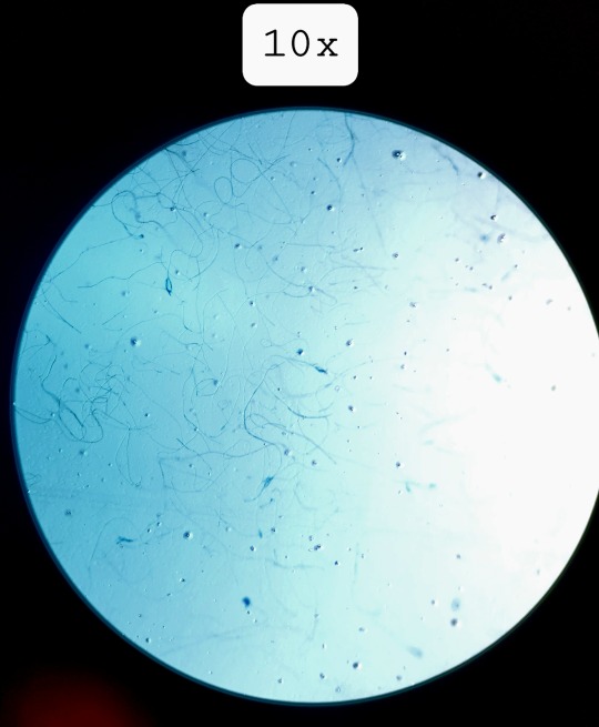
Fungal filaments at 10x (unknown sp.)
#studyblr#microbio#microbiology#microbio lab#microbiology lab#microbio laboratory#microbiology laboratory#fungal filaments#fungi#microscopy#microscope
3 notes
·
View notes
Text
Candida as Pathogens of Onychomycosis among Elderly Diabetic Patients_Crimson Publishers

Abstract:
Onychomycosis is a common fungal infection affecting nails. The primary cause for onychomycosis is Non-dermatophytes, while Candida species have emerged as second-line pathogens. Onychomycosis due to Candida [Candidal onychomycosis] is increasingly found in i patients with diabetes mellitus (T2DM). Diabetes can result in complications that affect all systems of the body. Of particular relevance to onychomycosis rates is the effect of (T2DM). on the lower extremities. (T2DM). can lead to lower extremity arterial disease; in turn this can predispose patients to onychomycosis of the toenails.
Materials and methods: Incidence were determined in 116 type 2 diabetes mellitus (T2DM) patients over from September2013 to January 2014 included direct microscopy and repeated cultures. A higher incidence of onychomycosis. who were registered at the Sedee Hussein Polyclinic of Benghazi city.
Results: The prevalence of onychomycosis among diabetics in our study was high, culture was positive in 84 of 116 patients [41%] patients with onychomycosis, laboratory culture identified yeasts as the pathogen in 22% of positive cases.
Conclusion: Onychomycosis is an important cause of morbidity in diabetic patients, increasing their risks for limb amputation and local and systemic secondary bacterial infections. Because onychomycosis is more common in diabetic patients and can complicate the disease, clinicians must be vigilant in its diagnosis and complete in its treatment.
Read More About this Article: https://crimsonpublishers.com/sbb/fulltext/SBB.000547.php
Read More Articles: https://crimsonpublishers.com/sbb/
#crimson publishers#open access journals in bioscience#crimson bioscience#crimson open access journals#bioengineering#crimson bioengineering#peer review journals#significances of bioscience and bioengineering
0 notes