#choanae
Explore tagged Tumblr posts
Text


Here’s a look at my updated rexy mask, the original one is from 2015 and styled from JP vs the present more scientifically accurate 2024 one. You can see a difference in my cardboard sculpting skills from 9 yrs ago.
Both done with a cardboard base and paper mache overlayed.
Also a better look at the choana and the glottis I added to the mouth.
Enjoy!
#art#dinosaur#paleoart#theropod#rexy#trex#tyrannosaurus#tyrannosaurus rex#tyrannosaur#mask#paper mache mask#paper mâché#papermache#paper mache sculpture#paper mache art#paper mache#cardboard sculpture#cardboard mask#cardboard art#animal art
118 notes
·
View notes
Text
Varanosuchus: First Fossil Croc of 2024
We are two weeks into the year and we already had a bunch of big croc papers, so today I'll cover the first of the two new genera named so far. Varanosuchus sakonnakhonensis (Monitor lizard crocodile from Sakon Nakhon) is a small atoposaurid neosuchian from the Early Cretaceous of Thailand, a country that has seen a virtual boom in croc papers this past year between the description of Alligator munensis and Antecrocodylus.
Varanosuchus was a small animal, maybe a meter in length if a little longer with a notably short and deep skull and long slender limbs revealing it to have been at least somewhat terrestrial. We actually have a decent amount of material of this guy. The holotype consists of a 3 dimensionally preserved skull as well as assorted postcranial remains (vertebrae, ribs, osteoderms and limbs), there is a second skull of whats likely to be a differently aged individual also showing a 3D skull and well the third ones just a skull table but 2/3 is still great.
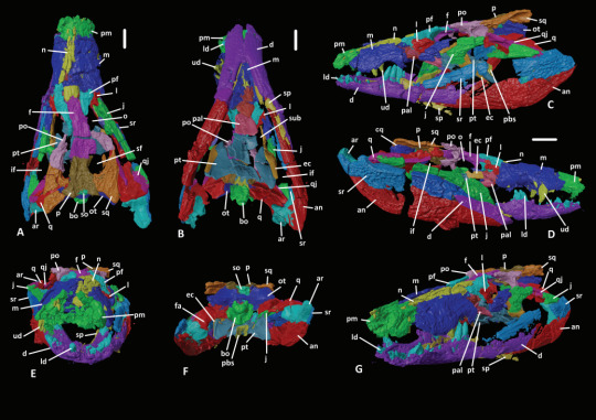
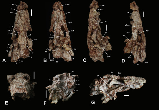
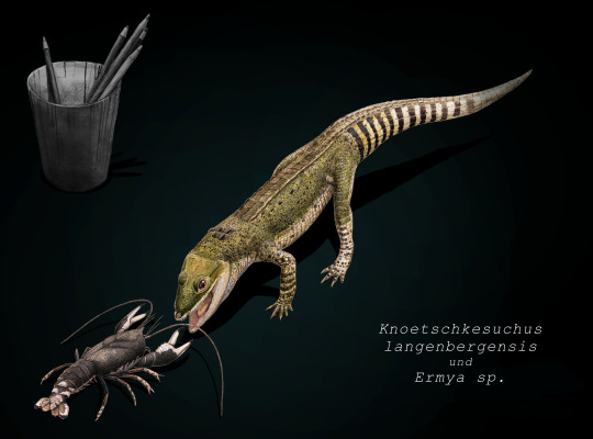
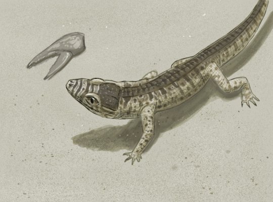
Now this guy was an atoposaurid, which is a group of crocodylomorphs that lived from the Jurassic to the end of the Cretaceous, their last members existing on the island of Hateg some 66 million years ago. Atopsaurids were generally small animals with short snouts and longish legs. Some examples of atoposaurids include Knoetschkesuchus from Germany, Aprosuchus from Romania and Alligatorellus from France and Germany, all three pictured below, art by @knuppitalism-with-ue
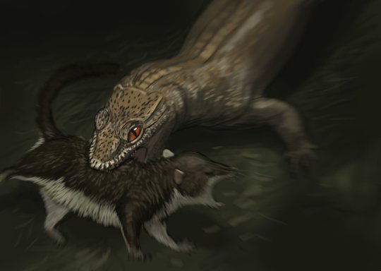
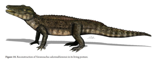
Now the matter of ecology for atoposaurids in general and Varanosuchus in particular is not clear. Altirostral skulls such as that of Varanosuchus are generally associated with terrestrial crocodylomorphs as best examplified by notosuchians. Their teeth and size both obviously speak against being shoreline ambush predators like modern crocs and their legs are straight and slender, suggesting they had an erect posture and not the more sprawling one seen in semi-aquatic forms. Though they could have still had some aquatic affinities. The authors for instance argue that the osteoderms, having plenty of pits, are more like those of an animal that spends time in the water and would thus use them in thermoregulation. So maybe they did enter water from time to time, somewhat like some modern lizards, tho I think its fairly certain that they spend a decent amount of time on land. The artwork below is the reconstruction from the paper itself.
Another matter discussed in the paper is phylogeny, more precisely the relationship of Neosuchians and how Eusuchia is defined. On the first front, its worth noting that the paper recovered both atoposaurids and paralligatorids as monophyletic groups and had them be each others closest relatives, a notion that has been recovered before. More interesting perhaps is the fact that the next closest relatives to these two were hylaeochampsids and Bernissartia, which are typically recovered closer to modern crocs. Which in fact form a separate branch that is the sister group to all the afforementioned clades and taxa. And then you got goniopholids, dyrosaurs and pholidosaurs which are all more basal than the paralligatorid+atoposaurid+crocodilian group, which is back to the ordinary really. The second thing is the definition of Eusuchia. So for the longest time Eusuchia has been defined to include those Neosuchians that have choanae that are fully enclosed by the pterygoid bones (I know I know a bunch of anatomy stuff bear with me). So if the choanae was surrounded by the pterygoid, its an Eusuchian, if not, its more basal. Well, atoposaurids don't have that....BUT VARANOSUCHUS DOES. This, coupled with hylaeochampsids also having this feature and being recovered closer to atoposaurids than to Crocodilians basically suggests that the feature is not diagnostic for Eusuchia and instead appeared multiple times independently.
Moving away from anatomy and phylogeny and all that stuff, I think its very cool that croc research in Thailand has kinda picked up this last year. And fittingly enough some people have even worked on a short documentary covering the known diversity of pseudosuchians from Thailand, giving an overview over the named forms from the Jurassic to today, from titans like Chalawan to even these newest dwarf forms. While the narration is obviously in Thai, there are English subs and I highly recommend looking into it (even if I disagree with their depiction of Varanosuchus as arboreal, its perhaps overshooting the goal a little bit).
youtube
Finally here's the paper itself (tho paywalled) New Cretaceous neosuchians (Crocodylomorpha) from Thailand bridge the evolutionary history of atoposaurids and paralligatorids | Zoological Journal of the Linnean Society | Oxford Academic (oup.com) and the wikipedia page I've been working on Varanosuchus - Wikipedia
I'll try to write up a post on the other new genus, Garzapelta, later this weekend so stay tuned for that.
#varanosuchus#atoposauridae#crocodylomorpha#neosuchia#thailand#cretaeous#pseudosuchia#croc#crocodile#land crocodile#prehistory#paleontology#palaeblr#long post#Youtube
195 notes
·
View notes
Text
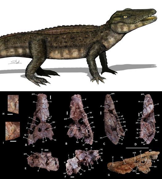
New Cretaceous neosuchians (Crocodylomorpha) from Thailand bridge the evolutionary history of atoposaurids and paralligatorids
Yohan Pochat-Cottilloux, Komsorn Lauprasert, Phornphen Chanthasit, Sita Manitkoon, Jérôme Adrien, Joël Lachambre, Romain Amiot, Jeremy E Martin
Abstract
The origin of modern crocodylians is rooted in the Cretaceous, but their evolutionary history is obscure because the relationships of outgroups and transitional forms are poorly resolved. Here, we describe a new form, Varanosuchus sakonnakhonensis gen. nov., sp. nov., from the Early Cretaceous of Thailand that fills an evolutionary gap between Paralligatoridae and Atoposauridae, two derived neosuchian lineages with previously unsettled phylogenetic relationships. Three individuals, including a complete skull and associated postcranial remains, allow for a detailed description and phylogenetic analysis. The new taxon is distinguished from all other crocodylomorphs by an association of features, including a narrow altirostral morphology, a dorsal part of the postorbital with an anterolaterally facing edge, a depression on the posterolateral surface of the maxilla, and fully pterygoid-bound choanae. A phylogenetic analysis confirms the monophyly and taxonomic content of Atoposauridae and Paralligatoridae, and we underline the difficulty in reaching a robust definition of Eusuchia. Furthermore, we put forward further arguments related to the putative terrestrial ecology with semi-aquatic affinities of atoposaurids based on their altirostral snout morphology and osteoderm ornamentation.
Read the paper here:
https://academic.oup.com/zoolinnean/advance-article-abstract/doi/10.1093/zoolinnean/zlad195/7513556
19 notes
·
View notes
Text

My take on the relatively uncommon (in the "I'd have two nickels" kind of way) but interesting spec bio trope of insect-like terrestrial fish, partially inspired by this thing I did earlier.
I'm not sure if their legs would be separate appendages (like, these critters would basically evolve from a bony fish version of Kathemacanthus) or the radialia (fingers) of a single limb pair, but what they definitely aren't is fin rays. Those aren't even part of the skeleton! They're still present in the wings and tail though.
Also these fish don't have choanae and instead developed novel orifices connecting nose to mouth, like Astroscopus, so they get to keep four external nostrils. The front pair is specialized for olfaction and the back pair for breathing. This would also mean their jaws can be not fused to the skull and protrusible, but I'm not sure a terrestrial animal would need that. Their eardrums are supported by derived operculum bones, though I didn't give it that much thought.
And by giving these bug-fish a neck and some more advanced external ears, you can easily turn them into dragons. Decrease the number of legs to get something similar to both certain medieval dragon depictions and the piscine wyverns of Monster Hunter.
1 note
·
View note
Text

Nasal Polyps - What Are They?
Persons who have chronic allergies of the nose associated with frequent colds or infections are prone to developing nasal polyps.
Nasal Polyps are like grape like clusters which accumulate inside the nasal passage and gradually grow with time.
TYPES
There are two main types of Nasal Polyps
1. Antro Choanal Polyp - This is usually a single polyp which arises from the Maxillary Sinus and grows backwards towards the back of the nose- hence the name 'Antro' - arising from the Maxillary Antrum and 'Choanal' - going towards the posterior Choana or the back of the nose. This polyp is usually present in younger individuals and grows to a large size before it causes symptoms. It does not recur once removed.
2. Ethmoid Polyps - These are multiple polyps that arise from the Ethmoid and other sinuses. They arise in older individuals and cause complications by blocking sinus openings as well as by growing to larger size and causing nasal obstruction. They usually arise later in life - mid thirties or so and are present simultaneously in both nasal fossae. They recur even after thorough removal at surgery.
SYMPTOMS
Nasal Polyps cause the following symptoms
- Nasal blockage
- Headache
- Thick nasal discharge
- Loss of sense of smell
- Post nasal drip
DIAGNOSIS

www.practostatic.com
(pic courtesy - caisinus.com)
Some blood tests may also be ordered.
TREATMENT
1. Antro Choanal Polyp - there is only one treatment and that is surgical removal by Endoscopic Sinus Surgery. Usually there is no recurrence
2. Ethmoid Polyps - Patients who have Ethmoid Polyps mus understand that their disease is of a chronic nature - there is no true cure - as per our current understanding of the disease. The treatment is a combination of medicines as well as surgery. Surgery is also followed by some medical therapy. The purpose of the surgery is to remove the polyps completely, open up the sinuses so that medication, specially nasal sprays can reach the deepest reaches of the Sinuses.
Alternative therapy like 'Jal Neti' - especially after a surgery - is helpful.
Patients must keep a positive attitude regarding the disease and seek a consultation as and when symptomatic. (top pic courtesy nycfacemd.com)
Video Link: https://youtu.be/djv-B06x-nc?feature=shared
#NasalPolyps #SinusHealth #ENTConditions #NasalHealth #NasalPolypAwareness #ENTCare #SinusProblems #NasalObstruction #BreatheEasy #NasalPolypRemoval #ENTSpecialist #HealthyBreathing #ChronicSinusitis #NasalPolypTreatment #ENTHealth #SinusRelief #NasalIssues #NasalHealthAwareness #SurgeryForPolyps #NasalPolypManagement
1 note
·
View note
Text
Choanal Atresia Repair, A Comparison Between Transnasal Puncture With Dilatation And Stentless Endoscopic Transnasal Drilling

Abstract
Background: in this study we present the outcome of surgical repair of choanal atresia of 33 patients underwent the surgery at Tripoli University hospital and Tripoli central hospital Objectives: The aim of this study to compare between two procedures used to repair choanal atresia. The first procedure is perforation and dilatation with stenting and the second procedure is endoscopic drilling without stenting. The comparison looked at the main complication which is restenosis of the choana. Method: A review the medical notes of 33patients diagnosed with choanal atresia and subjected to surgical repair. Results: 8 patients out of 13 developed restenosis after treatment with perforation and dilatation (61.5%), While 4 patients out of 20 treated with trans-nasal endoscopic drilling without stenting (20 %) Conclusion: The study showed that the risk of restenosis is significantly less in trans-nasal endoscopic drilling technique and the result of this procedure is comparable to the published data
Keywords: Choanal atresia; endoscopic repair; perforation and dilatation; restenosis
Read More About This Article Click on Below Link: https://lupinepublishers.com/otolaryngology-journal/fulltext/choanal-atresia-repair-a-comparison-between-transnasal-puncture-with-dilatation-and-stentless-endoscopic-transnasal-drilling.ID.000266.php
Read More about Lupine Publishers Google Scholar Articles: https://scholar.google.com/citations?view_op=view_citation&hl=en&user=dMOUw-wAAAAJ&cstart=100&pagesize=100&citation_for_view=dMOUw-wAAAAJ:4E1Y8I9HL1wC
#lupine publishers#lupine publishers group#scholarly journal of otolaryngology#journal of otolaryngology#sjo
0 notes
Text
I've been designing a lobe-finned mer species, but unfortunately have no art of them xP Their upper limbs developed from pectoral fins, obviously, and their pelvic fins are lobed, resembling those of coelacanths and australian lungfish. Their tail fin is not horizontal like those of aquatic mammals, but vertical, because their tail is formed out of, indeed, a tail, rather than fused hindlimbs. They bear live young like coelacanths, and have two dorsal fins like them, but have a distinct larval stage with external gills in the way of some lungfish, and both gills and lungs also in the way of lungfish. They also live in freshwater and brackish water like lungfish. They have a bone structure unlike that of humans, with coracoids and an interclavicle. They have no external ears, and their scales are cosmine scales. Their ear holes are above their eyes on the skull, as well. Like tetrapoda and lungfishes, they possess choanae.
Why are merfolk always based on ray-finned fish or cartilgenous fish?
I want to see a lobe-finned fish merman with some coelacanth features or an old looking merperson with hagfish inspiration would be such great characterization for their age. Come on Tumblr, there's so much untapped potential!
183 notes
·
View notes
Text
Choanae: Evolution
If you’ve read about anatomy, you’re likely to have encountered diagrams like this:
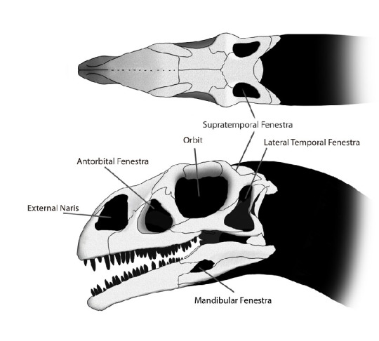
(Image: The skull of the Early Jurassic sauropodomorph Massospondylus, with labels pointing to the holes of the skull: Supratemporal fenestra, lateral temporal fenestra, mandibular fenestra, orbit, antorbital fenestra, external naris. [Source])
One thing that always struck me as peculiar about these diagrams is that they say “naris” instead of “nostril”, since they pretty much mean the same thing. Another thing that always struck me as peculiar as that they say “external” nostril. What, as if there’s an internal nostril?
There is. Welcome to the world of the choana.
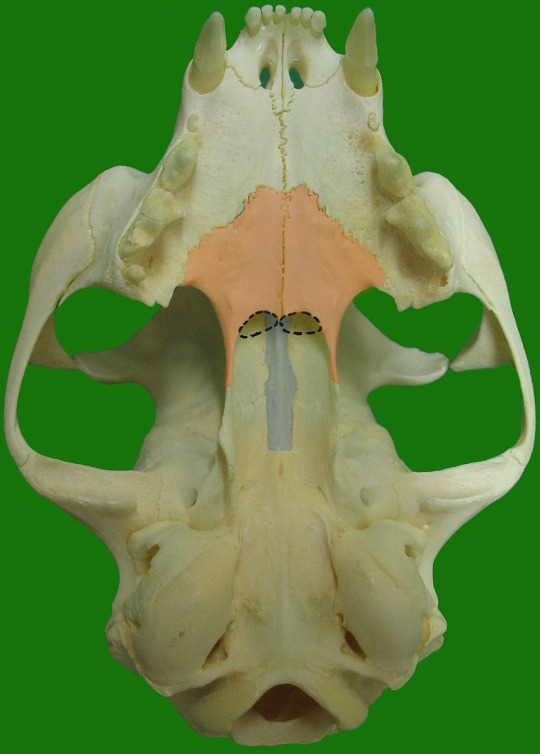
(Image: The skull of a cat in bottom view, showing the choanae (circled in dashed lines) at the back of the mouth, behind the palatine and below the vomer.)
The World of the Choana
The choanae (pronounced koh-ahn-ay, singular choanae/koh-ahn-ah) are openings at the other end of the nasal passage that connect the nostrils to the mouth. They’re what allows you to breath when your mouth is closed, unless you can’t do that, which is okay and I have trouble with it too.
From Whence Came the Choana?
The choanae evolved first not as internal nostrils, but as posterior nostrils.
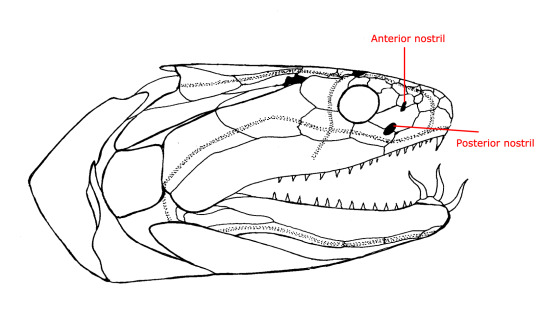
(Image: A diagram of the skull of Onychodus, a type of lobe-finned fish that lived in Australia during the Devonian (about 375 million years ago). Labels point out the anterior (front) and posterior (rear) nostril - both are on the outside. Modified from Long (2001).)
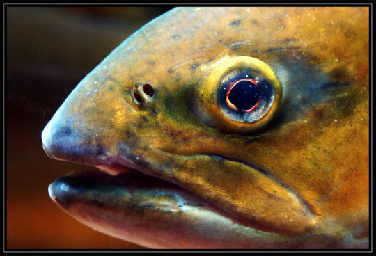
(Image: A photo of a living trout, showing the anterior and posterior nostrils. [Source])
In most fish – including those today! – there are two pairs of nostrils on each side: the anterior and posterior nostril, and both are on the outside of the skull. Water flows in through the anterior nostril, over the olfactory/smell sensors, and then back out the posterior nostril.
In the line that led towards modern tetrapods, however, something changed. Some of the earliest tetrapodomorphs (that is, everything closer related to tetrapods than to lungfish) started to move the posterior nostril lower. Why? I’m not entirely sure, but it may have something to do with improving the sense of smell. (These early tetrapodomorphs had not yet really begun to explore land.)
But in modern tetrapods, the choana is inside the mouth. In order for it to have moved from the outside to the inside, it had to cross the toothline. Is this right? Is this possible? This was a huge source of debate, and no fossils were showing up to prove it right or wrong. All fossils either had external posterior nostrils, or fully formed choanae.
This leads us to the discovery of, in my view, one of the most amazing transitional fossils ever found. Meet Kenichthys.
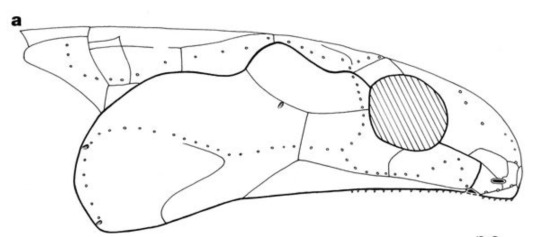
(Image: Reconstruction of the skull of Kenichthys. The posterior nostril is located riiiight at the edge of the mouth. Image from Zhu et al (2004).)
Take a closer look at that snout!
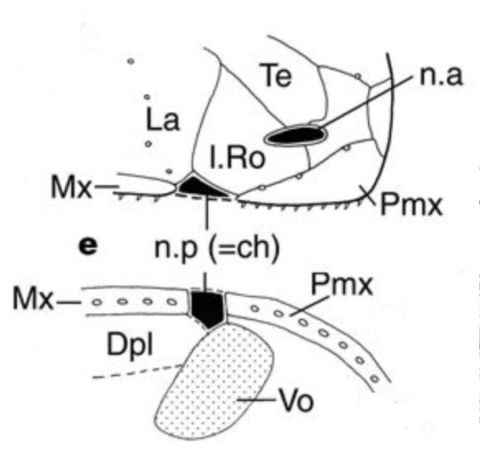
(Image: A closer look at that snout. Upper image shows a side view, and lower image shows a bottom view. I’ll explain exactly what’s going on here in the next paragraph.)
It turns out that the posterior nostril of Kenichthys is actually in line with the teeth! There is a line of teeth on the premaxilla, and a line of teeth on the maxilla, and then there’s a space in between the two bones. And that space is the posterior nostril! It’s an amazing, one-in-a-million transitional fossil that captures the stage in between the posterior nostril and the interior nostril.
Brief aside: The Choana Pretender
Before we go on, there’s something I should bring up. Modern lungfish – the closest relatives of tetrapods – also have choanae, and for a long time it was thought that this was something that evolved in their common ancestor. However, fossil lungfish and tetrapodomorphs both show that their ancestor had posterior nostrils, and each group evolved choanae independently.��
Next week we’ll be looking at choanae in living tetrapods, which I’ll link here, unless I decide not to, in which case I won’t.
913 notes
·
View notes
Note
Do you have any more info on choanal papillae? Specifically, pics of healthy vs unhealthy would be awesome! I haven't had any luck just googling it :/
Yes! (I am s l o w so for those of you who aren't familiar, this was in response to a post I reblogged many weeks back, so here's a quick little intro):
When avian veterinarians examine a bird's mouth, there is a slit-shaped opening in the roof of the mouth called a choana, and it connects the nasal and oral cavities. Mammals have one as well, but it's mainly in birds that this opening is narrow and slit-like, as opposed to wide and rounded. Bird choanae have small serrations on either side called choanal papillae, and blunting or shortening of these is a sign of vitamin A deficiency due to an improper diet. Not all species will have very obvious choanal papillae, however! For example, most pigeons have naturally short/barely visible choanal papillae, and that's completely normal.
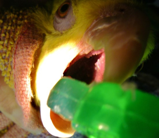
Some healthy lookin pointy bois (Photo from Hagen Avicultural Research Institute)

Pointy bois have mostly disappeared in this birb, likely because they were fed too much seeb (Photo from avianmedicine.net)
Hope this helps, thanks for the ask (and for the long wait sdfkljalsfzkd)!
74 notes
·
View notes
Note
Bunjywunjy pointed out that most cetaceans can't swallow large prey because their windpipe constricts their esophagus, and that sperm whales get around this because they eat squid, which are soft and boneless, but admitted that they had no idea how orcas are able to tackle large prey outside of "orcas don't care". Is there an actual scientific reason orcas can swallow larger prey than other cetaceans?
[clarification: in odontocetes, the trachea passes through the esophagus, and food has to pass around the trachea to get to the stomach]
Do they, though?

Macleod et al. 2007 looked into relative asymmetry of cetacean skulls relative to prey size. Basically, odontocetes have asymmetrical tracheae, so larger food can pass through the larger passageway by the trachea. The asymmetry of the trachea carries further across the respiratory tract wherever there isn't pressure to stay symmetrical; notably, the anterior choanae are also asymmetrical in odontocetes.
From stomach contents of stranded animals, orcas actually had the smallest relative prey size of the entire sample. This is because, although orcas take larger prey than other odontocetes, they can, will, and do tear larger prey into smaller pieces before swallowing them, so proportional to body size, the swallowed bits aren't that big (they only swallow small things like fish whole). Harbor porpoises actually had the largest relative prey size of the sample, and this is reflected by them having the most asymmetrical anterior choanae (=proportionally largest esophageal passage).
39 notes
·
View notes
Photo

This week's topic is CHARGE Syndrome, which stands for C_____, Heart defects, Atresia of the choanae, Restricted growth/development, Genitourinary abnormalities, and Ear abnormalities.
Choanal atresia is obstruction of the nasal passage(s). Infants with bilateral choanal atresia plus other birth defects tend to have poorer outcomes than those with unilateral obstruction of a choana.
Image: A: Unilateral choanal atresia; B: Bilateral choanal atresia. Contributed by Claudio Andaloro, MD. From: StatPearls Publishing LLC.
25 notes
·
View notes
Photo


Rotkehlchen is such a lovable character. Look at him, telling you to p*ss off one second, looking like he never hurt a fly the next.
Also look at him salivating. Look at his choana (I googled that). So much detail.

Rotkehlchen (European robin) im Botanischen Garten der Universität Hohenheim, Plieningen.
#rotkehlchen#european robin#robin#petirrojo#bird#birds#birding#bird watching#urban birding#ornithology#nature#wildlife#stuttgart#germany#deutschland#photographers on tumblr#my photography#wildlife photography#bird photography
73 notes
·
View notes
Note
Hey, what are choanal papillae for? Thanks!
So there isn't actually a ton of academic research about this!
The most recent academic mention of it I have seen is this paper: https://link.springer.com/article/10.1186/s41936-018-0026-6 which states "The anatomical information about the structure of the choana is lacking in literature, and its role in the olfactory and feeding mechanism is still unknown".
So in short, nobody really knows their function, but it is known that they are a good indication of the health of the bird. In starlings, sharp, spikey papillae indicated good diet and a healthy bird. Here's an article about it from an aviculture website: https://hari.ca/show-us-your-choanal-papillae/
14 notes
·
View notes
Photo

UPPER RESPIRATORY TRACT
Begins at the nose and ends with the pharynx.
Nose:
Opens the respiratory system to the external environment via the nostrils (aka, nares).
Comprises bone and cartilage.
Nasal cavity:
Posterior to the nose
Separated from the oral cavity by the hard and soft palates:
Olfactory cells line the superior part of the nasal cavity
The nasal septum divides the cavity into right and left sides.
3 nasal conchae: superior, middle, and inferior.
The hard palate comprises the maxillary and palatine bones.
The soft palate comprises soft tissues.
It comprises two vertical bony structures: the perpendicular plate of the ethmoid bone superiorly and the vomer, inferiorly.
A deviated nasal septum can obstruct airflow and cause sinusitis (sinus infection), epistaxis (nose bleeds), anosmia (inability to smell), and other health problems.
Bony projections that arise on the lateral walls of the nasal cavity.
The nasal conchae create meatuses (superior, middle, and inferior), which are small tunnels.
Movement of air around the conchae and through the meatuses creates turbulence, which helps to warm and humidify the air. Hence, the conchae are sometimes referred to as the "turbinate" bones.
Pharynx:
Descends posterior to the nasal cavity, oral cavity, and larynx, and is open to each of these structures.
Common passageway foods/liquid and air: it serves both the respiratory and digestive systems.
3 subdivisions:
The auditory tube (aka, Eustachian or pharyngotympanic tubes) opens into the nasopharynx
Nasopharynx: posterior to the nasal cavity (and receives air from the nasal cavity); The choanae are the openings between the nasal cavity and the nasopharynx. Oropharynx: posterior to the oral cavity (and receives foods and liquids from the oral cavity); The fauces is the opening between the oral cavity and the oropharynx.
Laryngopharynx: posterior to the larynx (it is the final common passageway for air and food/liquid).
Connects the ears and throat, which allows infection to pass between them.
Auditory tube inflammation occurs in otitis media (aka, ear infection), which is common amongst children.
#pharynx#respiratorysystem#humananatomy#anatomy#grossanatomy#anatomystudy#anatomynotes#medstudy#medstudyblr#usmle#comlex
5 notes
·
View notes
Text
Hypovitaminosis A (avian)
Vitamin A is an important component of avian nutrition. It is necessary for a healthy immune system and proper formation of membranes (including mucous membranes) throughout the body.
Inadequate vitamin A is unfortunately common in avian practice. Birds that are on all seed (or 1/2 seed or more) diets will likely suffer from deficiency as most seeds and grains contain no vitamin A. It is important to talk to owners about what they are feeding their birds, discussing the importance of balanced pellets and vitamin A rich fruits and vegetables (including carrots, sweet potatoes, mango, papaya, broccoli, and collard greens).
Hypovitaminosis A causes immune suppression, hyperkeratosis of the feet, and squamous metaplasia of epithelium within the oropharynx, choana, sinuses, GI tract, urogenital tract, reproductive tract, and uropygial gland. Squamous metaplasia is a benign change of regular lining epithelium to a squamous morphology.
Clinical signs may include: - nasal discharge - sneezing - periorbital swelling - conjunctivitis - feather picking/poor feather quality - anorexia - white plaques in/around mouth, eyes, and sinuses
Secondary infections are likely to develop with progression/chronicity: pustules/abscesses, respiratory or sinus infections.
Treatment: - treat secondary infections - supplement vitamin A - FIX DIET!
#vetblr#avain#exotic vet#VetMed#veterinarian#veterinary medicine#veterinary#vet student#avian vet#hypovitaminosis a
26 notes
·
View notes
Text
Lupine Publishers | Adenoid Hypertrophy in Adults- A Prospective Study

Lupine Publishers | Journal of Otolaryngology
Abstract
Adenoid enlargement is uncommon in adults and because examination of nasopharynx by indirect posterior rhinoscopy is inadequate, many cases of enlarged adenoid in adults are misdiagnosed and accordingly maltreated. Though adenoid hypertrophy is rare in adults, all patients presenting with nasal obstruction should undergo diagnostic nasal endoscopic examination. In the presence of hypertrophied adenoids, though the other causes of nasal obstruction are treated, symptoms may not be relieved unless patient undergoes adenoidectomy. In the present study, 36 cases of adenoid hypertrophy in adult patient is presented along with the review of literature.
Keywords: Adenoid; hypertrophy; adults
Introduction
The submucosal lymphoid tissue at the junction of roof and posterior wall of nasopharynx are known as adenoids. These are predominantly B cell organs amongst which the B cell lymphocytes comprise 50-65% of all adenoids and approximately 40% are T cell lymphocytes 3% are mature plasma cells [1]. Adenoids shows physiological enlargement up to 6 years of age and then they regress gradually during puberty and almost disappear completely by 16 years of age[2]. The enlargement of adenoid is uncommon in adults and most cases of enlarged adenoid in adults are misdiagnosed and they are maltreated[3]. Cases of adenoid hypertrophy in adults could be due to compromised immunity, especially those patients receiving organ transplants and in patients who are infected with human immunodeficiency virus (HIV)[2]. Enlarged adenoids can obstruct the airway resulting in nasal obstruction of varying degree, mouth breathing, snoring, nasal discharge, and nasal intonation of voice. Hypertrophied adenoids can also result in varied otologic symptoms due to blockage of Eustachian tube[4,5].
Objective
This descriptive study was done to evaluate the signs and symptoms associated with adenoid hypertrophy in adults.
Materials and Methods
This was a 3 years prospective descriptive study of adult patients, all aged more than 18 years with symptoms of nasal obstruction.
Study design: Prospective descriptive study
Inclusion Criteria
a) Patients who are willing to give an informed written consent. b) Patients with history of nasal obstruction with enlarged adenoids on investigations. c) Age of the patient more than 18 years. d) Patients who are willing for a regular follow up.
Exclusion criteria
a) Patients who are not willing to give informed written consent b) Patients who are not willing for a regular follow up
Methods
The present study was conducted in the Department of Otorhinolaryngology, Bangalore Medical College and Research Institute, between January 2014 to December 2018. All the patients fulfilling the inclusion/ exclusion criteria were enrolled in the study. Detailed medical history was taken. Relevant past and family history were also taken into consideration. Anterior rhinoscopy wad done to find out deviated nasal septum, septal spur, hypertrophied turbinate’s, nasal polyp, foreign body. Posterior rhinoscopy was done to examine the nasopharynx. The diagnosis of adenoid hypertrophy was made on the basis of the medical history, X-ray nasopharynx soft tissue lateral view, and endoscopy was obtained in an erect position with neck extended to visualize the shadow of the adenoid.Radiological assessment and grading of the size of adenoid was done accordingly [6]
a) Grade I: Soft tissue mass obstructing less than 25% of the nasopharyngeal airway. b) Grade II: Soft tissue mass obstructing 25- 50 % of the nasopharyngeal airway. c) Grade III: Soft tissue mass obstructing 50- 75 % of the nasopharyngeal airway. d) Grade IV : Soft tissue mass obstructing more than 75 % of the airway.
In endoscopic assessment, the size of the adenoid was determined according to Clemen’s et al. grading[6]
a. Grade I:Adenoid tissue filling 1/3rd of the vertical height of the choanae. b. Grade II: Filling 2/3rd of the vertical height of the choanae. c. Grade III: 2/3rd to nearly completely filling the choanae. d. Grade IV: Completely filling the choanae.
After confirmation of the mass, adenoidectomy was performed and then the mass was sent for histopathological examination. The other causes of nasal obstruction if found on clinical assessment, such as deviated nasal septum were also treated simultaneously.
Results
The study group consisted of 36 cases. The ages ranged from 20- 40 years. 25 patients were between 20- 30 years of age and 11 patients belonged to 30- 40 years (Table 1). Out of the 36 cases, 20 patients were males and 16 patients were female (Table 2& Figure1). Patients with adenoid hypertrophy were in 3rd decade of life and in our study we found that males were commonly affected than females in the ratio of 5:1.The presenting symptom was nasal obstruction in 34 patients, snoring was the main complaint in about 2 cases(Figures 2-4). 22 patients were diagnosed with adenoid hypertrophy during diagnostic nasal endoscopy for nasal obstruction, 14 patients were diagnosed with the adenoid hypertrophy with deviated nasal septum.Out of the 36 patients, 24 patients had Grade IV adenoid hypertrophy, 2 patients had grade 2 adenoid hypertrophy and in the remaining 10 patients adenoid tissue was found to fill more than 2/3rd of the vertical height of the choanae (Table 3& Figure5).
Table 1: Showing age distribution.
Table 2: Showing gender distribution.
Figure 1: Showing gender distribution.
Figure 2: Nasal endoscopic picture showing Grade IIadenoid hypertrophy [filling 2/3rd of the vertical height of the choanae].
Figure 3: Nasal endoscopic picture showing Grade IIIadenoid hypertrophy [ 2/3rd to nearly completely filling the choanae].
Figure 4: Nasal endoscopic picture showing Grade IVadenoid hypertrophy [completely filling the choanae].
Figure 5: Showing endoscopic grading distribution.
Table 3: Showing endoscopic grading distribution.
Discussion
The submucosal lymphoid tissue at the junction of roof and posterior wall of nasopharynx are called adenoids[6]. The nasopharyngeal lymphoid aggregate or Lushka’s tonsil was described by Santorini in 1724.Nasopharyngeal vegetations were then described by Wilhem as “Adenoids” in 1870. Adenoids along with palatine tonsil, lingual tonsils, tubal tonsils, and lateral pharyngeal bands forms the inner Waldeyer’s ring[6]. These are predominantly B cell organs which comprises of 50-65% of all adenoid lymphocyte and 40 % are T cell lymphocytes and about 3 % are mature plasma cells[1]. During the age of 3 and 10 years it shows greatest immunological activity and hence are most prominent during this period of childhood, and later shows involution[2].All the components of the Waldeyer’s ring play a very important role in the immunological memory. The epithelium contains a system of specialized channels lined by M cells that take up antigens into the vesicles and transport them to the intra- and subepithelial spaces. Here they are presented to lymphoid cells. After passing through the epithelium, inhaled or ingested antigens reach the extrafollicular region or the lymphoid follicles. In the extrafollicular region, interdigitating cells (IDC) and macrophages process the antigens and present them to CD4+ T lymphocytes. Helper T cells then stimulate the proliferation of follicular B lymphocytes and their development into either antibody-expressing B memory cells capable of migration to the nasopharynx and other sites, or plasma cells that produce antibodies, and release them. The contact of memory B cells in the lymphoid follicles with antigen is an essential part of the generation of a secondary immune response.
Among the Ig isotypes, IgA may be considered the most important product of the adeno tonsillar immune system. In its dimeric form, IgA can attach to the transmembrane secretory component (SC) to form secretory IgA (SIgA), which is a critical component of the mucosal immune system of the upper airway. This component is necessary for the binding of IgA monomers to each other and to the SC and is an important product of B cell activity in the tonsil follicles. While the tonsils produce immunocytes bearing the J (joining) chain carbohydrate, the SC is produced only in the adenoid and extra tonsillar epithelium, and therefore, only the adenoid possesses a local secretory immune system[6].Adenoid enlargement is uncommon in adults and many cases of adenoid hypertrophy in adults are misdiagnosed[3]. Involuted adenoidal tissue may re-proliferate in response to infections and irritants resulting in adenoid hypertrophy. Adenoid hypertrophy in adults may occur in immunocompromised state, especially in those patients receiving organ transplants and also in those patients infected with having human immunodeficiency virus[5].
Conclusion
Though adenoid hypertrophy is rare in adults, all patients presenting with nasal obstruction should undergo diagnostic nasal endoscopic examination. In the presence of hypertrophied adenoids, even though the other causes of nasal obstruction are treated, symptoms may not be relieved unless patient undergoes adenoidectomy.
For more Otolaryngology Journals please click on below link https://lupinepublishers.com/otolaryngology-journal/
For more Lupine Publishers Please click on below link https://lupinepublishers.com/
0 notes