#autoradiography
Explore tagged Tumblr posts
Text
hey tumblr this is a fairly niche topic but its been pressing on my mind recently so:
back in late march i went to the sackler gallery in DC and they had an exhibit for korean artist Park Chan-Kyong, centered on his film Gathering. My friend and I found the darkness and relatively empty area of the film Fukushima: Autoradiography and Child Soldier to be really nice so we ended up there for around 2 hours, during which the film(s) looped at least three times. At some point in the runtime of Child Soldier a song came on, which I found to be good and wanted to know what it's called. Unfortunately I did not get a recording of the song nor am I able to find ANY information about this one specific film outside of the website for the sackler gallery, which only lists it alongside the others and does no more. There are no online recordings of it, no audio clips, no wikipedia page, NOTHING. If any of you by chance are familiar with Park Chan-Kyong's work, please help me find the song. please. it was so good
#since the film is about a north korean child soldier i started by looking for north korean folk songs and there is. nothing that I can find#park chan kyong#park chankyong#sackler gallery#smithsonian institution#park chan-kyong#park chan-wook#park chan wook#park chanwoom#PARKing CHANce films#filmography#korean art#istg im going insane because this film does not seem to exist outside of the sackler gallery i literally cant find anything mentioning it#at least under the name “child soldier”#entirely possible they just. misnamed it but the smithsonian is such a reputable institution it seems unlikely
3 notes
·
View notes
Text
Exploring RNA Analysis Methods: Techniques for Comprehensive Understanding of RNA
RNA analysis is a cornerstone of molecular biology, enabling researchers to decode the various functions and regulatory mechanisms of RNA in cellular processes. With growing interest in transcriptomics, RNA analysis methods have evolved to offer more precise, high-throughput, and comprehensive insights into gene expression, alternative splicing, RNA modifications, and more. Here, we explore several RNA analysis methods that have become essential tools in biological and medical research.
Download PDF Brochure
1. RNA Sequencing (RNA-Seq)
RNA sequencing is the gold standard for transcriptome analysis. It allows researchers to examine both coding and non-coding RNA with high resolution. RNA-Seq provides quantitative data on gene expression levels, alternative splicing events, and even RNA-editing phenomena. This method has the advantage of being unbiased, offering a comprehensive snapshot of the entire transcriptome.
Steps Involved:
RNA extraction
cDNA synthesis
Sequencing via next-generation sequencing platforms
Data analysis using bioinformatics tools to map reads to reference genomes and quantify expression
2. Quantitative PCR (qPCR)
Quantitative PCR is a highly sensitive method to measure RNA expression levels. It is often used to validate results from RNA-Seq or microarray studies. By amplifying specific RNA sequences and using fluorescent probes, qPCR provides real-time quantification of RNA molecules, offering highly accurate and reproducible data.
Advantages:
High sensitivity
Quantitative results in real time
Often used for validation of gene expression studies
3. Microarrays
Microarray technology allows the simultaneous analysis of thousands of RNA molecules. Although it has been somewhat replaced by RNA-Seq due to the latter’s higher resolution and broader coverage, microarrays remain popular for focused studies on specific genes or pathways. They are relatively inexpensive and easy to use for researchers looking for rapid gene expression profiling.
Key Applications:
Gene expression profiling
Comparative studies across different samples or conditions
Focused analysis of known RNA sequences
4. Northern Blotting
Northern blotting is a classical technique used to detect specific RNA molecules within a mixture of RNA. While it is less commonly used today, northern blotting remains a reliable tool for detecting the presence and size of RNA molecules. This method is particularly useful for validating the results of RNA-Seq or qPCR.
Request Sample Pages
Process Overview:
RNA extraction and electrophoresis
Transfer of RNA onto a membrane
Hybridization with labeled probes specific to the RNA of interest
Detection via autoradiography or chemiluminescence
5. Single-Cell RNA Sequencing (scRNA-Seq)
Single-cell RNA sequencing is a cutting-edge technique that enables researchers to study gene expression at the resolution of individual cells. This method has revolutionized the field of transcriptomics by revealing cellular heterogeneity and identifying rare cell types that might be missed by bulk RNA-Seq.
Advantages:
High resolution for detecting cell-to-cell variability
Crucial for understanding complex tissues and diseases like cancer
Insights into cellular differentiation and development
6. RNA Immunoprecipitation (RIP)
RNA immunoprecipitation is used to study RNA-protein interactions. Researchers use specific antibodies to target RNA-binding proteins, isolating the associated RNA molecules. RIP is particularly valuable in studying RNA modifications, such as methylation, and understanding how RNA-protein complexes influence gene expression.
Applications:
Studying RNA modifications (e.g., m6A methylation)
Understanding the role of RNA-binding proteins in disease
Functional annotation of RNA molecules
7. In Situ Hybridization (ISH)
In situ hybridization is a method used to detect specific RNA sequences in fixed tissue sections or cells. This method provides spatial information about RNA localization within tissues, making it invaluable for developmental biology and cancer research.
Benefits:
Visualization of RNA expression patterns in intact tissues
High spatial resolution
Useful in identifying RNA localization in specific cell types
Conclusion
The diversity of RNA analysis methods allows researchers to study the complex roles of RNA in gene regulation, cellular function, and disease. While RNA-Seq remains the most comprehensive approach, each method offers distinct advantages depending on the research question and experimental needs. By combining these methods, scientists can gain a holistic view of RNA biology, paving the way for advancements in precision medicine and therapeutic development.
Whether it's detecting subtle changes in gene expression or unraveling RNA-protein interactions, these RNA analysis techniques continue to enhance our understanding of the molecular underpinnings of life.
Content Source:
0 notes
Quote
The inferior frontal sulcus is conceptualized as the landmark delineating ventro-from dorsolateral prefrontal cortex. Functional imaging studies report activations within the sulcus during tasks addressing cognitive control and verbal working memory, while their microstructural correlates are not well defined. Existing microstructural maps, e.g., Brodmann's map, do not distinguish separate areas within the sulcus. We identified six new areas in the inferior frontal sulcus and its junction to the precentral sulcus, ifs1-4, ifj1-ifj2, by combined cytoarchitectonic analysis and receptor autoradiography. A hierarchical cluster analysis of receptor densities of these and neighbouring prefrontal areas revealed that they form a distinct cluster within the prefrontal cortex. Major interhemispheric differences were found in both cyto- and receptorarchitecture. The function of cytoarchitectonically identified areas was explored by comparing probabilistic maps of the areas in stereotaxic space with their functions and co-activation patterns as analysed by means of a coordinate-based meta-analysis. We found a bilateral involvement in working memory, as well as a lateralization of different language-related processes to the left hemisphere, and of music processing and attention to the right-hemispheric areas. Particularly ifj2 might act as a functional hub between the networks. The cytoarchitectonic maps and receptor densities provide a powerful tool to further elucidate the function of these areas. The maps are available through the Human Brain Atlas of the Human Brain Project and serve in combination with the information on the cyto- and receptor architecture of the areas as a resource for brain models and simulations.
The inferior frontal sulcus: Cortical segregation, molecular architecture and function - ScienceDirect
0 notes
Link
0 notes
Text
Seasonal Fluctuations of Synechococcus spp. Abundance, Photo-assimilation, Growth Rate and Fluorescence Properties at the Individual Cell Level in the Sargasso Sea

Authored by: Rodolfo Iturriaga
Abstract
A seasonal study of Synechococcus spp. abundance, photo-assimilation, growth rate, and fluorescence properties were performed at the individual cell level during the BIOWATT-II Project in the Sargasso Sea. Investigations were carried out during four cruises in different seasons in 1987. Synechococcus spp., cell abundance, carbon photo-assimilation, growth rate, and fluorescence emission were higher in the upper 80 m during most of the seasonal periods observed with the exception of the summer cruise. A similar trend followed the nitrogenous nutrient (nitrite plus nitrate) concentrations and chlorophyll fluorescence patterns. Strong vertical mixing events are common during the spring, followed by an increase in solar irradiance, the development of a thermocline at 30-40m, and a drop in the nitrogenous nutrient concentrations in the upper 120m. These conditions intensified toward the summer when a nutrient depleted mixed layer occurred above a well-defined thermocline near 40m. Synechococcus spp. cell abundance, carbon photo-assimilation, growth rate and fluorescence intensity all declined over the summer months. During periods of higher solar irradiances and depleted nitrogenous nutrient concentrations, the fluorescence emission intensity was lower in cells collected from the upper 60-80 m of the water column. By comparison, higher fluorescence values were displayed by cells collected from the same cast at depths of 100-160 m. where nitrogen nutrient concentrations were detectable, but light limited growth rate. The fluorescence excitation spectra of Synechococcus spp. cells sampled at all depths and seasons, showed higher ratios of phycourobilin (PUB) to phycoerythrobilin (PEB) comparable to Synechococcus clone WH-8103. Synechococcus spp. population abundance, growth rate, and fluorescence intensity appeared to follow the seasonal trend of nitrogenous nutrient availability determined primarily by vertical mixing events.
Keywords: Synechococcus spp.; Abundance; Photo-assimilation; Growth Rates; Fluorescence; Individual cells study
Abbreviations: PUB: Phycourobilin; PEB: Phycoerythrobilin; CPS: Counts per seconds; PAR: photosynthetic available radiation
Introduction
One of the main objectives of the BIOWATT-II project was to investigate the seasonal changes on the physical forcing that set-in motion the bio-optical processes in the northern Sargasso Sea. The winter cooling and storms drive deep vertical mixing as well as nutrient replenishment. A phytoplankton bloom typically follows the re-stratification of the upper water column [1,2]. Synechococcus spp. have been found to occur abundantly and widely distributed in the ocean and its photosynthetic properties, growth rate, trophic fate and relevance in marine ecosystems have been described in the literature [3-18]. Several methods have been explored to determine Synechococcus spp. growth rates. Using techniques independent of grazing effects, such as the frequency of dividing cells, Campbell and Carpenter [8] reported growth rates between 0.42 and 0.86 d-1 in oceanic waters and 0.73 d-1 during February in the Sargasso Sea. Growth rates between 0.5 and 1.2d-1 determined by epifluorescence nuclear-track micro autoradiography were reported by Iturriaga and Marra [11] during a transect along the 700W between 320N and 260N in the North Atlantic and the Sargasso Sea. Other techniques, such as size-fractionation, dilution rates and eukaryotic cell inhibitors (to minimize grazing effects) have been used to determine picoplankton growth rates in the Sargasso Sea and other oligotrophic ocean regions [13,19,20-22].
Field and laboratory data have suggested that picoplankton photosynthesis, including coccoid cyanobacteria, saturate at low irradiance (Morris and Glover, 1981) [5,9]. However, observations with cultured Synechococcus clone WH-7803, indicated that photosynthetic activity was possible at high irradiance levels [23]. In a field study in the Sargasso Sea, Prézelin et al. [24], reported that the PE content of the Synechococcus populations increased with decreasing light and, or increasing nitrogen nutrient availability as their capability to harvest photons throughout a large portion of the photic zone with a maximal contribution near its base [14,20,24]. Other methodologies have used the fluorescence properties of Synechococcus spp. to estimate their concentrations, pigment composition, and photoadaptive state. Synechococcus have also been studied at the individual cell level by microphotometry (Campbell and Iturriaga, 1980), optical tweezers [25], and flow cytometry [12,26]. Fluorescence excitation spectra from microphotometry-enabled the fingerprinting of the phycobilliprotein chromophores of individual Synechococcus spp. cells from field samples with high precision, accuracy and spectral resolution. During the BIOWATT-II investigation, individual action spectra facilitated the sorting of the prevalent Synechococcus spp. cell type by their PUB:PEB ratios, as well as to determine the fluorescence emission intensity at the excitation maxima of these two chromophores on single cells from the surface to a depth of 160 m. Despite the abundant information on Synechococcus spp. in ocean waters, most of the observations have been conducted either on bulk populations and during sporadic spatial and temporal sampling. In this study, we examined seasonal fluctuations in Synechococcus spp. abundance, photo-assimilation, growth rate, and fluorescence properties at the same geographical site in the northern Sargasso Sea.
Methods
Water sampling and incubations were performed in the northwestern Atlantic Ocean at 720W, 340N during a series of four cruises: OCE-1 (March 4-8, 1987), OCE-2 (May 17-21, 1987), OCE- 3 (August 22-25, 1987), and OCE-4 (November 30-December 02, 1987) aboard the R/V Oceanus. Water samples were collected in the upper 300m with a rosette sampler equipped with a CTD, Go-Flo bottles, and bio-optical instrumentation. Coccoid cyanobacteria cell abundances were determined via direct cell counting using a Zeiss epifluorescence microscope as described by Watson et al. (1977) and Hobbie et al. [27]. Nitrogenous nutrient concentrations (NO-2 and NO-3) were determined using standard autoanalyzer methodology. Dawn-to-Dusk carbon fixation rates in the euphotic zone were measured in situ using the C14 technique. Water collection and sample preparation followed the trace-metal clean procedures described by Fitzwater et al. [28]. Synechococcus spp. carbon fixation and growth rates in individual cells were determined by epifluorescence nuclear-track autoradiography as described by Iturriaga & Marra [11].
The spectral fluorescence properties of Synechococcus spp. were determined on individual cells using a Zeiss Microphotometer system. A detailed description of this methodology is described in Campbell and Iturriaga [29] & Iturriaga and Bower [30]. An average of ten random individual excitation spectra from cells collected at each depth was utilized to determine the intensity of the fluorescence emission of the phycobiliproteins chromophores, as a function of the 490nm and 543nm excitation wavelengths respectively. These two wavelengths represent the maximal absorption of PUB centered at 490nm and PEB at 543nm. PUB does not fluoresce, but transfers excitation energy to PEB that fluoresces at 575nm. The excitation spectra were scanned continuously from 400nm to 600nm. The emission intensity of these two wavelengths was measured at 575nm and the outputs of the 490nm and 575nm excitation wavelengths were processed independently to calculate the PUB:PEB ratios. The microphotometer system is equipped with excitation and emission monochromators of high precision and spectral resolution. The fluorescence intensity of each action spectra was recorded continuously at a resolution of 1nm by the microscope photomultiplier and registered as counts per seconds (CPS) on a computer. The precision, spectral resolution and sensitivity of the system is comparable to a research benchtop spectral fluorometer.
Results and Discussion
Environmental Conditions
During the OCE-1 cruise in early March, surface water temperatures (20oC) were the lowest observed for the four cruises and no significant mixed layer was apparent in the upper 160m. The photosynthetic available radiation (PAR) levels of 1%, due to cloudiness, fluctuated between 80 and 100m, higher chlorophyll fluorescence values remained in the photic zone decreasing below 50m. Previous to the OCE-2 cruise in mid-May, an increase in solar irradiance, coupled to a rise of the upper surface water temperature initiated the formation of a mixed layer above 30- 40m depth, chlorophyll fluorescence decreased in the surface waters, as well as the nitrogen concentrations. The chlorophyll fluorescence maximum appeared between 90-105m, a depth where nitrogenous nutrients were more abundant and PAR was a growth rate limiting factor. By the time of the OCE-3 cruise in mid-August, the solar irradiance continued to warm the surface waters, reaching temperatures up to 27oC. A nitrogen depleted mixed layer set above a well-defined thermocline near 40m and a chlorophyll fluorescence maximum set at approximately 100m depth. During the OCE-4 cruise in early December, solar irradiance and PAR levels were the lowest observed in all cruises and nearly half of the values of those observed during OCE-1. An intermediate deep mixed layer located close to 150m with nitrogen replenished surface waters, temperatures reached 23oC and, chlorophyll fluorescence values followed a pattern similar to those observed during OCE-1.
The seasonal fluctuations of solar radiation and atmospheric forcing’s upon the upper ocean were the main criteria used to determine the BIOWATT-II study site [1,31]. The north Atlantic region corresponding to the BIOWATT-II study site is characterized by deep convective winter mixing that supplies nutrients to the surface waters. During the spring, the increase of solar irradiance and surface waters temperature induces stability to the water column, resulting in the annual spring phytoplankton bloom with the typical spring phytoplankton blooms, followed by a nutrient depleted well defined mixed layer and thermocline above 40m depth during the summer (Figures 1-2) (Table 1).
Synechococcus spp. cell abundance
The abundance of Synechococcus spp. ranges between approximately 103 to 105 cells mL-1 in oligotrophic and mesotrophic regions of the ocean. In stratified waters maximal numbers have been observed in the upper 100m depending on region and season. Coccoid cyanobacteria show a distinct seasonality in some temperate regions in the world’s ocean (Waterbury et al., 1987) [2,4,7,13,14,32]. During the BIOWATT-II investigations, higher cell concentrations were observed during OCE-1, OCE-2 and OCE- 4 in the upper 60-80m with values ranging from 1.0 to 2.7 x 10⁴ cells mL-1. Synechococcus abundances ranged from 1.0 to 0.4x10⁴ cells mL-1 at depths of 80m to 160m (Figure 3). During OCE-3 (summer) Synechococcus abundances dropped to approximately half that observed during the other seasons.
Seasonal carbon photo-assimilation and growth rate
Traditionally, the photosynthetic rates of Synechococcus spp. have been determined for the “picoplankton” fraction (<3 microns) via size fractionation analysis, this size fraction accounts for approximately 60% of the total primary production in oligotrophic waters with high specific growth rates [6,10,19,20,24,33]. However, specific growth rates of coccoid cyanobacteria and their contribution to the total primary production may be lower than previously thought, because the picoplankton fraction may contain variable amounts of other photoautotrophs, including Prochlorococcus spp. [8,11].
The environmental factors affecting the photo-assimilation rates within the picoplankton include spectral downwelling irradiance. Field and laboratory data have demonstrated that the picoplankton fraction, including coccoid cyanobacteria, efficiently utilize the blue to green regions of the visible light spectrum [14]. Based on individual cells in situ carbon-specific photo-assimilation and growth rates of Synechococcus spp. in the Sargasso Sea, Iturriaga and Marra [11] reported carbon fixation rates between 3.4 and 11.8 fgCcell-1h-1, with specific growth rates ranging between 0.5 to 1.2 d-1, with contributions to the total primary production ranging from 60% to 95%. The highest growth rates were observed in the upper 20m of the water column. During the OCE-1 and OCE-4 cruises, the highest carbon photo-assimilation and growth rates were observed in the upper 30-40m. These two sampling periods corresponded to the seasons when the deepening of the mixed layer was also associated with ventilation of nutrient-rich deep water (18oC isotherm) [2]. These two seasonal periods contrasted to the conditions observed during the spring (OCE-2) and summer (OCE-3), that were characterized by higher irradiances, higher water temperatures, water column stratification, and lower nitrogenous nutrient concentrations. During OCE-3, photo-assimilation and growth rates of cells located between 60 and 80 m were nearly double the rates of cells in the upper 40m (Figure 4).
The lower growth rates observed in nutrient-depleted surface waters during OCE-3, suggests that Synechococcus spp. cells have the capacity to contribute to carbon photo-assimilation processes at extremely low nitrogen concentration levels in the oligotrophic ocean. Utilizing a highly sensitive nitrite plus nitrate measurement technique [15], indicated that Synechococcus spp. have an affinity to extremely low nitrogenous nutrient concentrations and are able to utilize this resource at the nM concentration range. While the nitrate concentrations in the upper 40m were below the limit of detection, the Synechococcus cells were still capable of carrying out photo-assimilation processes as suggested by Glover’s (1988) findings.
Seasonal fluorescence properties of Synechococcus spp.
Fluorescence excitation is a powerful technique that is commonly used to determine chromophore identities and their cellular concentrations. This technique has found wide applications in chemical and biological studies [34,35]. During this investigation several hundred fluorescence excitation spectra were collected on individual Synechococcus spp. cells sampled from the upper 160m. Each one of the Synechococcus spp. spectra analyzed displayed higher PUB:PEB ratios, a distinctive characteristic of the Synechococcus WH8103 clone. Similar PUB:PEB ratios have been reported on individual cells sampled from the Sargasso Sea by microphotometry [29] and dual beam flow cytometry [12] (Figure 5).
During OCE-1 and OCE-4, the fluorescence intensity output and the PUB:PEB ratios displayed smaller variations from surface to depth than those observed during the OCE-2 and OCE-3. The seasons corresponding to the (spring and summer cruises) were characterized by higher irradiances as well as depleted nitrogenous nutrient concentrations in the upper water column. The lower fluorescence output and PUB:PEB ratios observed for cells collected in the upper 40m during OCE-3, as compared to the higher fluorescence output of cells collected from 100-160m during the same cast could be interpreted as a photo-adaptive response to higher irradiance levels at the surface and lower at depth. However, the lower fluorescence intensity output and lower PUB:PEB ratios observed for cells in the upper 100m during the spring (OCE2) and summer (OCE-3) could also be attributed to photoadaptation to higher irradiances and/or a reduction in cellular pigments caused by cell nutrient starvation. In a study conducted in the Sargasso Sea, Prézelin et al. [24] reported that the PE content of Synechococcus populations increased with decreasing light and/or increasing nitrogen availability. Using a technique capable of measuring nitrite plus nitrate at nM levels Glover et al. [18,24] reported a relationship between nitrogenous nutrient concentration, fluorescence response, and photo assimilation of Synechococcus at very low levels. Studies performed with Synechococcus cultures and natural populations suggest that changes in phycobiliprotein pigments are a consequence of photoadaptation (Palenik, 2001) [12,24,29,36]. The capacity of Synechococcus to utilize some of their phycobilliprotein pigments as a nitrogen reserve has also been debated [37,38]. This is an interesting aspect to be considered, however it was beyond the scope of this investigation.
Summary
In the Sargasso Sea, high wind stress regimes are common events during the late fall and winter seasons in the geographical area of the BIOWATT-II study site. These events deepen the mixed layer to approximately 200m depth providing nitrogenous nutrients to the photic zone. During OCE-1 (early March) and OCE-4 (late November early December) cruises, higher nitrogenous nutrient concentrations were found throughout the water column. By comparison, lower concentrations observed in the upper water column during OCE-2 (mid May) and OCE-3 (mid- August). In general, higher Synechococcus spp. abundances, photoassimilation, growth rates and fluorescence yields were observed in samples collected from the upper 80-90m when nitrogenous nutrient concentrations were detectable. During the spring and summer periods, both characterized by higher solar irradiance and lower nutrient levels, Synechococcus spp. abundances, photoassimilation, growth rates and fluorescence yields declined in the upper ocean. These observations suggest that the Synechococcus spp. abundances appeared to be regulated by nitrogenous nutrient availability rather than by temperature or solar irradiance.
Despite the abundant information regarding Synechococcus spp. in oceanic waters, most of the data has been collected on bulk populations. The observations performed during BIOWATT-II were conducted at the cellular level and expand the knowledge of Synechococcus spp. population abundances, photo-assimilation, growth rates and fluorescence yields in the oligotrophic ocean.
To Know More About Oceanography & Fisheries Open Access Journal Please click on: https://juniperpublishers.com/ofoaj/index.php
For more Open Access Journals in Juniper Publishers please click on: https://juniperpublishers.com/index.php
1 note
·
View note
Text
New Gel Documentation Mechanism to actually purchase
Another example of innovation helping medicine is "gel documentation," also known as "gel doc" or "gel imaging system." In polyacrylamide or agarose gels stained with ethidium bromide or other fluorophores like SYBR Green, it is typically used in sub-atomic research to record nucleic acids and proteins. Today, gel documentation systems are a need for every extremely sophisticated and top-tier clinical research centre. Prior to then, it merely contained a Polaroid print of a reversed pyramid's highest point. It was placed over a gel, and a photograph was taken, but it wasn't good enough to guide a point-by-point examination of the needed component or clear enough to call for pictures of temporary DNA and protein gels.
The gel records of today are very different from all that nonsense. The current gel documentation system uses PC-based excitation and discovery systems, multipurpose nooks, and programming that enables a point-by-point examination and analysis of the captured images. They can handle a variety of things, from DNA and protein gels to deeply confused autoradiography, and they are either fully or partially robotized.
Today, if you were able to get a gel documentation system, you would discover its utility and expansion. Given that, the value attained would also change. You may look for systems that would capture images and documentation of only DNA and protein gels or more sophisticated ones that would deal with things like autoradiography, fluorescence, and chemiluminescent Western smears. The type of camera and its lenses, the product that comes with the system, and the level of computerization it offers are further criteria for determining the system's cost.
A highly sophisticated gel documentation system would make it simple for even a beginner to take images. The bulk of the methods would be automated and humanised, but they would still let the client choose the image's brightness and zoom for the person who has to capture it. In other circumstances, you can discover that some companies sell the system with the testing software as an additional option, while others would include everything, clearly at a more expensive price.
By breaking down the programming, you would be able to pay close attention to the image and add more services to the system. It is preferable to have suggestions and assistance from others when you are looking to get the gel documentation system, especially if you are unfamiliar with biomedical innovation. Get a good system, and success can appear out of nowhere.
For More Info:-
Gel Doc India
Gel Doc System India
0 notes
Text
DNA - FINGERPRINTING
DNA fingerprinting is also know as DNA profiling is a process of determining an individuals DNA characteristics. This is a forensic technique in criminal investigations, comparing criminal suspect's profiles to DNA to identify the like hood of their involvement in the crime. Also can be used for paternity tests. 99 % of human DNA sequences are the same in every person. Remaining sequences are different . It makes every individual unique.

This is used to distinguish and identify the individuals of a same species by using their DNA samples. These biological samples may be blood, hair, saliva, semen, body tissue cells. The DNA samples are isolated first. Then digested by restriction endonucleases. After this electrophoretic separation of fragments are done. These fragments are transferred into nylon or nitrocellulose membrane. This is followed by probing, hybridization and autoradiography. From this banding pattern is analyzed.
youtube
FOR FURTHER INFORMATION VISIT : -
https://readitupwithswathy.blogspot.com/2022/04/dna-fingerprinting.html
#dna#dna fingerprinting#dna profiling#forensic science#crime#suspect#dna sequence#restriction endonucleases#nylon#nitrocellulose membrane#probe#hybridization#autoradiography#banding pattern#genetics#genome
1 note
·
View note
Link
Autoradiography Films Market Report is expected to grow at CAGR XX% by 2028. Autoradiography Films Market Report was $XX Million in 2020 and is expected to reach $XX Billion by the forecasted period 2021 to 2028. The Autoradiography Films Market rep
0 notes
Text
Global Autoradiography Films Market Report by types, applications, players and regions,gross, share, cagr ,outlook 2026
Global Autoradiography Films Market Report by types, applications, players and regions,gross, share, cagr ,outlook 2026
The Autoradiography Films Market Report Research Industry, 2021″ report has been added to Marketdesk.us offering. The Global Autoradiography Films Market is set up in a joint exertion with the main business specialists and dedicated assessment investigator group to give an endeavour inside and out market experiences and help them to take pivotal business choices. This Autoradiography Films report…
View On WordPress
0 notes
Text
Autoradiography
BY- Ezhuthachan Mithu Mohanan (MSIWM043) It is a bioanalytical technique. Autoradiography is used for the detection of components or materials that have radioactive properties. The film obtained is known as autoradiograph. Radioactive materials can emit ionizing radiation or particles. Using X-ray, relative bands on gels can be obtained by radioactive emission. The first emission of…

View On WordPress
#alpha rays#applications of autoradiogarphy#autoradiogaraphy#autoradiograph.#beta rays#bioanalytical technique#Factors for efficiency of autoradiography#Fluorography#gamma rays#gieger muller counter#mechanism of autoradiography#scintillation counter#Steps of autoradiography#types of radiation
0 notes
Text
Global Autoradiography Films Market Growth, Analysis and Industry Forecast (2019-2025): Carestream, GE Healthcare, Fujifilm, Thermo Fisher Scientific, etc.

Autoradiography Films market research delivers real-world and industry intelligence of the market to support your idea with research-based facts. It provides deep understanding, clarifies diversities of the market to help you decide not only the succeeding strategy but also to achieve the desired market position in Power sector. This market research is a combined result of inputs from business professionals with awareness, the experience of Autoradiography Films industry and qualitative and quantitative synthesis of the market. Request for Sample Report Here @ https://www.acquiremarketresearch.com/sample-request/9256/ This report presents a detailed study of the global market for Autoradiography Films by evaluating the growth drivers, restraining factors, and opportunities at length. The examination of the prominent trends, driving forces, and the challenges assist the market participants and stakeholders to understand the issues they will have to face while operating in the worldwide market for Autoradiography Films in the long run. This report provides in depth study of “Autoradiography Films” using SWOT analysis i.e. Strength, Weakness, Opportunities and Threat to the organization. The Autoradiography Films report also provides an in-depth survey of key players in the market which is based on the various objectives of an organization such as profiling, the product outline, the quantity of production, required raw material, and the financial health of the organization. For More Information On This Report, Please Visit @ https://www.acquiremarketresearch.com/industry-reports/autoradiography-films-market/9256/
It further maps the competitive landscape of this Autoradiography Films market by evaluating the company profiles of the leading market players, such as Carestream, GE Healthcare, Fujifilm, Thermo Fisher Scientific, Santa Cruz Biotechnology, MIDSCI, Diamed, LabScientific, Harvard Bioscience On the basis of the product, the market has been classified into: Nuclear Emulsion, X-ray Film, Others Based on the application, the market has been categorized into: Blotting, Sequencing, Others Any query?Inquire Here For Discount Or Report Customization (Use Corporate email ID to Get Higher Priority): https://www.acquiremarketresearch.com/enquire-before/9256/ Global Autoradiography Films Report mainly covers the following Chapters: => Chapter 1: Autoradiography Films Industry Overview. => Chapter 2: Autoradiography Films Region and Country Market Analysis. => Chapter 3: Autoradiography Films Technical Data and Manufacturing Plants Analysis. => Chapter 4: Autoradiography Films Production by Regions by Technology by Applications. => Chapter 5: Autoradiography Films Manufacturing Process and Cost Structure. => Chapter 6: Autoradiography Films Productions Supply Sales Demand Market Status and Forecast. => Chapter 7: Autoradiography Films Key success factors and Market Overview. => Chapter 8: Autoradiography Films Research Methodology and About Us. Please note Chapters four, five and six data will depend on the feasibility of the Autoradiography Films market.
0 notes
Text
How Do We Analyze Cell Membranes?
Organelles have very unique and distinct membrane composition from each other in the cell, so if we wish to analyze them separately, how can we achieve this?
First, we need to figure out how to separate the membranes from the organelles themselves. And in cell biology, there are quite a few techniques we can use to do this.
Before we can separate cell components based on their size, density, or protein composition, we first need to homogenize the cells. Homogenization is typically a very gentle mechanical procedure that breaks up the cells, however the membranes, nuclei and organelles stay intact. (The exception is the endoplasmic reticulum, due to its large size.) This can be done in a variety of ways, such as high frequency sound, using mild detergents, or physically lysing the cell mechanically.

Following this, there are a few methods of separating the cell components depending on what factor of separation you are aiming for.
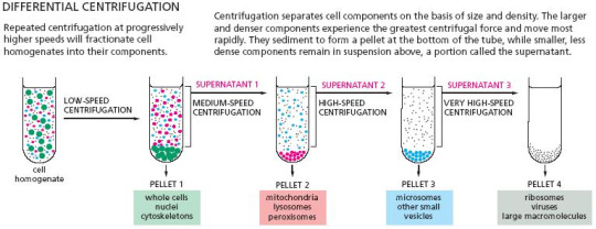
Differential centrifugation is where the cell components are separated in different fractions by spinning the fractions at different speeds for different amounts of time. This technique does not separate the organelles to purity, as the various fractions are separated on their various sizes. As such, cell components that are of similar size, for example mitochondria, lysosomes and peroxisomes, will all end up in the same fraction.

Velocity centrifugation, also known as rate-zonal sedimentation, separates organelles based on their different sizes and relative masses. A cell homogenate would be poured as a thin layer over top a shallow sucrose gradient (5-20%), and centrifuged as such so larger organelles sink to the bottom of the fraction faster than the smaller organelles. The resulting fractions would be collected at the bottom of the tube.

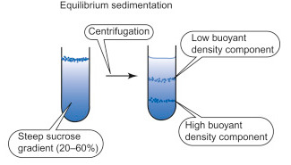
Equilibrium density centrifugation is a technique where recovered organelles are separated based on their different densities. Similar to velocity centrifugation, the sample would be added to a gradient. However the sucrose gradient is much steeper (20-70%) and the homogenate would not be poured as a thin layer, but instead mixed in with the gradient. The fraction would then be centrifuged to equilibrium, and the low density components would collect higher on the fraction, and the higher density components would collect near of the bottom of the fraction.

And lastly, there is also immuno-isolation. Instead of spinning the cell fractions by size, the fractions would be mixed with targeted antibodies coated with protein-A beads. Protein-A is a bacterial protein that binds and sticks to antibodies, and as such is used as a tool that marks targeted organelles that antibodies stick to. The beads can then be recovered by centrifugation, and the targeted proteins can be analyzed.

Once the targeted components are isolated, we can analyze them. Depending on the type of analysis, we can test a variety of characteristics about cell membranes, like their lipid composition, properties, cytoskeleton, and protein-protein interactions. However one of the most common methods of protein analysis is SDS gel electrophoresis.
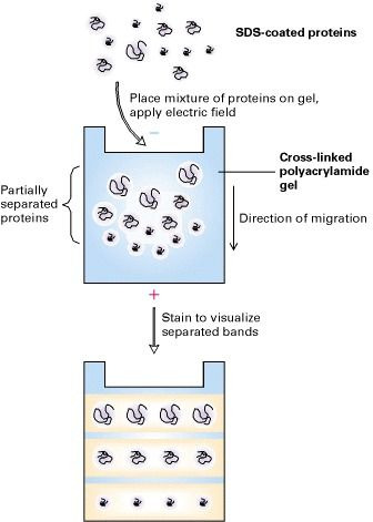
The isolated proteins would essentially be denatured with SDS and heat, and loaded on a gel with a running voltage. Because the proteins treated with SDS have a negative charge, they are pulled through the gel towards the positive electrode. The smaller proteins will migrate through the gel faster, and be closer to the bottom of the gel. Afterwards we can visualize and analyze the proteins via staining with Coomassie blue or autoradiography.


4 notes
·
View notes
Text
A dopamine gradient controls access to distributed working memory in the large-scale monkey cortex
https://www.cell.com/neuron/fulltext/S0896-6273(21)00621-8
Summary
Dopamine is required for working memory, but how it modulates the large-scale cortex is unknown. Here, we report that dopamine receptor density per neuron, measured by autoradiography, displays a macroscopic gradient along the macaque cortical hierarchy. This gradient is incorporated in a connectome-based large-scale cortex model endowed with multiple neuron types. The model captures an inverted U-shaped dependence of working memory on dopamine and spatial patterns of persistent activity observed in over 90 experimental studies. Moreover, we show that dopamine is crucial for filtering out irrelevant stimuli by enhancing inhibition from dendrite-targeting interneurons. Our model revealed that an activity-silent memory trace can be realized by facilitation of inter-areal connections and that adjusting cortical dopamine induces a switch from this internal memory state to distributed persistent activity. Our work represents a cross-level understanding from molecules and cell types to recurrent circuit dynamics underlying a core cognitive function distributed across the primate cortex.
#dopamine#working memory#parvalbumin#calbindin#somatostatin#calretinin#VIP#activity-silent#short-term synaptic plasticity#persistent activity#shaunice k. grier#shaunice the scientist#shaunice grier#neuron#stem#neurons#cell
1 note
·
View note
Text
Buying a Gel Documentation System
Otherwise called "gel doc" or "gel imaging system," "gel documentation" is one more illustration of innovation supporting medication. It is normally utilized in sub-atomic science for recording nucleic acids and proteins in polyacrylamide or agarose gels that are stained with ethidium bromide or other fluorophores like SYBR Green. Gel documentation systems are now an essential component of any exceptionally advanced and high-level clinical research facility. Before that, it included only a Polaroid print of the highest point of a reversed pyramid. It was set over a gel, and the picture was taken, which was neither adequate to direct a point-by-point investigation of the part required nor sufficiently clear to require photos of momentary DNA and protein gels.
The present gel records are a long way from all that nonsense. What the gel documentation system involves today are excitation and discovery systems that are constrained by PCs, multifunctional nooks, and programming that empower a point-by-point study and analysis of the pictures taken and recorded. They are either semi- or completely robotized and can deal with a scope of things, from DNA and protein gels to profoundly muddled autoradiography.

If you somehow managed to buy a gel documentation system today, you would find its extension and usefulness The value reached would likewise vary in light of that. You could track down systems to get pictures and documentation of simply the DNA and protein gels or the more developed ones that would deal with things like chemiluminescent Western smears, fluorescence, and autoradiography. The nature of the camera and its focal points, the product that accompanies the system, and the degree of computerization it provides are likewise standards for deciding the expense of the system.
A truly advanced gel documentation system would permit even a novice to take pictures easily. The majority of the means would be mechanized and humanized; however, in any case, it would allow the client to pick the brilliance and zoom of the image the person in question needs to take. In some cases, you would find that a few brands offer the system with the examination programming as an extra choice, though some would incorporate everything, obviously at a more exorbitant cost.
Breaking down the programming would allow you to focus on the image in depth, thereby providing additional services within the system. When you are on a mission to purchase the gel documentation system, it is better to seek advice and ideas from others, particularly if you are new to biomedical innovation. Get a quality system and you could out of nowhere.
For More Info:-
Shop Gel Documentation System
Gel Documentation System Procedure India
1 note
·
View note
Text
Immunostaining - Overview
Immunostaining is the use of an antibody-based method to detect a specific protein in a sample. This post is an overview / contents post from which to link my other posts and try to put some order in this blog!
Immunohistochemistry (IHC)
Immunostaining of cells of fixed or frozen cells (see my IHC post)
A primary antibody is added which sticks to the protein that’s being investigated.
A secondary antibody, linked to an enzyme, is added which then sticks to the primary antibody .
A chromogen is added, which the enzyme reacts with to deposit an insoluble coloured compound onto the cell.
This colour is detectable by light microscopy.
Therefore, the areas where that protein is present can be deduced by coloured areas visible down the microscope.
Alternatively, radioactive elements can be used as labels, and the immunoreaction can be visualized by autoradiography.
Tissue is fixed to preserve cell morphology and tissue architecture (see my post on this process).
Some antigens don’t survive formalin fixation. For samples needing these immuno tests, the tissue is rapidly fresh frozen in liquid nitrogen and cut with a cryostat before IHC.
The disadvantages of frozen sections include poor morphology, poor resolution at higher magnifications, difficulty in cutting over paraffin sections, and the need for frozen storage.
Western blotting
(See my Western blot post) Western blotting allows the detection of specific proteins from extracts made from cells or tissues.
Proteins are separated by size using gel electrophoresis
Smaller molecules move through the gel faster than larger molecules, and so travel further.
The separated proteins are transferred to a synthetic membrane via dry, semi-dry, or wet blotting methods.
Antibodies, linked to something that will allow visualisation eg enzymes that produce a coloured product, are added to the membrane.
Enzyme-linked immunosorbent assay (ELISA)
(See my ELISA post) The ELISA quantitatively or semi-quantitatively determines protein concentrations from blood plasma, serum or cell/tissue extracts in a multi-well plate format (usually 96-wells per plate).
An antigen is immobilized on a solid surface
It is complexed with an antibody that is linked to an enzyme.
The conjugated enzyme activity is assessed via incubation with a substrate.
Which produces a product that can be measured.
Immuno-electron microscopy
Electron microscopy or EM studies detailed microarchitecture of tissues or cells.
Immuno-EM allows the detection of specific proteins in ultrathin tissue sections.
Antibodies labelled with heavy metal particles (e.g. gold) can be directly visualised using transmission electron microscopy.
While powerful, it is technically challenging, expensive, and requires rigorous optimisation.
#medicine#biomed#biomedicine#notes#medblr#studyblr#sciblr#premed#nursing#biology#human biology#med#biomedical science#science#immunology#immuno#immunostaining#clinical#2#3#overview#contents#ihc
58 notes
·
View notes
