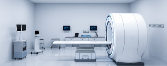
#MedicalImaging...
Explore tagged Tumblr posts
Text
X-Ray Technician: A Quick Guide to a Vital Healthcare Role
What Is an X-Ray Technician?
An X-ray technician, also known as a radiologic technologist, operates imaging equipment to capture internal body images, aiding in the diagnosis of medical conditions. These professionals are key in healthcare, providing essential support to doctors and radiologists.
Key Responsibilities
Patient Preparation: Explain procedures and position patients for accurate imaging.
Image Capture: Operate X-ray machines to create clear diagnostic images.
Safety: Use protective measures to minimize radiation exposure.
Image Processing: Analyze images and assist in diagnosing conditions.
Equipment Maintenance: Ensure imaging machines are in good working order.
Educational Path and Certification
Education
To become an X-ray technician, an Associate’s degree in Radiologic Technology is typically required, though some pursue a Bachelor’s degree for advancement.
Certification
Certification involves passing a comprehensive exam. Licensing requirements vary by state but generally include continuing education.
Skills and Qualities Needed
Technical Skills: Proficiency in operating complex imaging equipment.
Attention to Detail: Accuracy in capturing high-quality images.
Communication: Ability to explain procedures and interact with patients.
Physical Stamina: Capability to stand for long periods and assist patients.
Opportunities for Advancement
Specializations: Technicians can specialize in areas like CT or mammography for higher salaries.
Further Education: Continuing education and additional certifications can lead to advanced roles in management or specialized fields.
Challenges in the Field
Radiation Exposure: Managing and minimizing exposure risks is crucial.
Physical Demands: The job requires significant physical effort.
Technological Advances: Staying updated with new technology and procedures is necessary for career longevity.
Conclusion
Becoming an X-ray technician offers a rewarding career with a strong job outlook and opportunities for growth. This role is essential in providing high-quality patient care and supporting the medical field.
#XRayTechnician#RadiologicTechnologist#MedicalImaging#HealthcareCareer#XRayTech#Radiology#ImagingTech#RadiologyTechnologist#MedicalDiagnostics#TechInHealthcare#MedicalTechnician
7 notes
·
View notes
Text

Unlocking the secrets of MRI scans in Ahmedabad! Discover the best center for MRI scans and cost-effective solutions at Usmanpura Imaging. Your path to precise diagnosis begins here. 🧲💡
#MRI#mri scan#diagnosis#mri centre#AhmedabadHealthcare#MedicalImaging#AffordableHealthcare#mri scan cost#usmanpuraimaging
2 notes
·
View notes
Text
B.VOC - RADIOLOGY & MEDICAL IMAGING TECHNOLOGY
Radiology is a professional course in the paramedical field, offered to help individuals become professional in performing diagnostic tests related to various medical treatments by using radiation technology. In simpler terms, radiology involves using different scientific equipment to photograph the human body’s hidden and internal portions. These images are required in the process of identifying alignments and diseases.

2 notes
·
View notes
Text
Our Sonography Services at Care & Cure Hospital are state-of-the-art to deliver accurate imaging, and hence accurate diagnosis and appropriate planning for treatment. Our board-certified radiologists conduct complete scans for pregnancy monitoring, abdominal scan, heart testing, muscular skeletal disease, and numerous other diseases with the latest ultrasound technology. From the routine ultrasound to Doppler test, and 3D/4D imaging, we take extra care to make our patients feel comfortable, secure, and get an accurate diagnosis. By virtue of non-invasive technology and real-time imaging, sonography is a valuable tool for the early diagnosis and medical evaluation. Leave precise and effective ultrasound diagnostics to our expert personnel!
0 notes
Text
𝐏𝐡𝐚𝐧𝐭𝐨𝐦 𝐇𝐞𝐚𝐥𝐭𝐡𝐜𝐚𝐫𝐞 𝐚𝐭 𝐌𝐈𝐃-𝐓𝐄𝐑𝐌 𝐂𝐌𝐄 𝟐𝟎𝟐𝟓, 𝐁𝐡𝐨𝐩𝐚𝐥!
We are proud to have participated in MID-TERM CME 2025, organized by the Musculoskeletal Society of India in collaboration with the Indian Radiological & Imaging Association- IRIA M.P. Chapter. This prestigious event took place on Sunday, 23rd March 2025, at Chirayu Medical College & Hospital, Bhopal (Chirayu University).

With the theme "Imaging and Interventions in Musculoskeletal Infections & Inflammation," the conference brought together leading radiologists, musculoskeletal imaging specialists, and healthcare professionals to discuss cutting-edge techniques in diagnostics and interventions for musculoskeletal disorders.
At Phantom Healthcare, we are dedicated to empowering the radiology community with refurbished MRI, CT, PET-CT, and Cath Lab systems, ensuring affordable, high-quality diagnostic imaging solutions for healthcare providers across India.
A heartfelt thank you to the Musculoskeletal Society of India and the Indian Radiological & Imaging Association (IRIA) M.P. Chapter for organizing such an insightful and engaging conference. We look forward to continuing our journey of advancing radiology and medical imaging across the country!


#PhantomHealthcare#MIDTERMCME2025#Musculoskeletal#MIDTERMCME#BHOPAL#radiology#MusculoskeletalRadiology#IRIA#IRIA2025#IndianRadiologicalAndImagingAssociation#RadiologyInnovation#MedicalImaging#HealthcareExcellence#MRI#CT#RadiologyConference#AdvancingDiagnostics
0 notes
Text

❓💡 What if AI could detect breast cancer risk earlier than ever before? ❓💡 Early detection saves lives, but many women still face delayed diagnoses due to limited access to advanced screening tools. What if AI could change that? Recently, Segmed’s real-world imaging data was used to clinically validate an AI algorithm designed to assess breast cancer risk. This breakthrough technology could help expedite the identification of at-risk women, leading to earlier intervention and better outcomes. At Segmed, we: ✅ Source diverse, real-world imaging data from healthcare providers ✅ De-identify and structure it for research use ✅ Empower AI and healthcare innovators with high-quality datasets AI in healthcare is only as strong as the data it learns from. Let’s work together to unlock new possibilities in early disease detection. 💡 How do you see AI transforming cancer detection in the next decade? Drop your thoughts in the comments! 👉 Want to collaborate? Learn more by reaching to us out here: https://www.segmed.ai/
#AIinHealthcare#MedicalAI#HealthcareInnovation#MedicalImaging#RadiologyAI#RealWorldData#PrecisionMedicine#ClinicalTrials#HealthTech#DataDrivenHealthcare#AIforGood#MedTech#DigitalHealth#HealthcareAI#FutureofMedicine#TeamSegmed#MedicalDataPartnership
0 notes
Text
Tele Radiology Services in India – Radblox
Tele radiology services in India are transforming the healthcare industry by providing remote diagnostic imaging solutions with accuracy and efficiency. Radblox provides advanced tele radiology solutions, ensuring seamless image transmission, precise diagnostics, and 24/7 radiology support. With a team of highly skilled radiologists, we deliver timely and reliable interpretations, improving patient outcomes and reducing delays in treatment.
Key advantages of tele radiology services in India with Radblox:
Round-the-clock radiology reporting for emergency and routine cases.
Faster diagnosis and reduced turnaround time for medical imaging.
Seamless integration with hospital PACS and radiology workflows.
Access to expert radiologists, eliminating geographical limitations.
Cost-effective solutions for hospitals and diagnostic centers.
Radblox enhances healthcare accessibility by bridging the gap between medical imaging and expert interpretation. Our tele radiology services in India cater to hospitals, standalone diagnostic centers, and telemedicine providers, ensuring high-quality reports with accuracy and speed. With cutting-edge technology, secure data transmission, and compliance with international healthcare standards, we provide a trusted solution for medical professionals. Partner with Radblox for efficient and scalable tele radiology services in India.

0 notes
Text
What advanced diagnostic services are available at Altamash Hospital?
Altamash Hospital in Karachi offers a comprehensive range of advanced diagnostic services to ensure accurate and timely medical evaluations. Their state-of-the-art facilities include:
Clinical Laboratory: Equipped with the latest automated analyzers, the laboratory operates 24/7 under the supervision of qualified pathologists and trained technicians, fulfilling modern diagnostic requirements.
Radiology Services:
X-Ray and Orthopantomogram (OPG): Utilizes computerized radiography for detailed imaging.
CT Scan: Employs computerized tomography to create cross-sectional images of the body.
MRI: Uses powerful magnets and radio waves to produce detailed internal body images.
Ultrasound: Generates images of soft tissue structures like the gallbladder, liver, kidneys, and pancreas.
Mammography: Specialized imaging for breast tissue examination.
Endoscopy: A non-surgical procedure to examine the digestive tract using a flexible tube with a light and camera.
These services are supported by a dedicated team of radiologists and skilled technicians, ensuring precise diagnostics for effective patient care.
For more detailed information, you can visit their official website:altamashhospital.com
#AltamashHospital#DiagnosticServices#KarachiHealthcare#AdvancedDiagnostics#MedicalImaging#ClinicalLaboratory#Radiology#CTScan#MRI#Ultrasound#Endoscopy
0 notes
Text
Halide Scintillator Crystals Market, Global Outlook and Forecast 2025-2032
Halide scintillator crystals are materials used in detecting radiation by emitting visible light when exposed to high-energy particles. Common types include sodium iodide (NaI) doped with thallium and cesium iodide (CsI), used in applications like medical imaging and radiation monitoring.
Market Size
Download FREE Sample of this Report
The global Halide Scintillator Crystals market size was estimated at USD 197 million in 2023, with a projected growth to USD 282.83 million by 2032, exhibiting a CAGR of 4.10% during the forecast period.
Global Halide Scintillator Crystals Market: Market Segmentation Analysis
This report provides a deep insight into the global Halide Scintillator Crystals market covering all its essential aspects. This ranges from a macro overview of the market to micro details of the market size, competitive landscape, development trend, niche market, key market drivers and challenges, SWOT analysis, value chain analysis, etc.
The analysis helps the reader to shape the competition within the industries and strategies for the competitive environment to enhance the potential profit. Furthermore, it provides a simple framework for evaluating and accessing the position of the business organization. The report structure also focuses on the competitive landscape of the Global Halide Scintillator Crystals Market. This report introduces in detail the market share, market performance, product situation, operation situation, etc. of the main players, which helps the readers in the industry to identify the main competitors and deeply understand the competition pattern of the market.
In a word, this report is a must-read for industry players, investors, researchers, consultants, business strategists, and all those who have any kind of stake or are planning to foray into the Halide Scintillator Crystals market in any manner.
Market Segmentation (by Application)
Medical & Healthcare
Industrial Applications
Military & Defense
Others
Market Segmentation (by Type)
NaI
CsI
LaBr3
Others
Key Company
Luxium Solutions (Saint-Gobain Crystals)
Dynasil
Shanghai SICCAS
Rexon Components
EPIC Crystal
Shanghai EBO
Beijing Scitlion Technology
Alpha Spectra
Scionix
FAQ
01. What is the current market size of Halide Scintillator Crystals Market?
The global Halide Scintillator Crystals market was estimated at USD 197 million in 2023, projected to reach USD 282.83 million by 2032 with a CAGR of 4.10%.
02. Which key companies operate in the Halide Scintillator Crystals Market?
Key companies in the Halide Scintillator Crystals Market include Luxium Solutions (Saint-Gobain Crystals), Dynasil, Shanghai SICCAS, Rexon Components, EPIC Crystal, Shanghai EBO, Beijing Scitlion Technology, Alpha Spectra, and Scionix.
03. What are the key growth drivers in the Halide Scintillator Crystals Market?
Main growth drivers in the Halide Scintillator Crystals Market include advancements in medical imaging technologies, increasing use in industrial applications, growing demand in military & defense sectors, and expanding applications in radiation monitoring.
04. Which regions dominate the Halide Scintillator Crystals Market?
The Halide Scintillator Crystals Market is dominated by regions like North America, Europe, Asia-Pacific, South America, and the Middle East and Africa. These regions exhibit significant demand, supply, and market share in the Halide Scintillator Crystals Market.
05. What are the emerging trends in the Halide Scintillator Crystals Market?
Emerging trends in the Halide Scintillator Crystals Market include technological advancements in scintillator crystals, the development of new crystal types like LaBr3, increasing adoption in medical & healthcare sectors, and the rising focus on improving detection accuracy in radiation monitoring.
Get the Complete Report & TOC
CONTACT US: 203A, City Vista, Fountain Road, Kharadi, Pune, India - 411014 International: +1(332) 2424 294 Asia: +91 9169162030
Follow Us On linkedin :-https://www.linkedin.com/company/24chemicalresearch/
About 24Chemical Research: 24chemicalresearch was founded in 2015 and has quickly established itself as a leader in the chemical industry segment, delivering comprehensive market research reports to clients. Our reports have consistently provided valuable insights, aiding our clients, including over 30 Fortune 500 companies, in achieving significant business growth.
#HalideScintillatorCrystals#ScintillatorCrystals#RadiationDetection#MedicalImaging#NuclearMedicine#MarketForecast#CrystalTechnology#GlobalMarketTrends#CAGR#ScientificResearch
0 notes
Text
🌟 Your Health, Our Priority at A'Care Medical Imaging 🌟
At A'Care Medical Imaging, we believe that your health should always come first. That's why we provide the highest quality, non-invasive, and safe imaging services to ensure accurate results for your well-being.
Whether you're undergoing a routine check-up or need a specialized diagnostic procedure, our team is dedicated to offering you the best care every step of the way. From ultrasounds to advanced imaging, we’ve got you covered. 📅
✨ Why Choose Us? ✔️ Trusted Experts ✔️ Safe & Painless Procedures ✔️ Fast, Reliable Results ✔️ Patient-Centered Care
Book your appointment with us today and take the first step towards better health! 🏥💙
#A'CareMedicalImaging#HealthFirst#PatientCare#MedicalImaging#DiagnosticServices#HealthyLiving#YourHealthMatters
0 notes
Text
instagram
#ShivamHospital 🏥#Sonography 🩺#ColorDoppler 🌟#AdvancedImaging 📡#ExpertCare 👨⚕️👩⚕️#DombivliHealthcare 📍#DiagnosticCenter 🔬#HealthcareFirst ❤️#MedicalImaging#HealthMatters#AccurateDiagnosis ✅#WellnessCare 🌿#StayHealthy 💙#BookYourTest 📅#Instagram
0 notes
Video
youtube
Telemedicine 2.0: How AR/VR Integration is Revolutionizing Virtual Healt...
#youtube#Telemedicine2.0 ARVRinHealthcare VirtualHealthcare FutureOfMedicine HealthcareInnovation RemoteSurgery MedicalImaging VirtualRehabilitation
0 notes
Text
Successful De-installation Project in Qatar - Phantom Healthcare
Phantom Healthcare recently completed a successful de-installation project in 𝐐𝐚𝐭𝐚𝐫, showcasing its expertise in handling complex medical imaging equipment.
Our dedicated team of professionals ensured MRI scanners' safe and efficient removal while adhering to international standards and protocols. Through our meticulous approach, we minimized downtime and ensured that the equipment remained in optimal condition for future use.
As a trusted leader in refurbished radiology solutions, Phantom Healthcare continues to deliver seamless de-installation, relocation, and maintenance services worldwide, reinforcing our commitment to quality and excellence in medical imaging.
Visit Website: https://www.phantomhealthcare.com/ E-Commerce Website: https://phantomhealthcare.shop/ Youtube: www.youtube.com/@phantomhealthcare
Let's Discuss Your Requirement: +𝟗𝟏-𝟗𝟖𝟗𝟗𝟗𝟔𝟑𝟔𝟎𝟏, 𝟖𝟑𝟖𝟒𝟎𝟑𝟕𝟎𝟕𝟑
𝐖𝐞 𝐚𝐫𝐞 𝐨𝐧𝐞 𝐨𝐟 𝐭𝐡𝐞 𝐥𝐚𝐫𝐠𝐞𝐬𝐭 𝟑𝐫𝐝 𝐩𝐚𝐫𝐭𝐲 𝐯𝐞𝐧𝐝𝐨𝐫𝐬 𝐨𝐟 𝐌𝐑𝐈 𝐌𝐚𝐜𝐡𝐢𝐧𝐞, 𝐂𝐓 𝐒𝐜𝐚𝐧𝐧𝐞𝐫, 𝐂𝐚𝐭𝐡 𝐋𝐚𝐛 𝐒𝐜𝐚𝐧𝐧𝐞𝐫, 𝐁𝐨𝐧𝐞 𝐃𝐞𝐧𝐬𝐢𝐭𝐨𝐦𝐞𝐭𝐞𝐫 𝐌𝐚𝐜𝐡𝐢𝐧𝐞 𝐚𝐧𝐝 𝐏𝐄𝐓 𝐂𝐓 𝐒𝐜𝐚𝐧𝐧𝐞𝐫.


#deinstallation#Qatar#MRI#MRIScan#MRIScanner#CTScan#CT#radiology#radiographer#radiography#medicalimaging#medicalequipment#medicaldevice#medicalequipments#medicaldevices#medicaldevicemanufacturing#medicalequipmentforsale#medicalequipmentsupplier#phantom#phantomhealthcare#relocating#relocationservices
0 notes
Text
Accelerate Life Sciences Research with Segmed's Real-World Imaging Data Solutions
Segmed offers life sciences researchers access to extensive longitudinal disease data, featuring regulatory-grade multimodal datasets and millions of high-quality, diverse, tokenized imaging studies. Our platform supports various R&D stages, including biomarker discovery, clinical trial design, patient recruitment, and post-marketing surveillance, facilitating rapid and informed decision-making.
#life science#rwid#medical imaging#real world data#real world imaging#medicalimaging#radiology#real world evidence#healthcareinnovation#segmed
0 notes