#AFM imaging
Explore tagged Tumblr posts
Visit Tumblr Blog
Explore Tumblr blogs with no restrictions, modern design and the best experience.
Last Seen Tumblr Blogs


Fun Fact
Mobile US users spent an average of 115.8 minutes on Tumblr app monthly.
Text
youtube
#Atomic Force Microscopy#nanomechanical tool#cancer liquid biopsy#cancer diagnostics#extracellular vesicles#circulating tumor cells#nanotechnology#cell elasticity#non-invasive biopsy#cancer biomarkers#early cancer detection#precision oncology#AFM imaging#tumor analysis#biomechanics#personalized cancer treatment#cancer research#molecular profiling#cellular adhesion#liquid biopsy techniques.#Youtube
0 notes
Text
A typical AFM image is shown in figure 1.6.

"Chemistry" 2e - Blackman, A., Bottle, S., Schmid, S., Mocerino, M., Wille, U.
1 note
·
View note
Text
My artistic side is not lost in the world of science!
My art featuring antibiotics that slow down or dissolve domains in bacteria membranes was selected as the cover of the latest Nano Letters issue! The cover art combines hand-drawn bacteria and 3D rendered atomic force microscopy images of bacteria membranes.
My process of drawing and shading the coccus bacteria:

And the final cover:

I had a great time thinking about the concept of this cover, drawing the bacteria, making the 3D bacteria membrane views from my AFM data, and putting everything together. It was a great creative work, although it was under a stressful deadline for the manuscript resubmission including the cover art suggestion.
The Nano Letters issue and the article here: https://pubs.acs.org/toc/nalefd/24/38
#science#art#postdoc#research#bacteria#antibiotics#original content#cover art#journal cover#Nano Letters#publication#scientific illustration#scientific publication
27 notes
·
View notes
Text
X-Men 2 fans: First Look at Nightcrawler and Stryker as brothers in Glenrothan

Brian Cox‘s feature directorial debut “Glenrothan” — billed as his “love letter to Scotland” — has wrapped production, with a first look revealed showing the “Succession” star alongside Alan Cumming in a room full of whisky barrels.
Alongside revealing a first look from the feature, Protagonist Pictures has come aboard for worldwide sales and is set to screen exclusive footage for buyers at the upcoming American Film Market.
Lionsgate, which co-developed the project with London based studio Nevision, has taken U.K. rights on the family drama, which shot in Scotland.
In “Glenrothan,” Donal (Cumming) reluctantly returns to the Scottish Highlands after 35 years in Chicago to make amends with his estranged older brother, Sandy (Cox). Sandy needs Donal to take over the family’s whisky distillery or he will be forced to sell and give up on the family’s legacy. But their reunion forces the brothers to confront the past and the real reason Donal left Glenrothan. Shirley Henderson (“Harry Potter and the Goblet of Fire,” “See How They Run”) and Alexandra Shipp (“Barbie,” “Anyone But You”) also star in the film.
“Glenrothan” is produced by Cox, Neil Zeiger, Crystine Zhang (“Prisoner’s Daughter,” “Lee”), Phin Glynn (“Argentina 1985,” “Kill The Jockey”), Nic Crum (“Book of Love”), James Cabourne and Vladimir Zemtsov (“Club Zero”). The film is co-produced by Nevision and Blazing Griffin Pictures in association with Screen Scotland, Oval-5, Gold Rush Pictures, Head Gear, Infinity Hill and Lionsgate.
“’Glenrothan’ is a poignant, funny and powerful story about the universal themes of family and heritage, set against the stunning backdrops of Scotland and steeped in the world of whisky,” said Protagonist CEO Dave Bishop. “Brian is an undeniable talent, both behind and in front of the camera, and is joined by the exceptional Alan Cumming — as such we are delighted to introduce buyers to this special film when we debut exclusive footage at the upcoming AFM.”
Neil Zeiger, lead producer added: “I have thoroughly enjoyed helping bring ‘Glenrothan,’ Brian’s ‘love letter to Scotland’, to the screen. Working with our incredible cast led by Alan, Shirley and Alexandra, Brian directed the film brilliantly. A universal story, wonderful performances and stunning images define the film. I am confident Glenrothan will compel and captivate audiences wherever in the world they are.”
Protagonist’s current slate includes the Venice, TIFF and San Sebastian selection “Kill the Jockey,” directed by Luis Ortega and recently selected as Argentina’s feature film contender for the 2025 Academy Awards; Samir Oliveros’ TIFF title, “The Luckiest Man in America,” starring Emmy and Golden Globe winner Paul Walter Hauser; the genre-bending comedy “Sister Midnight,” which recently screened at the London Film Festival and was nominated for Cannes’ Camera D’Or; and from directors Guy Maddin, Evan Johnson, and Galen Johnson, the political horror-comedy “Rumours,” which debuted at Cannes before going on to screen at Toronto, San Sebastian, New York and London Film Festivals.
13 notes
·
View notes
Text

Researchers explain the imaging mechanisms of atomic force microscopy in 3D
Researchers at Nano Life Science Institute (WPI-NanoLSI), Kanazawa University report the 3D imaging of a suspended nanostructure. The technique used is an extension of atomic force microscopy and is a promising approach for visualizing various 3D biological systems. Atomic force microscopy (AFM) was originally invented for visualizing surfaces with nanoscale resolution. Its basic working principle is to move an ultrathin tip over a sample's surface. During this xy-scanning motion, the tip's position in the direction perpendicular to the xy-plane follows the sample's height profile, resulting in a height map of the surface.
Read more.
#Materials Science#Science#Atomic force microscopy#Materials characterization#Nanotechnology#Kanazawa University
16 notes
·
View notes
Text
Atomic force microscopy, or AFM, is a widely used technique that can quantitatively map material surfaces in three dimensions, but its accuracy is limited by the size of the microscope's probe. A new AI technique overcomes this limitation and allows microscopes to resolve material features smaller than the probe's tip. The deep learning algorithm developed by researchers at the University of Illinois Urbana-Champaign is trained to remove the effects of the probe's width from AFM microscope images. As reported in the journal Nano Letters, the algorithm surpasses other methods in giving the first true three-dimensional surface profiles at resolutions below the width of the microscope probe tip.
Continue Reading.
46 notes
·
View notes
Text
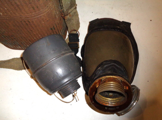

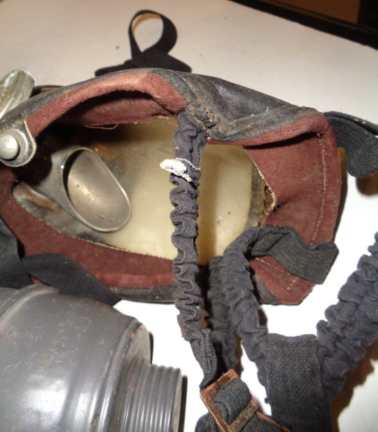
I will not comment on the French beyond saying that some of their 1930's-40's developments in chemical protection come off as almost Baroque in nature. This is a variant of the GEP (Gaz Et Protection) with an added speech diaphragm. The large metal cone in the 3rd image is to direct the user's voice into said cone. Placement on the side of the mask like this aids in the use of a standard telephone handset.
GEPs were one of many, many, many private market civilian respirators manufactured in Europe during the decades surrounding World War 2. Both the carrying case and speech diaphragm on this example appear very visually similar to the respective components of the AFM-34 kit. Not entirely sure if this is because GEP made AFM-34s, or because the patterns and shapes were readily available from a common source and simply purchased by GEP as stock for assembling their masks.
15 notes
·
View notes
Text

SUBJECT
The superior physicochemical ambiance provided by Chitin/Carboxylated Chitosan for the formation of hydroxyapatite film
DEPARTMENT
Department of Chemical Engineering
PROFESSOR AND STUDENT
Professor:Ten-Chin Wen
Student:Wei-Cheng Li
ABSTRACT
Chitosan (CS) and chitin (CH) are two natural polysaccharides. Chitosan with different carboxylation degrees rendering specific zwitterionic properties. In this study, carboxylated chitosan (CCS) and CH polymeric matrix was mineralized to form an hydroxyapatite film.
CS grafted carboxylated group at pH 6, 8, and 10 for products denoted as CCS6, CCS8, CCS10. It was the better degrees of carboxylated, the higher ion conductivity. CCS with zwitterion helped ions movement. Based on thermogravimetric analysis, thermal cracking temperature of the amide group on chitin increased after mineralization. In the TEM image, the PILP behavior was found, and formed hexagonal hydroxyapatite beside chitin. The Young's modulus of hydroxyapatite evaluated by AFM was 5.19±0.06GPa. Meanwhile, Ca/P ratio by EDX analysis was 1.65, similar to bones.
Original URL: 15-Student:Department of Chemical Engineering【Wei-Cheng Li】-Undergraduate Research , NCKU https://en.ur.ncku.edu.tw/book/15-Student:Department+of+Chemical+Engineering%E3%80%90Wei-Cheng+Li%E3%80%91/
The copyright belongs to the author. For commercial reprints, please contact the author for authorization, and for non-commercial reprints, please indicate the source.
2 notes
·
View notes
Note
So on the whole nano vehicles thing!
This is something people are researching in the field of nanotechnology! It's a field that combines chemistry, biology, physics (and maths/computer science in some ways but that makes it even more complicated) on the nano scale! Now first what even is nano? Nano refers to the scale of something of 10^-9 in this case it's about sizes but nano is a prefix so eh.
Regardless nanotech is a relatively new field (if you're interested in it there's a lecture called "there's plenty of room at the bottom" by Richard P. Feynman who is often considered the parent of nanotechnology) having only been brought up by the previously mentioned, R. Feynman in 1959 and only really sparking the research field to start in the 1980s.
Anyways so yay small scale science! So nanovehicles: nanovehicles are often bottom up (see: bottom-up = using bricks to build a house vs. top down = carving a house out of rock) produced small "vehicles". They came about because we're looking for ways to transport cargos in a more controlled method than we've been able to mimicking certain biological functions. But the way we've being going about it is by making tiny little car shaped molecules. As you can see in the image below just car shaped with Buckminster fullerene (not actually buckminster fullerene but spherical carbon structures) type wheels.

So basically just like cars work, movement is perpendicular to the axle(usually), however you cannot change the positioning of the axles so this thing can only move forwards and backwards.
Oh quick little side note to even analyse the motion of these things, we have to put them on a surface and depending on the type of motor that can lead to either a caterpillar like motion or just increasingly long tails. But what we can also do is use the tips AFM (atomic force microscopy)or SPM (scanning probe microscopy) and move them with that however that's incredibly expensive Sheesh. We have to do this on a surface because of Brownian motion because these things are so small they'll just move around and about randomly in solution.
Okay back to vehicles. So before I showed a 2 directional transverse moving nanocar. But in the previous ask I said there's nanovehicles that can only spin around themselves. And that's because the Axel's are thusly oriented that any transverse motion it wants to make would be obstructed by another axle. So then it can only do the spinny thing.
See below:
a) shows translational motion such as a nanocar can make.
b) shows rotational motion, such as the nanovehicle I was so excited about.
As well as these images having microscopy which shows how they have moved and where the vehicles are and it's very cool.
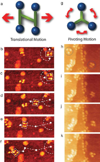
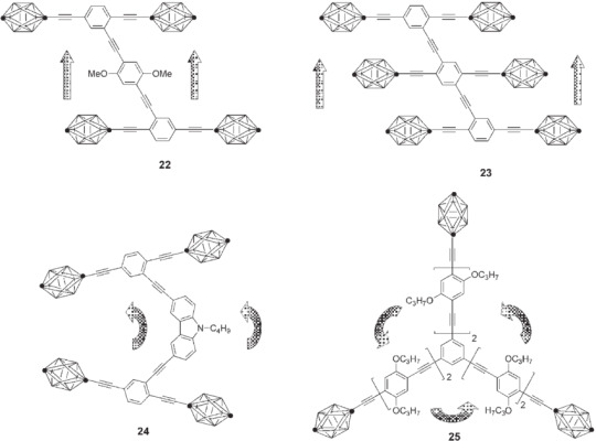
And below you can see different types of nanovehicles, 22 & 23 can do translational motion forwards and backwards. 24 can move in a slight circle around a middle point that is not its own center point.
And then you have 25, my beloved, it can only spin in circles <3 its sole purpose is to just spin around and around and around and I think that's somehow so very poetic, the fact you've been made in such a way that your sole purpose cannot be fulfilled but you did prove that yes we can move nanovehicles in more than just the two directions. Also that molecule is a blorbo to me.
i forgot all about this and i have still never been so interested in something i understood next to nothing about thank you for infodumping lulu i heart you
2 notes
·
View notes
Text
The Revolutionary History of Nanotechnology

Nanotechnology, a groundbreaking field that has revolutionized numerous industries, continues to shape the world as we know it. In this article, we delve into the rich history of nanotechnology, exploring its origins, major milestones, and transformative applications. Join us on this captivating journey through the nano realm and discover how this remarkable technology has reshaped various sectors, from healthcare and electronics to energy and materials science.
Origins of Nanotechnology
Unveiling the Nanoscale
Nanotechnology finds its roots in the exploration of the minuscule world at the nanoscale. The concept of nanoscale was first introduced by physicist Richard Feynman in his visionary lecture in 1959, where he discussed the potential for manipulating matter at the atomic and molecular levels. This groundbreaking concept laid the foundation for the birth of nanotechnology.
The Birth of Nanotechnology
In 1981, the term "nanotechnology" was officially coined by engineer K. Eric Drexler in his influential book, "Engines of Creation." Drexler envisioned a future where nanomachines could manipulate matter at the atomic scale, leading to remarkable advancements in various fields. His work served as a catalyst for the rapid development of nanotechnology research and applications.

Major Milestones in Nanotechnology
Scanning Probe Microscopy
In the early 1980s, the invention of scanning probe microscopy revolutionized nanotechnology research. The scanning tunneling microscope (STM) and atomic force microscope (AFM) allowed scientists to visualize and manipulate individual atoms and molecules with unprecedented precision. These breakthroughs opened up new possibilities for studying nanoscale phenomena and laid the groundwork for further advancements in the field.
Fullerenes and Nanotubes
In 1985, a significant discovery shook the scientific community—the identification of fullerenes. Researchers Robert Curl, Harold Kroto, and Richard Smalley stumbled upon these unique carbon molecules, marking the birth of a new class of nanomaterials. Fullerenes paved the way for the development of carbon nanotubes, cylindrical structures with remarkable strength and conductivity. These nanotubes would go on to become key building blocks in various nanotechnology applications.
Nanotechnology in Medicine
Nanotechnology's potential to revolutionize healthcare became evident with the advent of targeted drug delivery systems. Nanoparticles, such as liposomes and polymeric nanoparticles, can be designed to encapsulate drugs and deliver them precisely to targeted cells or tissues. This approach minimizes side effects and maximizes therapeutic efficacy. Additionally, nanotechnology plays a vital role in imaging techniques, enabling highly sensitive and precise detection of diseases at the molecular level.
Nanoelectronics and Quantum Computing
The relentless pursuit of smaller, faster, and more energy-efficient electronics led to the emergence of nanoelectronics. By utilizing nanoscale materials and devices, researchers have pushed the boundaries of traditional silicon-based technology. Nanoscale transistors, quantum dots, and nanowires have paved the way for advancements in computing power, memory storage, and energy efficiency. Furthermore, the field of quantum computing, which harnesses quantum phenomena at the nanoscale, holds the promise of solving complex problems that are currently beyond the capabilities of classical computers.
Nanomaterials and Energy
Nanotechnology has also played a significant role in addressing global energy challenges. By developing advanced nanomaterials, scientists have made strides in enhancing solar cell efficiency, enabling the production of clean and renewable energy. Nanomaterials have also been employed in energy storage devices, such as batteries and supercapacitors, to improve their performance and longevity. Additionally, nanotechnology has opened up avenues for energy harvesting and energy conversion, contributing to a more sustainable future.

Transformative Applications of Nanotechnology
Nanomedicine and Disease Treatment
Nanotechnology has revolutionized medicine, offering innovative solutions for disease diagnosis, treatment, and prevention. Targeted drug delivery systems, nanoscale imaging techniques, and nanobiosensors have transformed the landscape of healthcare, enabling personalized and precise interventions. From cancer therapy to regenerative medicine, nanotechnology has the potential to revolutionize patient care and improve outcomes.
Nanoelectronics and Wearable Technology
The marriage of nanotechnology and electronics has given rise to the era of wearable technology. Nanoscale sensors, flexible displays, and energy-efficient components have paved the way for smartwatches, fitness trackers, and augmented reality devices. These advancements in nanoelectronics have made it possible to integrate technology seamlessly into our everyday lives, enhancing convenience and connectivity.
Nanomaterials and Advanced Manufacturing
Nanotechnology has propelled advancements in materials science and manufacturing. Nanomaterials with tailored properties and enhanced performance characteristics have found applications in aerospace, automotive, and construction industries. From lightweight and high-strength composites to self-cleaning surfaces and energy-efficient coatings, nanomaterials have revolutionized product design, durability, and sustainability.
In Conclusion
Nanotechnology's journey from its conceptualization to its present-day applications has been nothing short of extraordinary. The field's remarkable achievements in diverse domains, including medicine, electronics, and energy, continue to drive innovation and shape the future. As we delve deeper into the nanoscale world, the possibilities seem boundless. With ongoing research and collaboration, nanotechnology will undoubtedly unlock new frontiers, leading to breakthroughs that will reshape industries and improve lives across the globe.
#history of nanotechnology#richard feynman#chemestry#green chemistry#nanotechnology#science#nanomaterials#nanocoating#nanomedicine#probe microscopy#revolutionary
3 notes
·
View notes
Text
Powerhouse of Precision: Exploring the Key Features of Piezo Stacks
Piezo stacks are innovative devices that utilize the piezoelectric effect to achieve precise and controlled movements. These stacks consist of multiple layers of piezoelectric ceramic materials stacked together, enabling them to generate high forces and displacements with nanoscale precision. In this infographic, we explore the key features, working principle, and applications of piezo stacks.
For More Information Please visit, pzt stacks
Key Features of Piezo Stacks:
Nanoscale Precision: Piezo stacks offer exceptional precision, allowing for nanometer-level movements and control. This precision is crucial in applications such as microscopy, semiconductor manufacturing, and optical alignment.
High Force Output: Despite their compact size, piezo stacks can generate significant amounts of force. The stacked design allows for the amplification of force, enabling precise control even in applications that require high force requirements.
Rapid Response: Piezo stacks can respond to electrical signals in microseconds, enabling fast and dynamic adjustments. Their rapid response is vital in applications that require quick positioning or movement changes.
Direct Drive and Non-Magnetic Operation: Piezo stacks operate without the need for mechanical gears or magnetic components. This direct drive mechanism eliminates backlash and allows for precise control, even in non-magnetic environments.
Working Principle of Piezo Stacks:
Piezoelectric Effect: Piezo stacks utilize the piezoelectric effect, which refers to the ability of certain materials to generate an electric charge when subjected to mechanical stress. This effect allows the stack to convert electrical signals into precise mechanical movements.
Layered Structure: Piezo stacks consist of multiple thin layers of piezoelectric ceramics. Each layer is carefully engineered to maximize the piezoelectric effect.
Voltage Application: When a voltage is applied to the stack, an electric field is created within the piezoelectric layers. This field causes the layers to expand or contract, resulting in precise mechanical displacements.
Applications of Piezo Stacks:
Precision Positioning: Piezo stacks are extensively used in positioning systems for microscopy, atomic force microscopy (AFM), lithography, and optical alignment. Their precise control allows for accurate positioning and scanning.
Micro-dispensing and Jetting: Piezo stacks find application in micro-dispensing and jetting systems, enabling precise control of fluid flow. They are utilized in industries such as inkjet printing, pharmaceuticals, and biotechnology.

Active Damping and Vibration Control: Piezo stacks are employed in active damping and vibration control systems to minimize unwanted vibrations. They find use in precision manufacturing equipment, semiconductor fabrication, and optical instruments.
Adaptive Optics: In astronomy and laser applications, piezo stacks are used in adaptive optics systems to compensate for atmospheric distortions. They allow for real-time adjustments of deformable mirrors, improving image quality and resolution.
Conclusion:
Piezo stacks provide an effective solution for achieving precision and control in various industries. Their nanoscale precision, high force output, rapid response, and direct drive operation make them invaluable in applications requiring accurate positioning, micro-dispensing, vibration control, and adaptive optics. With ongoing advancements in technology, piezo stacks are poised to continue empowering innovation and driving progress in precision engineering and control systems.
2 notes
·
View notes
Text
Atomic force microscope operates at very close range and without a lens. There are several different types of atomic force microscope, and they all operate by measuring a local property, whether height, optical absorption, or magnetism, using a probe placed very close to the sample. This probe makes it possible to measure qualities over a small area, and an image of that area can be produced that resembles an image on a television screen, consisting of many rows or lines of information placed one above the other. The size of the probe is what generally limits resolution, as opposed to a traditional microscope, where the limitation derives from diffraction effects. When brought close to the sample, the probe measures attractive or repulsive forces between the tip and the sample. The "contact" mode is also called the repulsive mode in which the instrument lightly touches a tip at the end of a leaf spring or "cantilever" to the sample. A raster-scan drags the tip over the sample, and as this occurs, the detection apparatus measures the vertical deflection of the cantilever, indicating the local sample height. This means that in contact mode, the AFM measures hard-sphere repulsion forces between the tip and sample. The probe also works in noncontact mode to derive topographic images from measurements of attractive forces, and in this approach, the tip does not touch the sample. This device is able to produce a resolution of 10 pm. It is superior to electron microscopes in that it can image samples in air and under liquids (Baselt paras. 2-4). The first such device was created "by meticulously gluing a tiny shard of diamond onto one end of a tiny strip of gold foil" (Hong-Qiang Li para. 1). This was in the fall of 1985 as Gerd Binnig and Christoph Gerber used the cantilever to examine insulating surfaces, so that the small hook at the end of the cantilever pressed against the surface while the sample was scanned as the force between tip and sample was measured by tracking the deflection of the cantilever: This was done by monitoring the tunneling current to a second tip positioned above the cantilever. They could delineate lateral features as small as 300 A. The force microscope emerged in this way. In fact, without the breakthrough in tip manufacture, the AFM probably would have remained a curiosity in many research groups. It was Albrecht, a fresh graduate student, who fabricated the first silicon microcantilever and measured the atomic structure of boron nitride. (Hong-Qiang Li para. 1) The tip-cantilever assembly today is usually microfabricated from Si or Si3N4. With further developments, the microcantilevers were perfected. The development of the AFM is part of an ongoing process whereby scientists are trying to analyze smaller and smaller spaces, and the AFM offers many advantages: Scientists are thus gaining new knowledge about how matter operates and interacts at the atomic and molecular level. This means that they can now begin connecting different molecules to one another -- molecules that nature might never have been able to put together. The result will be the creation of entirely new materials, such as a material 100 times stronger than steel but weighing only one-sixth as much. (Uldritch para. 11) Philip Ball emphasizes the importance of the AFM in molecular studies, noting that the AFM "allows researchers to probe the mechanical properties of molecules - how stiff or stretchy they are, for instance. A molecule can literally be grasped at one end by the AFM and pulled like a piece of elastic" (Ball 107). The AFM has its limitations a swell. It is used in the analysis of proteins in medical research, but it cannot provide all the data needed: "The atomic force microscope has resolution sharp enough to see individual atoms but is unable to penetrate below the surface" (Dyson 44). The atomic force microscope has found many uses in different fields. Brian Kooyman notes one use for the AFM in archaeological studies when he writes, The use of the Atomic Force Microscope has allowed Kimball and colleagues to produce textural analysis surface plots that allow them to assess the differences in polish in high and low areas of topography which is critical to success in differentiation of polishes. (Kooyman 159) Ruth Kavenoff points out the use of the AFM in studying the genome, stating that the AFM "can visualize fine details like the two strands of the double helix in small segments of DNA, but they are not suited to DNA molecules as large as the bacterial chromosome" (Kavenoff 37). Another form of this microscope is called the scanning tunneling microscope (STM), which also provides pictures of atoms on or in surfaces. Both types have been used for a variety of purposes, including "to solve processing and materials problems in a wide range of technologies affecting the electronics, telecommunications, biological, chemical, automotive, aerospace, and energy industries. The materials being investigated include thin and thick film coatings, ceramics, composites, glasses, synthetic and biological membranes, metals, polymers, and semiconductors. The AFM is being applied to studies of phenomena such as abrasion, adhesion, cleaning, corrosion, etching, friction, lubrication, plating, and polishing" ("What is an Atomic Force Microscope?" para. 4). The AFM has a laser beam detection system to monitor the bending of the tip, and by this means a topographical image is generated. This imafge is third-dimensional and allows for the measurement of surface features and the generation of surface statistics. One company uses the AFM "to generate pore size distribution data for filtration membranes which is then used in process prediction and optimization. Different AFM imaging modes can be used to optimize the study of different surfaces increasing resolution or accessing further data. Thus the non-contact AFM mode, as its name suggests, allows the imaging of soft easily damaged samples without contact" ("Atomic Force Microscopy" para. 2). Francesc Perez-Murano writes about the use of AFM in the process of nanolithography. This method has been used for two decades in order to define nanometer scale structures and devices: The most common method is based on applying a voltage between the AFM tip and the surface: the presence of humidity in the air induces local oxidation of the surface. The resulting thin oxide layer forms itself into a nanostructure, or can serve as a mask for subsequent selective etching of the surface. (Perez-Murano para. 1). The author notes a new but related use of AFM for a process known as PMMA Polymethyl-methacrylate). The general ability of AFM to oxidize materials has been used to fabricate structures on many materials: Now we have applied the same technique to thin layers of PMMA. PMMA is particularly relevant to nanotechnology as it's used as a resist material for electron beam lithography. The PMMA is locally exposed to a beam of electrons, changing its properties and making it solvent in a convenient developer. With AFM nanolithography, we have obtained at least the same resolution as with electron beam lithography systems, which are not as readily available as AFM in research laboratories. What's more, there is no need for a development process since the PMMA is directly eliminated. (Perez-Murano para. 2) Other methods have been used for scratching the PMMA by exerting a high force with the AFM tip, but this new approach involves a new mechanism responsible for eliminating the PMMA: "As confirmed by electrical measurements, the process involves an electrochemical reaction that causes the PMMA to dissolve" (Perez-Murano para. 3). The work is of interest both from a practical point-of-view - combination with electron beam lithography is already demonstrated - as well as for fundamental reasons - it identifies a new mechanism of surface modification. Because AFM operates without damaging the material being analyzed, this method has been widely used for analyzing biological materials. In order to understand biological systems, their structure must be understood, and structural biology is the study of the structure and function of components of living systems. The AFM is a vital tool for analyzing the surface topography of native biomolecules at subnanometer resolution: Unlike X-ray crystallography and electron microscopy (EM), the AFM allows biomolecules to be imaged not only under physiological conditions, but also while biological processes are at work. Because of the high signal-to-noise (S/N) ratio, the detailed topological information is not restricted to crystalline specimens. Hence single biomolecules without inherent symmetry can be directly monitored in their native environment. (Miller, Aebi, and Engel para. 1) The AFM can also offer data on the binding properties of biological systems, such as the specific interaction between two kinds of molecules. This can be accomplished by binding one kind of molecule to the top of the cantilever and the other on the surface of the sample support: The adhesion force upon separation is then a measure of the binding strength. This method allowed the intermolecular forces between individual ligand-receptor pairs... complementary DNA strands... cell adhesion proteoglycans... And the specific antigen-antibody interaction... To be determined. (Miller, Aebi, and Engel para. 2) An advantage of the AFM is that it can analyze native tissue directly without prior dehydration, useful for such investigations as that of articular cartilage which has to be kept in physiological buffer to preserve its ultrastructure. Surface irregularities are often seen when using the scanning electron microscope, but these are absent using the AFM. One such analysis is described below: Occasionally, the cartilage surface exhibits local discontinuities where an underlying fibrous network is distinguishable. Digestion of the cartilage surface with chondroitinase AC exposes this fibrous network more systematically so that the individual fibers are visualized with great clarity by AFM. When imaged at higher magnification, these distinct fibers exhibit a 60nm repeat, indicating that they are assembled from collagen fibrils. (Miller, Aebi, and Engel para. 4) The AFM has been shown to be valuable in similar analyses of biological materials and processes. While AFM images also offer a view of the atomic detail of solids, the process is not useful for analyzing biomolecules such as proteins because they are designed to undergo conformational changes and form flexible supermolecular assemblies, meaning they are mechanically "soft" so that the surface cannot be probed for atomic detail. However, as Miller, Aebi, and Engel note, "state-of-the-art specimen preparation and instrumentation now allow the surface topography of native proteins to be imaged at subnanometer resolution" (para. 5). A recent example of biological research using the AFM comes from Santa Barbara, California, where researchers used the AFM to discern unique properties of bone: Collagen, the most abundant protein in the human body -- serving as a structural component of a variety of tissues including bone, tendon and skin -- reveals special properties which allow it to "bounce back" when pulled or stressed in laboratory experiments. The AFM operates by tapping and pulling with a tiny needle. ("Bone Strength Probed by Scientists" para. 1) This research shows that the collagen in bone contains sacrificial bonds that rupture as the collagen is stretched, and these ruptures then heal. The purpose of these bonds is to provide a means for dissipating mechanical energy in collagen molecules (("Bone Strength Probed by Scientists" para. 2). AFMs constitute a subset within the larger group known as scanning-probe microscopes, which can utilize many different types of tips to measure electrical, mechanical, or magnetic properties. Tips exist that can perform simultaneous dimensional and electrical measurements. It is when a scanning-probe microscope uses a tip that can discern properties at the atomic level that the instrument becomes an atomic-force microscope. Such devices can measure features within a few Angstroms and do so without harming the sample. As on scientist notes, "Manufacturers need to perform nondestructive measurements in all three dimensions to ensure their device geometries fall within ever-smaller tolerances" (Titus para. 4). Another company uses AFM to detect flaws in surface-acoustic-wave devices, defects that cannot be seen with an optical microscope. The same company uses AFM to check the results of steps in photolithograpy (Titus paras 5-6). Kevin Kjoller considers how to measure the true resolving power of an atomic force microscope, noting first that resolution means the same as it does for an optical microscope, meaning the minimum distance between two adjacent objects that a microscope can identify as separate. Kjoller complains that most manufacturers substitute some meaningless term for resolution and ignore the reality. Several issues need to be considered, such as the size of the probe and three types of limiting noise, electical, mechanical, and acoustic. Kjoller defines electtical noise as "the sum of the thermal and operating noise from all components inside the AFM electronics, including any noise due to cross-talk and less-than-optimal grouping" (Kjoller para. 7). Mechanical noise can be particularly harmful and may derive from the mechanical path length between the probe and the sample. Acoustic noise means that the system generates noise, and this can be eliminated with a well-designed and well-implemented acoustic isolation environment" (Kjoller paras. 8-9). The accuracy of the AFM depends on the state of the tip, and a worn tip can produce blurred images ("V-shaped Tips Blur Microscopy" paras. 1-7). Efforts to control for this include the recent development of an automatic tip evaluation system showing when the tip needs to be replaced ("Automatic Tip Evaluation Broadens AFM Applications" para. 1). More and more applications for the AFM are developed all the time. Scientists have found many ways to make use of the unique capabilities of this system to analyze different substances down to the atomic level. References Automatic Tip Evaluation Broadens AFM Applications." R & D (1 July 1998). September 15, 2005. http://www.highbeam.com/library/doc3.asp?DOCID=1G1:21007984&num=31&ctrlInfo=Round17ProdSRResult&ao=&FreePremium=BOTH. Atomic Force Microscopy." 2005. September 16, 2005. Ball, Philip. Molecules. Oxford: Oxford University Press, 2003. Baselt, David. "How AFM Works." Atomic Force Microscopy. 1993. September 16, 2005. http://stm2.nrl.navy.mil/how-afm/how-afm.html#Generalconcept. Bone Strength Probed by Scientists: Atomic Force Microscope Reveals 'Sacrificial Bonds' in Bone Collagen." Ascribe Higher Education News Service (12 Dec 2001). September 16, 2005. http://www.highbeam.com/library/doc3.asp?DOCID=1G1:80759635&num=27&ctrlInfo=Round17ProdSRResult&ao=&FreePremium=BOTH. Dyson, Freeman J. The Sun, the Genome & the Internet: Tools of Scientific Revolutions. New York: Oxford University Press, 1999. Hong-Quiang Li. "Introduction." Atomic Force Microscopy Student Module. 1997. September 15, 2005. http://www.chembio.uoguelph.ca/educmat/chm729/afm/introdn.htm. Kjoller, Kevin. "Judging AFM Performance." R & D (1 Aug 2000). http://www.highbeam.com/library/doc3.asp?DOCID=1G1:65189799&num=24&ctrlInfo=Round17ProdSRResult&ao=&FreePremium=BOTH. Kooyman, Brian P. Understanding Stone Tools and Archaeological Sites. Calgary, Alberta: University of Calgary Press, 2000. Muller, Daniel J., Ueli Aebi, and Andreas Engel. "Imaging, Measuring and Manipulating Native Biomolecular Systems with the Atomic Force Microscope." 1996. September 16, 2005. http://www.mih.unibas.ch/Booklet/Booklet96/Chapter3/Chapter3.html. Perez-Murano, Francesc. "Nanolithography of PMMA Using Atomic Force Microscopy." (4 July 2005). September 16, 2005. http://nanotechweb.org/articles/journal/4/7/1/1. Ravenoff, Ruth. "Portrait of a Genome." Art Journal, 55(1)(1996), 37. Titus, Jon. "AFMs Pinpoint Surface Defects: When Optical Inspection Fails to Uncover Defects, You Can Turn to an Atomic Force Microscope to Scan a Sample's Surface." Test & Measurement World (2004, March 1). September 17, 2005. http://www.highbeam.com/library/doc3.asp?DOCID=1G1:118793765&num=2&ctrlInfo=Round17ProdSRResult&ao=&FreePremium=BOTH. Uldrich, Jack. "Why Nanotechnology Will Arrive Sooner Than Expected." The Futurist.36(2)(March-April 2002). September 16, 2005. http://www.questia.com/PM.qst?a=o&d=5000698146. V-shaped Tips Blur Microscopy." Chemistry and Industry (17 March 2003). September 15, 2005. http://www.highbeam.com/library/doc3.asp?DOCID=1G1:99773234&num=37&ctrlInfo=Round17ProdSRResult&ao=&FreePremium=BOTH. What is an Atomic Force Microscope?" The University of Toledo College of Engineering (2005). September 16, 2005. http://www.che.utoledo.edu/nadarajah/webpages/whatsafm.html. Read the full article
0 notes
Text
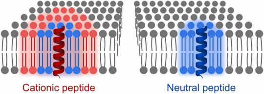
New publication out!
We were curious what happens with the lipid membrane if proteins are inside. As a model of membrane proteins we use transmembrane peptides with neutral and positive charge. The charges turn out to be quite crucial in the peptides influence on the lipids.
publication link: https://doi.org/10.1016/j.colsurfb.2024.113765…
All transmembrane peptides hinder mobility of lipids around. The positive charges in the peptide make this hindering a long range influence.
The membrane with the positive peptides then has heterogeneities in the lipid mobility. This hampers free rearrangement of lipids and leads to lower ability to seal ruptures. You can see below how the membrane stays fragmented after indentations with AFM (atomic force microscope), which is basically a very tiny tip that scans the surface of the membrane for images and can also push through it to test mechanical stability.
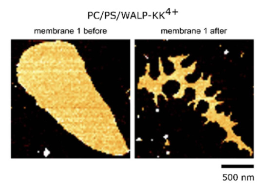
For comparison this is how it looks like with a membrane without any peptides. The mebrane seals the ruptures induced by the AFM tip and recovers its round shape. The same happens if we have a neutral, non-charged, peptide.
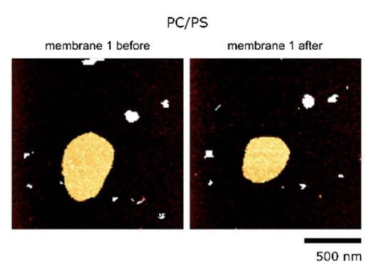
With this work we highlight the importance of including charges of proteins and peptides in membrane model studies.
And last but not least...
This is the very first time I am the last corresponding author of a publication. I am immensely proud on myself!
#science#women in science#research#biophysics#afm#fluorescence#simulations#peptides#proteins#cell membranes#publication#collaboration#corresponding author#proud#original content
16 notes
·
View notes
Text
Innovations and Techniques in Analytical Sciences
Analytical sciences are constantly evolving, driven by the need for more precise, efficient, and cost-effective methods to analyze and characterize substances across various industries. From pharmaceuticals to environmental monitoring, innovations in analytical techniques have revolutionized the way we understand materials, chemicals, and biological systems. This article explores the latest advancements in analytical sciences, highlighting innovative techniques that are shaping the future of research, development, and quality control.
The Growing Importance of Analytical Sciences
Analytical sciences are pivotal in multiple sectors, from healthcare to manufacturing, as they provide the tools to measure, identify, and analyze chemical compositions with remarkable precision. With the increasing complexity of research and industry needs, analytical methods must continuously adapt to address challenges such as real-time monitoring, data volume, and miniaturization of tools for portable applications.
Recent breakthroughs in analytical techniques promise to enhance the speed, accuracy, and reliability of scientific discoveries, impacting everything from drug development to environmental conservation. Let’s explore some of the key innovations and cutting-edge techniques in analytical sciences.

Innovations in Analytical Techniques
1. High-Resolution Mass Spectrometry (HRMS)
Mass spectrometry (MS) is already a cornerstone of analytical chemistry, but high-resolution mass spectrometry (HRMS) has taken this technique to new heights. HRMS allows for the detection and characterization of ions with unparalleled accuracy and resolution, making it indispensable in complex molecular analyses, such as proteomics, metabolomics, and drug testing.
Benefits:
Enhanced sensitivity for detecting low-abundance compounds.
High resolution allows for the identification of structural isomers with minimal interference.
Essential for analyzing complex mixtures in pharmaceuticals and environmental testing.
2. Raman Spectroscopy for Real-Time Analysis
Raman spectroscopy is a non-destructive technique that provides rapid, real-time analysis of molecular structures. Recent advancements in Raman spectrometers have made them more portable and accessible for in-field applications. This innovation is crucial in industries like pharmaceuticals, food safety, and environmental monitoring, where timely data is essential.
Applications:
Pharmaceuticals: Verifying the chemical composition of drugs in real time during production.
Food: Detecting contaminants or adulterants without the need for complex sample preparation.
Environmental Science: Monitoring air and water quality by analyzing molecular fingerprints of pollutants.
3. Nano-Analytical Tools
Nanotechnology is expanding the boundaries of analytical sciences by providing tools that can analyze materials at the molecular and atomic scale. Techniques such as atomic force microscopy (AFM) and scanning tunneling microscopy (STM) allow scientists to directly observe surfaces, structures, and even individual atoms with incredible precision.
Benefits:
Detailed imaging and analysis of nanoparticles, which are essential in drug delivery and materials science.
Understanding surface properties at the atomic level, crucial for designing new materials and enhancing the performance of electronic devices.
Unlocking new possibilities in biomolecular research, including protein folding and molecular interactions.
4. Integration of Artificial Intelligence (AI) and Machine Learning (ML)
The integration of AI and machine learning (ML) in analytical sciences has opened new frontiers in data interpretation and analysis. With the ability to process vast datasets and recognize complex patterns, AI-driven approaches significantly improve the accuracy and efficiency of analytical techniques.
Applications:
Pharmaceuticals: Predicting drug interactions and identifying potential drug candidates using large chemical datasets.
Environmental Monitoring: Analyzing environmental data to detect subtle patterns in air and water quality that may indicate emerging risks.
Manufacturing: Predicting material failure or optimizing production processes based on data analytics.
AI and ML algorithms can also aid in automating tedious and repetitive tasks, reducing human error and speeding up analytical workflows.
5. Portable Analytical Devices
One of the most transformative innovations in analytical sciences is the development of portable devices that allow for in-field analysis. These handheld instruments are compact, easy to use, and capable of providing real-time results, making them invaluable in environmental, forensic, and pharmaceutical applications.
Examples:
Portable Raman Spectrometers: For field-based chemical analysis, especially in remote areas.
Handheld X-ray Fluorescence (XRF) Spectrometers: Used in geological surveys and material science to analyze elemental composition on-site.
Portable DNA Analyzers: For quick genetic analysis in medical diagnostics or wildlife research.
These devices enable researchers and professionals to make critical decisions without the need for laboratory settings, improving the speed and efficiency of data collection.
Future Trends in Analytical Sciences
1. Sustainability in Analytical Chemistry
As environmental concerns grow, the need for sustainable analytical practices has become more pressing. Innovations in green chemistry and eco-friendly analytical methods are now taking center stage. Techniques such as green solvents, energy-efficient instruments, and non-toxic reagents are increasingly being incorporated into laboratory practices to reduce waste and energy consumption.
Green Analytical Chemistry: Emphasizes the use of renewable materials and eco-friendly methodologies, helping to minimize environmental impact while maintaining analytical precision.
2. Miniaturization and Lab-on-a-Chip
The miniaturization of analytical instruments, especially in the form of lab-on-a-chip devices, promises to revolutionize fields like medical diagnostics and environmental monitoring. These micro-scale devices integrate multiple analytical functions on a single chip, making them portable, affordable, and capable of performing complex analyses quickly.
Applications:
Medical Diagnostics: Rapid point-of-care testing for disease markers and pathogens.
Environmental Monitoring: Real-time pollution detection and analysis in air and water systems.
3. 3D Imaging and Spectroscopy
3D imaging techniques are gaining popularity in analytical sciences, particularly in material science and biology. These methods, including 3D electron microscopy and 3D Raman spectroscopy, enable researchers to study samples in three dimensions, providing more detailed and accurate insights into structures at the molecular level.
Applications:
Drug Development: Mapping the structure of biological targets and evaluating how drug molecules interact with them.
Material Science: Investigating the properties of complex materials and composites in 3D, leading to better product designs.
Conclusion
Innovations in analytical sciences are continuously pushing the boundaries of what is possible in research and industry. From high-resolution mass spectrometry to AI-driven analysis, these advancements are revolutionizing how we approach complex scientific challenges. With growing demand for precision, efficiency, and sustainability, the future of analytical sciences promises even more transformative techniques and applications across diverse fields, including pharmaceuticals, environmental monitoring, and materials science. The ongoing developments in this field are not just improving existing processes but also creating new possibilities for scientific discovery and innovation.
1 note
·
View note
Text

Imaging advance creates clearer picture of organic solar cells' molecular structure
Research on organic solar cells has been conducted for a long time. Recent advancements in understanding their molecular structures are now paving the way for the development of highly efficient solar cells. "By using atomic force microscopy-infrared spectroscopy, AFM-IR, we've been able to create clearer images of the morphology or structure of the material," says Ishita Jalan, postdoc in physical chemistry and main author of a recently published article in ACS Applied Polymer Materials. This structure and how it can be controlled determines the effect of the solar cell.
Read more.
#Materials Science#Science#Solar power#Materials characterization#Structures#Atomic force microscopy#Polymers#Karlstad University
4 notes
·
View notes