#taphozous
Explore tagged Tumblr posts
Visit Tumblr Blog
Explore Tumblr blogs with no restrictions, modern design and the best experience.
Last Seen Tumblr Blogs


Fun Fact
Tumblr was created by web developers David Karp and Marco Arment.
Text

Black-Bearded Tomb Bat, photographed by Lars Petersson, (source)
#Black-Bearded Tomb Bat#taphozous melanopogon#bat#bats#cute bats#daily bats#bats of asia#batposting#chiroptera#microbat#microchiroptera#taphozous#tomb bat#emballonuridae#yangochiroptera
144 notes
·
View notes
Text
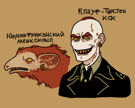
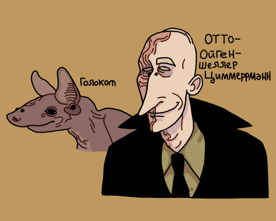
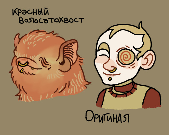
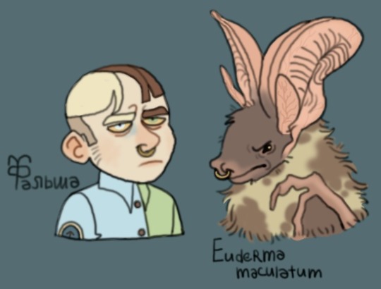
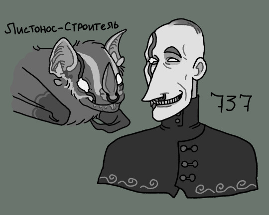
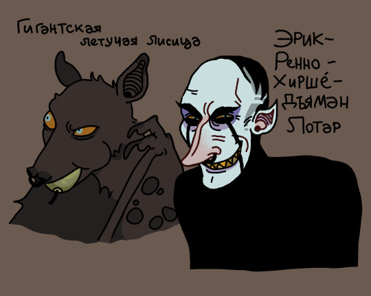
If my OCs were bats: Part 1
Taphozous mauritianus (Klauf Torsten Koch)
Cheiromeles torquatus (Otto-Eugern-Scherller Zimmerrmann)
Lasiurus borealis (Original)
Euderma maculatum (False Original)
Uroderma bilobatum (737)
Pteropus vampyrus (Erick-Renno-Hirshé-Djaman Lothar)
#my ocs#my art#shitpost#bat#bats#ocs#OC: Klauf Torsten Koch#OC: Otto-Eugern-Scherller Zimmerrmann#OC: Original#OC: False Original#OC: 737#OC: Erick-Renno-Hirshé-Djaman Lothar#art#animals#flying mice hehe#Taphozous mauritianus#Cheiromeles torquatus#Lasiurus borealis#Euderma maculatum#Uroderma bilobatum#Pteropus vampyrus
40 notes
·
View notes
Text

A Children's python (Antaresia childreni) eating a common sheath-tailed bat (Taphozous georgianus) in the Pilbara, Australia
by Jordan Vos
#childrens python#stimsons python#pythons#snakes#reptiles#predation#animal death#antaresia childreni#antaresia#pythonidae#serpentes#squamata#reptilia#chordata#wildlife: australia#wildlife: oceania
73 notes
·
View notes
Text
Bat Profile: Hildegarde's Tomb Bat

Taphozous hildegardeae
Source & Photo credit: Dr. Paul Webala
RANGE: Africa (coastal Tanzania and Kenya)
CONSERVATION STATUS: Endangered
HABITAT & DIET: These bats require coral caves to thrive, but there is much to learn about why exactly that is. They are insectivores who likely forage in mangroves as well as forests.
Fun Fact: Their namesake is the English linguist, Hildegarde Beatrice Hinde, who wrote multiple books about East African languages and the Maasai people. She also sent collections of bats and rodents to the British Museum. Not only is the Hildegarde's tomb bat named in her honor, but so are TWO rodent species (Hildegarde's shrew and Hildegarde's broad-headed mouse)
14 notes
·
View notes
Photo


My attempt to do a portrait of an ARPG critter I own for each day of October is so far going well! Almost done! Only about a week more to go!
First four are included in this post here. Links to their official in-game sheets below the cut, because there’s a lot of kids in here I have a hoarding OCs problem
Xaica and Joaquin are adoptive mom-dog and adopted crow-son and are on their way to crash a party
Cake and Mazin are siblings; Cake just wants to gift you twigs and stuff and Mazin is watching you warily
Aurelia just wants to sing you a very loud song
Finlay and Rufo are bitter rivals and are engaged in a fight at the moment
Wyn is soft and probably tripped on his own feet on his way to greet you
Wandering Soot made eye contact with you for a moment as you glimpsed her on a hike
Taphozous froze in place and looked alarmed when he noticed you’d seen him
Ghost Pepper won’t stop barking at you
Trudi came by to say hello
Machaeron passes by on his way to hunt mammoths
Sandokan enjoys the action-packed story you’re telling him
Sauvignon accepts no excuses
Rubeus hopes you’re gonna pet him
Plague is trying to be scary by showing off his 80-foot wingspan and setting houses on fire; It’s working
Wyandotte is being exceedingly vain today and is waiting (impatiently) for your expected compliment
Hanako is asking you for food today
Tam was coming up to say hi but then got distracted
Sifiso chirped at you in greeting as he raced by
Solstice is happy to help you carry your groceries today
#art#my art#ocs#arpg animals#tokotas#dracostryx#kukuri#xaica#joaquin#cake#mazin#aurelia#finlay#rufo#wyn#wandering soot#taphozous#ghost pepper#trudi#machaeron#sandokan#sauvignon#rubeus#plague#wyandotte#principe cioccolato wyandotte#hanako#sifiso#tam#solstice
9 notes
·
View notes
Text

Taphozous australis | John Gould (1804-1881) | The mammals of Australia. v.3 (1863) | Flickr (Biodiversity Heritage Library)
0 notes
Text

Grzimek's Animal Life Encyclopedia, vol. 11, Mammals II. 1972.
1.) Greater mouse-tailed bat (Rhinopoma microphyllum)
2.) Balantiopteryx sp.
3.) Coleura sp.
4.) Emballonura sp.
5.) Taphozous sp.
6.) Greater sac-winged bat (Saccopteryx bilineata)
351 notes
·
View notes
Text
Год пандемии COVID-19 и почему мы сами ответственны за неё
Сегодня мы можем “отметить” годовщину с официального начала пандемии COVID-19, когда гендиректор ВОЗ Тедрос Аданом Гебреисус объявил об этом на пресс-конференции в Женеве 11.03.2020г.
Мы пришли к выводу, что ситуацию с COVID-19 можно охарактеризовать как пандемию..И мы крайне обеспокоены как пугающими темпами распространения вируса, так и пугающим бездействием... В ближайшие дни и недели мы ожидаем, что количество случаев заражения COVID-19, количество вызванных им смертей и количество охваченных вспышкой заболевания стран продолжат увеличиваться...

Я специально не писал в течении года об этом заболевании, т.к. очень многие моменты были специально раздуты или, наоборот, скрывались от общественности, тогда как полезной информации о свойствах возбудителя и путях его передачи было мало. Пробовались различные схемы лечения, из запасников извлекались давно позабытые препараты, которые могли потенциально оказать положительный эффект, что в дальнейшем во многих случаях не оправдалось и, наконец, на рынок вышло множество вакцин с различным механизмом действия, которые мы фактически тестируем на огромной популяции добровольцев и ждём, что из этого выйдет. Медлить нельзя, но и спешка может оказаться фатальной, ведь на кону стоят наши жизни.
В этой статье я буду в основном писать об истоках. Ничего не меняется. Наше потребительское отношение к природе приносит свои плоды, и, к сожалению, они поражены порчей.
Как известно, это вирусное заболевание относится к зоонозным инфекциям, т.е. передаваемым от животных к человеку. Животные служат естественным резервуаром (источником) возбудителей, а человек может заразиться от животных, например, при близком контакте с ними, или употребляя в пищу их мясо, или при использовании предметов, изготовленных из кожи или шерсти больных животных. О некоторых зоонозах, таких как чума, бешенство, сибирская язва, мы с вами хорошо знаем, но большинство из них по-прежнему остаются неизученными достаточно. А ведь вирусы и бактерии интенсивно мутируют. Откуда ждать следующего “удара”, мы можем только догадываться.
Переносят инфекции обычно представленные ниже животные
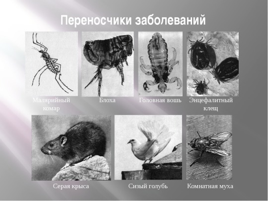
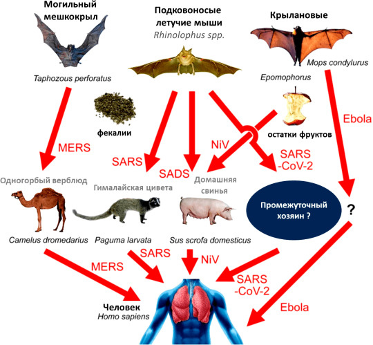
однако есть и более специфические варианты, как в случае с коронавирусом, - летучие мыши. И в данном случае именно большой подковонос из рода Rhinolophus. SARS-CoV-2 был впервые зарегистрирован осенью 2019 года в китайском Ухане (провинция Хубэй).
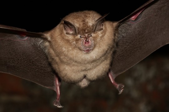
Ну не красавчик ли?
Ещё в 2013 г. было проведено исследование о сравнении обычных грызунов с летучими мышами в плане распространения вирусных инфекций. Используя обширную базу данных, авторы постарались учесть множество факторов в пределах каждой группы переносчиков вирусов, в частности процент видов, имеющих перекрывающиеся ареалы, размер и плотность колоний, длительность жизненных циклов, массу тела, количество приплода в год, продолжительность спячки и степень миграции. Итог работы: летучие мыши обыграли своих “коллег” как по количеству (и опасности) переносимых возбудителей, так и по потенциальным возможностям в их распространении. Но есть одно “но”: шанс встретиться с грызунами у человека намного выше, чем с тем же подковоносом. Летучие мыши хоть и распространены в мире, но их колонии расположены в пещерах, вдали от мест обитания людей. По крайней мере, так оно и было до недавнего времени.
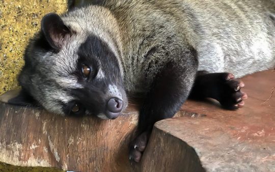
Вирус SARS-Cov-2, возбудитель COVID-19, на 96% идентичен вирусу подковоносых летучих мышей, однако эволюционно отделился от него еще 40-70 лет назад, иными словами, если коронавирус и произошел от летучих мышей, для того, чтобы вирус получил способность передаваться человеку, его строение должно было видоизмениться в организме какого-то другого животного, сыгравшего роль посредника. Вероятнее всего ими стали гималайские циветы (Paguma larvata). Идентичность белка-шипа S коронавируса гималайской циветы и SARS-CoV, выделенного у человека, составляет 98%, отчетливо указывая на то, что именно циветы были промежуточными хозяевами этого вируса.
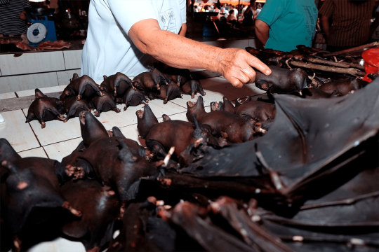
Этих зверьков используют в пищу, они продаются на влажных рынках и в качестве блюда подаются в ресторанах

Хотя и летучих мышей также активно употребляют в пищу
Кроме коронавирусов летучие мыши являются природным резервуаром для большого числа других опасных для человека вирусов. Вирус Эбола, относящийся к семейству филовирусов (Filoviridae), вызывает одноименную геморрагическую лихорадку у высших приматов и происхоит от ангольского складчатогуба (Mops condylurus), его промежуточный хозяин до сих пор неизвестен. Вирусы Хендра и Нипах, относящиеся к роду Henipavirus, перешли к нам от представителей семейства крылановых (Pteropodidae). Вирус Хендра поражает людей и лошадей, вызывая кровоизлияния, отек легких и в тяжелых случаях вирусный менингит, а вирус Нипах у людей приводит к респираторным заболеваниям и нередко тяжелому воспалению головного мозга (энцефалиту). Вирус, вызывающий ближневосточный респираторный синдром (MERS), пришел от представителя футлярохвостых летучих мышей, предположительно от могильного мешкокрыла (Taphozous perforatus). Здесь промежуточный хозяин - одногорбый верблюд, дромадер (Camelus dromedarius).
Благодаря адаптивным особенностям своего иммунитета, в подробности функционирования которого мы с вами не будем вдаваться в этой статье, летучие мыши стали хорошо приспособленными к патогенам, это настоящие передвижные лаборатории по культивированию возбудителей, и они представляют опасность как для других видов животных, так и для человека.
Однако истребление летучих мышей не спасет нас от инфекций. Напротив, массовое уничтожение и их изгнание из привычной среды обитания приведет к еще более тяжелым последствиям. Около 70% из 14 тысяч особей летучих мышей насекомоядны, а многие крылатые или ночные насекомые, поедаемые мышами, в свою очередь сами являются разносчиками болезней, к примеру, малярии или лихорадки денге. Летучие мыши также - активные опылители для цветов.
Именно вторжение человека в естественную среду обитания летучих мышей, их появление в больших городах, распространение их продуктов жизнедеятельности рядом с людьми, привело к тому, что мы имеем на данный момент - вспышку нового заболевания, буквально парализовавшего мировую экономику. Надеюсь, что после этой небольшой статьи, вы пришли к пониманию того, что эта тенденция продолжится, если мы не внесём коррективы в свой образ жизни.
Спасибо за внимание! Буду благодарен за репост ;)
20 notes
·
View notes
Photo

🦇Bat Fact! Do you know of the Coastal Sheathtail Bat (Taphozous australis)? First described in a paper in 1854, this insectivorous microbat can be found in Australia and Papua New Guinea. This bat is also known as the “Coastal Tomb Bat” and the “Southern Sheathtail Bat.” Habitat usually consists of coastal dune scrublands and heathlands, open forests, grasslands, swamps and more. This bat eats mostly beetles and brings their food back to their roost site to eat, roosts usually being in caves, cracks, under rock ledges and piles of rocks. This bat will also share large roosts with other bat species such as Troughton's Sheathtaill Bat and Beccari's Freetail Bat. This bat is listed as “Near Threatened” by the IUCN🦇
📸Photo by G.B. Baker/Australian Museum📸
#batfacts #bats #bat #akhyls #education
⬇️Follow Bat Facts⬇️ https://akhylsthebat.tumblr.com/ https://www.minds.com/akhylsthebat/ https://twitter.com/AkhylsBatFacts https://t.me/AkhylsBatFacts https://www.facebook.com/groups/137858924078846/ Disclaimer: All images used here are for educational purposes and are not used in any way for profit or to promote any products or services. Copyright Disclaimer under section 107 of the Copyright Act 1976, allowance is made for “fair use” for purposes such as criticism, comment, news reporting, teaching, scholarship, education and research. Fair use is a use permitted by copyright statute that might otherwise be infringing
3 notes
·
View notes
Text
Fine structure of Somatotrophs in Pars Distalis of the Indian wild Caught Female Bat, Taphozous nudiventris kachhensis (Dobson)

Fine structure of Somatotrophs in Pars Distalis of the Indian wild Caught Female Bat, Taphozous nudiventris kachhensis (Dobson) by Pankaj R Chavhan in Biomedical Journal of Scientific & Technical Research (BJSTR) https://biomedres.us/fulltexts/BJSTR.MS.ID.000227.php
0 notes
Text
Biomed Grid | Histological and Ultra Structure Observations of the Adrenal Gland of Fruit Eating Bat (Rousettus Aegyptiacus)
Introduction
As general in all mammals, adrenal gland is composed of two distinct portions: an outer cortex and an inner medulla [1- 5] paired right and left flat adrenal glands located to the craniomedial of the kidneys were observed in porcupine (Hystrixcristata). In human, the adrenal glands (suprarenal glands) associated with the kidneys. A gland present top on each kidney like a cap and is embedded in the mass of adipose tissue that encloses the kidney. The adrenal glands are shaped like pyramids; each gland is very vascular and consists of two parts. The central portion is the adrenal medulla and the outer part is the adrenal cortex [6]. Adrenal cortex is subdivided into three zones; the outermost zona glomerulosa that contained cells formed cords and contained lipid vacuoles, the middle thickness and palest zona fasciculata formed of large polyhedral spongiocytes which arranged in longitudinal straight cords separated by straight blood capillaries and the last third narrow zona reticularis formed of small polyhedral deeply stained cells arranged in branching anastmosing cords separated by irregular blood capillaries [7]. Adrenal medulla formed of chromaffin cells that are large polygonal and arranged either in groups around blood vessels or in branching cords. These cells secrete catecholamines (adrenaline and nor-adrenaline). One pair of adrenal glands was found in the Emballonurid bat, Taphozous longimanus against the lateral kidney, left one was pyramidal, but the right was oval. The gland has two layers, inner medulla and outer cortex which have zona glomerulosa and zona fasciculata while the third zone zona reticularis is absent [8]. Adrenal gland considered as a key organ in bat because it plays an important role in metabolism and homeostasis of animals, it protects the organism against acute and chronic stress; Catecholamine’s of the medulla mobilize glucose and fatty acids for energy, muscles and lung for action in acute actress. There is a little information available on anatomy of adrenal gland of Egyptian bats. The present report reveals the anatomy of adrenal glands in Rousettus eagyptiacus.
Materials-and-Methods
The specimen used was females non pregnant adult Frugivorous bat Rousettus eagyptiacus. Each animal was euthanized by chloroform, dissected and the adrenal gland go out, thin fixed with 10% formalin for general histology and stained with Hematoxylin and eosin stain [9].
Transmission Electron Microscopy:
For transmission electron microscope, small pieces of 1 x 1 x 1 mm of pituitary gland of bats were obtained and were rapidly processed as follow: -
a. Fixation:
Small pieces of fresh specimens were fixed in a mixture of Formaldehyde / Glutraldehyde (4:1) at PH 7.4 at room temperature for 4 hours, and then rinsed twice in 0.1 M phosphate buffer (15 minutes for each).
b. Post-Fixation Treatment:
Specimens were post fixed in 2.0% buffered Osmic acid for half an hour at 4˚C. The tissue specimens were then washed twice in phosphate buffer for 30 min.
c. Dehydration:
The tissues were dehydrated in ascending grades of ethyl alcohol (50, 70, 80, 90 and 100%) for two changes each of 15 minutes (2 x 15 minutes) in each grade. Specimens then cleared in propylene oxide for 2 changes 5 min, each.
d. Infiltration:
Dehydrated tissues were initiated in 1: 1 solution of propylene oxide and epon mixture. Infiltration was continued with 1: 3 (propylene oxide: epon mixture) overnight at room temperature.
e. Embedding:
Embedding was carried out using freshly prepared araldite epon mixture in capsules pre-dried for 1-3 hours.
f. Polymerization:
The capsules were polymerized at 60˚C and then the polymerized capsules were cured at room temperature for at least a day before attempting to section.
g. Sectioning and Staining of Semithin Sections for Light Microscopy:
Blocks were trimmed under the binocular microscope of ultra-cut Reichert jung ultramicrotome. Semithin sections of 1 μm thickness were obtained with the aid of glass knives made on Leica EM KMR (Knife marker). Semi-thin sections were stained by toludine blue and examined for general orientation with the light microscope.
h. Sectioning and Staining of Ultrathin Sections for Electron Microscopy:
Specimens were then retrimmed to the selected region and ultra-thin section 60 nm thickness were cut and picked up on copper grids. Sections mounted on grids were double stained using uranyl acetate and lead citrate. Seven percent uranyl acetate solution in methyl alcohol was prepared and centrifuged at 5000 rpm for 10 minutes. A drop of uranyl acetate solution was placed on a dental wax sheet placed in a Petri-dish. Grids were then placed on the uranyl acetate drop (the surface grids loaded with section being facing the stain). Staining was done in the dark for about 20 mins. Grids were washed in three successive glass bottles containing distilled water. After the last bath, grids were dried on a filter paper. Sections were then stained with freshly prepared lead citrate that centrifuged at 5000 rpm for 10 minutes and used for staining for 10 minutes as in uranyl acetate, then washed with 0.02 N Noah and finally with freshly distilled water. The grids dried on filter paper and examined with electron microscope (TEM Philips 400 T at 80 Kv). Photos were made on Kodak EM sheet films; developed then enlarged and printed and investigated.
ResultsThe Histological Structure
In R. aegyptiacus the pair glands appear embedded at the lateral surface of each kidney covered by thick connective tissue capsule. It is oval and has two zones an outer cortex and an inner medulla (plate: 1 Figure 1 & 2 ). The thickness of cortex surrounding the medulla different from region to region, so the gland oval. The cortex divided into three major zones; zona glomerulosa, zona fasciculata and zona reticularis (plate: 1 Figure 3 & 4 ). The medulla is present centrally and surrounded by cortex.
Zona Glomerulosa
Zona glomerulosa is the smallest zone of the adrenal cortex present beneath the capsule. This zone consists of irregular glomerular cells arranged in rods and generally found in group of cells. These cells have darkly stained nucleus, either centrally or eccentrically placed. The cytoplasm takes acidic stain and eosinophilic in nature. Numbers of lipid vacuoles are observed in the cytoplasmic matrix and blood capillaries are also observed between the clusters of acini. Zona glomerulosa is compactly associated with zona fasciculata and hence no identifying separation is observed in between these two zones (plate: 1 Figure 1, 2).
Zona Fasciculata
It is an intermediate zone present beneath the zona glomerulosa and is the largest zone of the cortex present between the zona glomerulosa and zona reticularis. This zone consists of small columns of single or double rows. These cells are cubical and low columnar types with large numbers of lipid vacuoles. Nucleus is centrally situated, spherical in shape, darkly stained with welldeveloped nuclear membrane. Each cell shows spongy cytoplasm due to the presence of large number of lipid vacuoles (plate: 1 Figure 1, 2).
Zona Reticularis
It is the innermost zone of the cortex without any clear demarcation. The zona reticularis is present below the zona fasciculata and above the medulla. This zone possesses irregular cells with lightly stained cytoplasm and vesicular nuclei. The cytoplasm shows presence of few lipid vacuoles. Few blood capillaries are also observed in this zone (plate: 1 Figure 1, 2).
Medulla
Medulla is the innermost region of the adrenal gland. The layer has two types of cells epinepheric and non epinepheric cells (plate:1 continue Figure 6, 7 & 8 ).
The Fine StructureThe Zona Glomerulosa
The cells have circular or oval nuclei bounded by a plasma membrane of uniform thickness. The lipid droplets are few from secreted type and large numbers from vacuolated types. These droplets are variable in size, mainly round to ovoid in shape. Mitochondria are many, round, some of them hypertrophy. The smooth endoplasmic reticulum is in the form of small vesicles scattered throughout the cytoplasm. (Plate: 2 Figure 1-6 ).
The Zona Fasciculate
This layer constitutes the major portion of the adrenal cortex. These cells are large and arranged in radial cords, one or two cells thick and lie between vascular channels which converge towards the medulla. Mitochondria are Present. The cells of zona fasciculata contain a greater number of lipid droplets than those observed in the cells of glomerulosa. These droplets from secreted type and large numbers from vacuolated types. Smooth endoplasmic reticulum in the form of vesicles scattered throughout the cytoplasm (Plate: 3 Figure 1-6). In contrast to the zona glomeruolosa the cells of the zona fasciculata are rich in smooth endoplasmic reticulum. Free ribosomes are present in the cytoplasm. The cells of fasciculata contain large dense bodies (lysosomal bodies) not observed in the cells of glomerulosa.
The Zona Reticulosa
This layer constitutes the last of the adrenal cortex. These cells are large, one or two cells thick and lie between vascular channels which converge towards the medulla. Mitochondria are Present small in numbers than zona fasciculata. The cells contain a greater number of dark lipid droplets than those observed in the cells of zona fasciculata. The nucleus small and spherical. Large number of mitochondria present. Smooth endoplasmic reticulum in the form of vesicles scattered throughout the cytoplasm (Plate: 4 Figure 1-6 ).
The Medulla
The, medulla consists of chromaffin cells arranged in small groups or short cords surrounded by blood capillaries and connective tissue. Some chromaffin cells have dense and large secretary granules and others have small. The nucleus small with nucleous. Mitochondia are round scattered throughout the cytoplasm and smooth endoplasmic reticulum present. The distinctive features of modularly cells are the chromaffin granules which appear as a membrane bound body of variable electron density. Some cells contain predominantly dense black granules called epinephrine cells, while others show scattered empty vesicles with small amounts of granular material lining to the inner layer or present in the lumen of the vesicles called norepinephrine cells (Plate: 5 & 6 Figure 1-6 ).
Explanation of Plates
Plate: -1 (Figure 1 & 1(Continues))
Figure 1: The structure of adrenal gland of adult Rousettus Aegyptiacus. Figures 1a, 2a: Light micrographs of transverse section of adrenal gland shows the presence of capsule (ca) covering the gland, outer cortex (Ac) and inner medulla (AM) with Blood sinusoids (Bs) H&E X 100. Figures 3a-5a-6a-7a- 8a: Light micrographs of transverse section of adrenal gland shows the outermost capsule (CP). Note the cortex differentiated into outer small zone, zona glomerulosa (ZG), middle long cell cords of zona fasciculate (ZF) and inner zona reticularis (ZR), (L) lipid vacuoles, (N) nucleus H&E X 400. Figure 4a: Light micrograph of transverse section of adrenal gland shows the epinephrine cell of Medulla (E) and non-epinephrine (NE) H&E X 400.
Plate: -2 (Figure 2)
Figure 2:Ultrastructure of Zona glomerulosa of adult Rousettus Aegyptiacus. Figure 1b: Electron micrograph of the cell of zona glomerulosa showing (N) nucleus, (M) mitochondria, (SL) saturated lipids, (V) Vacuoles, (R) ribosomes. Figure 2b: Electron micrograph of the cell of zona glomerulosa showing (N) nucleus, (M) mitochondria, (HM) hypertrophy mitochondria, (SL) saturated lipids, (V) Vacuoles, (R) ribosome’s, (PM) plasma membrane. Figures 3b, 4b: Electron micrographs of the cells of zona glomerulosa showing enormous lipids droplets (L), (N) nucleus, (M) mitochondria and (HM) hypertrophied mitochondria, (SL) saturated lipids, (V) Vacuoles, (R) ribosome’s and (PM) plasma membrane.
Plate: -3 (Figure 3)
Figure 3:Ultrastructure of Zona glomerulosa of Rousettus Aegyptiacus. Figures.1c-2c-3c-4c-5c-6c: Electron micrographs of the cells of zona fasciculata showing enormous lipids droplets (L) and lipid vacuoles(LV), (SER) smooth endoplasmic reticulum, (N) nucleus, (M) mitochondria, (SL) saturated lipids, (V) Vacuoles, (R) ribosome’s, (PM) plasma membrane, (L) lysosomes.
Plate: -4 (Figure 4)
Figure 4:Ultrastructure of Zona reticularis of adult Rousettus Aegyptiacus. Figures.1d-2d-3d- 4d: Electron micrographs of the cells of zona reticulosa showing enormous lipids droplets ( L) and saturated droplet (SL), (v) vacuoles, (SER) smooth endoplasmic reticulum, (N) nucleus with nucleolus (Nu), (M) mitochondria, (V) Vacuoles, (R) ribosome’s, (PM) plasma membrane, (L) lysosomes and (HC) heterochromatin.
Plate: -5 (Figure 5)
Figure 5: Ultrastructure of Norepinephrine cells of adult Rousettus Aegyptiacus. Figures.1e-2e-3e-4e: Electron micrographs of the cells of modularly cell showing nor Epinephrine (SG) granules, rough endoplasmic reticulum (RER), ( SER) smooth endoplasmic reticulum, small and large secretary granules, (N) nucleus with nucleolus (Nu), (L) lipid vacuoles and (LY) lysosomes
(Figure 6)
Figure 6:Ultrastructure of Epinephrine cells of adult Rousettus Aegyptiacus. Figure 1f: Electron micrograph of modularly cells showing dark Epinephrine and light non-Epinephrine cells (SG) granules, small and large secretary granules (SG). Figures 2f-3f: Electron micrographs of the cells of modularly cell showing Epinephrine cells (SG) granules, rough endoplasmic reticulum (RER), (SER) smooth endoplasmic reticulum, small and large secretary granules (N) nucleus, (L) lipid vacuoles, (LY) lysosomes and (HM) hypertrophied mitochondria.
Discussion
The study aims to describe the histological and the ultrastructure of the adrenal gland of Rousettus aegyptiacus. The result showed that the left and right gland oval different from Emballonurid bat, Taphozous longimanus in which the left adrenal gland is pyramidal, and the right adrenal gland is oval [8]. Adrenal gland divided into cortex and medulla, the cortex surrounding the medulla in different thickness as same to other species of mammals. According to [10], the duck cortex constitutes 68.2%, the medulla 28.6% and vascular space 3.2% of the total area. The adrenal cortex is divided into three distinct zones, zona glomerulosa, zona fasciculata and zona reticularis. A distinct zone of the cortex is observed in M schereibersii [11], M lyra lyra [12], V pipistrellus [9], P giganteus giganteus and R leschenaultia [13], Cynopterus sphinx, H lankadiva [14] and P giganteus giganteus [15]. In the present species, zona reticulosa is distinct but in some species, it is present in the form of islets of cortical cells in the medullary region in T melanopogon [16]. In the present study, zona glomerulosa consists of polyhedral glomerular cells which appear group of 3-7 acini or cells. Similar structure is also observed in the zona glomerulosa of Taphozous Kachhensis [17] in T longimanus, Hipposideros lankadiva [18]. In Cynopterus sphinx the cells are arranged in groups. Whereas, in Rousettus leschenaulti and Pteropus giganteus the cells are arranging in columns [19] and P giganteus giganteus [20]. The ultrastructural characteristics of the cells of zona glomerulosa of present bat adrenal gland show low numbers of vacuolated lipids and secreted lipids, circular and small nucleus. In Taphozous longimanus [21] observed some striking differences during estrus and pregnancy; Golgi apparatus is inconspicuous during estrus but well developed during late pregnancy, mitochondria show vesicular cristae during estrus and vesicular and lamellar cristae during pregnancy, moderate to high amounts of tubular profiles of smooth endoplasmic reticulum is seen during estrus. However, there is extensive development of tubular profiles of smooth endoplasmic reticulum during pregnancy. A few lipid droplets are observed in the cytoplasm during estrus, but large numbers of lipid droplets of various size and electron density are seen in the cytoplasm during pregnancy. Similar observations are reported in the glomerulosa cells of bat, H lankadiva during estrus and pregnancy [22] and during hibernation and arousing of dormouse, Muscardinus avellanarius [23] supporting present observations. Ultra-structure of the zona fasciculata of present animal characterized by secreted lipid droplet and sooth endoplasmic reticulum but in bat, T longimanus shows presence of vesicular smooth endoplasmic reticulum, vacuolated lipid droplet [21]. The modularly cells of the present animal characterized by large numbers of dens large secretary granules and dense small secretary granules, rough and smooth endoplasmic reticulum in both nepherin and epinepherin cells. Different from the case of T longimanus, the medullary cells are characterized by the presence of well-developed Golgi apparatus, rough endoplasmic reticulum, large number of mitochondria and chromaffin granules and similar in rat shows two types of chromaffin cells as described in rat medulla [24]. Nor epinephrine cells are predominant in the medulla of pregnant bat while epinephrine cells are predominant in the medulla of estrus bat. The present observations suggest that two cell types are present in bat species like that observed in mammals [25-28].

Read More About this Article: https://biomedgrid.com/fulltext/volume5/histological-and-ultra-structure-observations-of-the-adrenal-gland-of-fruit-eating-bat.000961.php
For more about: Journals on Biomedical Science :Biomed Grid | Current Issue
0 notes
Link
0 notes
Photo

The naked-rumped tomb bat (Taphozous nudiventris) is a species of sac-winged bat in the family Emballonuridae. Found in northern Africa, the Middle East, and southeastern Asia, its natural habitats are dry savanna, subtropical or tropical dry shrubland and forests, caves, and arid areas.
https://en.wikipedia.org/wiki/Naked-rumped_tomb_bat
0 notes
Text
अब चमगादड़ों में मिला कोरोनावायरस
कोरोनावायरस इंसानों तक कैसे पहुंचा, इसका पता लगाने के लिए दुनिया भर के वैज्ञानिक जुटे हुए हैं। इस बीच भारतीय चिकित्सा अनुसंधान परिषद (आईसीएमआर) ने एक अध्ययन में कहा है कि भारतीय चमगादड़ों में रोगजनक कोरोनावायरस पाए जाने से उनमें नोवेल वायरस के लिए स्क्रीनिंग किए जाने की आवश्यकता रेखांकित होती है।
यह अध्ययन भारतीय चमगादड़ों में कोरोनावायरस परिसंचरण को समझने की दिशा में एक महत्वपूर्ण कदम है। इंडियन…
View On WordPress
0 notes
Text
Molecular detection of viruses in Kenyan bats and discovery of novel astroviruses, caliciviruses and rotaviruses
Abstract
This is the first country-wide surveillance of bat-borne viruses in Kenya spanning from 2012–2015 covering sites perceived to have medium to high level bat-human interaction. The objective of this surveillance study was to apply a non-invasive approach using fresh feces to detect viruses circulating within the diverse species of Kenyan bats. We screened for both DNA and RNA viruses; specifically, astroviruses (AstVs), adenoviruses (ADVs), caliciviruses (CalVs), coronaviruses (CoVs), flaviviruses, filoviruses, paramyxoviruses (PMVs), polyomaviruses (PYVs) and rotaviruses. We used family-specific primers, amplicon sequencing and further characterization by phylogenetic analysis. Except for filoviruses, eight virus families were detected with varying distributions and positive rates across the five regions (former provinces) studied. AstVs (12.83%), CoVs (3.97%), PMV (2.4%), ADV (2.26%), PYV (1.65%), CalVs (0.29%), rotavirus (0.19%) and flavivirus (0.19%). Novel CalVs were detected in Rousettus aegyptiacus and Mops condylurus while novel Rotavirus-A-related viruses were detected in Taphozous bats and R. aegyptiacus. The two Rotavirus A (RVA) strains detected were highly related to human strains with VP6 genotypes I2 and I16. Genotype I16 has previously been assigned to human RVA-strain B10 from Kenya only, which raises public health concern, particularly considering increased human-bat interaction. Additionally, 229E-like bat CoVs were detected in samples originating from Hipposideros bats roosting in sites with high human activity. Our findings confirm the presence of diverse viruses in Kenyan bats while providing extended knowledge on bat virus distribution. The detection of viruses highly related to human strains and hence of public health concern, underscores the importance of continuous surveillance.
http://ift.tt/2oYyg3t
0 notes
Photo
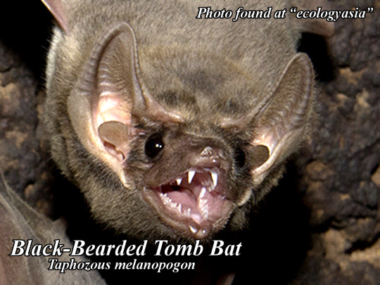
🦇Bat Fact! Do you know of the Black-Bearded Tomb Bat (Taphozous melanopogon)? This bat is named after the black fur under it’s chin, which appears in males around 5-6 months of age. This insectivorous microbat can be found in South and SE Asia from China to India, Sri Lanka to Thailand, to Malaysia and Vietnam and beyond! This bat is usually 9-10cm in size and can be found in colonies of up to 4000 (in Thailand)! This bat is listed as “Least Concern” by the IUCN🦇
#batfacts #bats #bat #akhyls #education
📸Photo found at “ecologyasia”📸
⬇️Follow Bat Facts⬇️ https://akhylsthebat.tumblr.com/ https://www.minds.com/akhylsthebat/ https://twitter.com/AkhylsBatFacts https://www.facebook.com/groups/137858924078846/ https://t.me/AkhylsBatFacts Disclaimer: All images used here are for educational purposes and are not used in any way for profit or to promote any products or services. Copyright Disclaimer under section 107 of the Copyright Act 1976, allowance is made for “fair use” for purposes such as criticism, comment, news reporting, teaching, scholarship, education and research. Fair use is a use permitted by copyright statute that might otherwise be infringing
3 notes
·
View notes