#pedunculate
Explore tagged Tumblr posts
Visit Tumblr Blog
Explore Tumblr blogs with no restrictions, modern design and the best experience.
Last Seen Tumblr Blogs


Fun Fact
Tumblr has 16.74 million mobile monthly users in the US.
Text
I should've taken a picture but I had one of these guys in my house today! The little guy got taken back outside.

(Source: BugGuide.net)
#Scarites subterraneus#big-headed ground beetle#pedunculate ground beetle#ground beetle#beetle#beetles#bugs#insects#bug#animals
28 notes
·
View notes
Text


No tree of the day today, because I want to spend some time researching oak galls. The top two pictures are of a sessile oak in Fife, and the bottom two are a pedunculate oak near Gretna Green. I don't know enough to know the different types of oak gall yet, but to my untutored eye I assume the growths in the top left and bottom right are both examples, and anyone who has more information, please let me know.
0 notes
Text


Mist and pedunculate oak forests
Photographed by Freddie Ardley
#photographers on tumblr#landscape#photography#artists on tumblr#travel#nature#art#photographer#beauty#forest#green#dark#horror#halloween#uk#fog#misty#spooky
2K notes
·
View notes
Text


Among the pedunculate oaks or English, common, European oaks (lat. Quércus róbur).
Среди дубов чере́шчатых или дубов обыкновенных, также называемых европейскими (лат. Quércus róbur).
#noseysilverfox#photography#nature#naturecore#autumn#atmospheric#green aesthetic#nature aesthetic#autumn aesthetic#trees#forest#forest photography#in the forest#love life#interesting places#tumblr#photographers on tumblr#фотографии природы#природа#фотоблог#лес#деревья#растения#флора#фотографии на тумблере#турумбочка#осеннее настроение#autumn mood#лесная эстетика#лесной пейзаж
142 notes
·
View notes
Text
Tiny book of bill thing. (Most of this information comes from the butterfly-conservation webpage)
Imagine Ford DOES show you his moth collection and he goes and gives you a fun fact on the ones he likes the most:
Hummingbird hawk moth: often found in a variety of habitats such as costal areas, gardens, urban areas and more.
Brimstone moth: often seen before dusk and regularly attracted to light. Have a wing span range of 28-42mm and main habitats are hedgerows, gardens, scrub and woodlands.
Bordered gothic moth: found throughout Europe/ Central Asia but two subspecies have been found in Britain; Marginosa (light brown) and Hibernica (darker grey colour) however both have pale veins and cross-lines.
Merveille du jour moth: light green in colour with black markings, some of which are edged in white. They feed on leaves and immature flowers on Pedunculate oak, turkey oak and sessile oak.
Peach blossom moth; adults fly at night and are attracted to light, in day they hide in ground cover. Their wing span ranges from 32-38mm
And here’s the thing; you don’t think it’s boring at all nor want to throw yourself over a bridge! Everything that Ford said has you hooked on wanting to know more about moths, much to Ford’s surprise, considering how the last person he shown his collection to (bill) wanted to throw himself off of a bridge.
‘You don’t find this boring?’ He’d ask you.
‘No, not at all and besides the gleam in your eye when you talk about something you love only makes this spot more special to you.’ You tell him. ‘I want to care about the things you care about Ford. I might not be as smart as you, I still want to show that I care about you regardless.’ You add.
Poor Ford has never hugged you tighter than that day. (I just want to listen to him talk about his moth collection. IS THAT TOO MUCH TO ASK!? MOVE ASIDE BILL I’LL TREAT FORD LIKE A PRINCESS!!!)
#gravity falls x reader#gravity falls imagine#gravity falls imagines#gravity falls#stanford pines x you#stanford pines imagines#stanford pines imagine#stanford pines x reader#ford pines x you#ford pines imagines#ford pines imagine#ford pines x reader#the book of bill
314 notes
·
View notes
Text

Quercus robur — English oak a.k.a. pedunculate oak
10 notes
·
View notes
Text
Yet More shenanigans of Mer!AU
Screw it, tentacles on the back, note tentacles not arms- cephalopod definition of arms and tentacles here with suckers only on parts of the limb and pedunculated for tentacles
Now, on to Scott’s new animal obsession.
Scott’s genuinely interested in critters but is also trying to make a niche for himself that covers for his declining mobility. He sees the writing on the wall at this point, he’s becoming a mer. After Submerion and the interaction with the Tainted Mass he’s started growing scales on his torso and he’s so thirsty all the time and his tail nub is getting harder and harder to hide. He’s not going to be able to keep his changes quiet for much longer; and even though he trusts some of the other pirates on the Isle, he knows he’s a target. The animals help him keep doing his missions and offer a deterrent to would be Scott- nappers. It’s good to have friends that don’t care how much Mer and mer body parts sell for and don’t mind scales.
27 notes
·
View notes
Text
Giant Cell Tumor of the Infrapatellar Fat Pad of the Knee: A Case Report by Ahmad Jiblawi in Journal of Clinical Case Reports Medical Images and Health Sciences
Abstract
Giant Cell Tumor is a rare benign soft tissue tumor occurring in two forms: localized and diffuse. The two subtypes differ in their location at presentation, shape, recurrence after treatment and prognosis. MRI is still essential in the diagnosis, however pathology remains the gold standard for the final diagnosis. In this article, we report a case of Giant Cell Tumor involving a very rare location with very few reports in the literature; the infrapatellar (Hoffa’s) fat pad of the knee. We discuss its keen clinical and radiological features. The tumor was managed with arthroscopic resection. Confirmation of the diagnosis was done by pathology. Our case is the first to be reported in Lebanon.
Keywords: GCT; Hoffa’s fat pad; STIR
Introduction
First described by Chassaignac in 1852, Giant Cell Tumor (GCT) is a benign soft tissue tumor [1]. It is a rare disease, associated with synovial inflammation due to hemosiderin deposition. GCT occurs in two forms: localized GCT and diffuse formerly known as pigmented villonodular synovitis. The former typically consists of small well circumscribed, nodule or pedunculated mass that might be intra- or extra-articular, most commonly (85%) in the small joints (ex: hands and feet) while the latter is typically intra-articular with an infiltrative growth pattern commonly occurring in large joints (ex: ankles and knees) [2–4]. Both share similar histologic features; however they have different biological behavior, treatment outcome and prognosis. Thus the importance of differentiating between the two entities [5,6].
MRI is considered essential for the diagnosis, staging, preoperative planning and clinical follow-up of GCT. The mass appears of iso/low signal intensity on T1 and T2 weighted images. In addition to joint effusion and synovial proliferation. Some “blooming” artifact of low signal might be noted on echo-gradient because of the magnetic susceptibility from hemosiderin deposition [1,2].
In this article, we report the first case in Lebanon (to our best knowledge) of a rare, localized Giant Cell Tumor originating in the infrapatellar (Hoffa’s) fat pad, emphasizing on its radiologic manifestation.
Case report
We report a case of a 35-year-old gentleman, previously healthy, complaining of a 4-month history of recurrent and painful left knee locking. The patient denies any trauma, any recent surgery, no accompanying systemic symptoms as of fever, rash, diffuse arthralgia, or myopathy. His presentation was mimicking that of a meniscal tear injury.
An MRI of the left knee was performed using 1.5 Tesla Philips Ingenia Unit, manufactured in the Netherlands. The following planes and sequences: A sagittal T1 weighted (T1W), proton density (PD) and STIR image, a coronal STIR and an axial STIR image. Result showed the presence of a soft tissue-like lesion arising directly anterior to the anterior cruciate ligament in between both femoral condyles estimated to be 3 cm in its transverse diameter, 2.7 cm in its antero-posterior diameter and 1.2 cm in its supero-inferior diameter. The lesion showed iso-intensity to the cartilage on T1W as well as on PD but showed an increase signal intensity on STIR weighted images. The lesion relaxes directly on the ACL posteriorly which is of adequate continuity and signal. Minimal associated excess of joint fluid filling the supra-patellar bursa. Both menisci, anterior cruciate ligament, posterior cruciate ligament and medial and lateral collateral were normal. No capsule-meniscal separation is seen. The overall radiologic impression was for a Cyclops lesion or a soft tissue tumor such as Giant Cell Tumor.
The patient underwent an arthroscopic excision of the soft tissue tumor. Procedure went uneventful. The tissue was sent to pathology. Microscopic examination showed fragments of fibrous tissue involved by sheets of fibro-elastic to epithelioid cells with band nuclei and moderately abundant cytoplasm. They are intermixed with osteoclast-like giant cells and foamy histiocytes. There was no evidence of malignancy. Findings suggestive of Giant Cell Tumor of the Tendon Sheath. Unfortunately, the patient was lost to follow up, thus recurrence could not be reassessed.
AT1 weighted image, sagittal plane: showing a soft tissue-like lesion iso-intense to the cartilage measuring 2.7 cm in its antero-posterior diameter relaxing directly on the anterior cruciate ligament posteriorly which is of adequate continuity and signal B: Proton density weighted image, sagittal plane: showing a soft tissue-like lesion iso-intense to the cartilage measuring 2.7 cm in its antero-posterior diameter relaxing directly on the anterior cruciate ligament posteriorly which is of adequate continuity and signal.
C: Short T1-Inversion Recovery weighted image, sagittal plane: showing a hyperintense soft tissue-like lesion measuring 2.7 cm in its antero-posterior diameter. D: Short T1-Inversion Recovery weighted image, coronal plane: showing a hyperintense soft tissue-like lesion measuring 1.2 cm in its supero-inferior diameter. E: Short T1-Inversion Recovery weighted image, transverse plane: showing a hyperintense soft tissue-like lesion measuring 3 cm in its transverse plane.
Discussion
Giant Cell Tumor is a rare benign soft tissue tumor arising from the synovial tissue of the joints, tendon sheath, mucosal bursas, and fibrous tissues adjacent to tendons. Multiple terms are found in the literature to describe this entity; pigmented nodular tenosynovitis, fibrous xanthoma of synovium, benign synovioma, xanthogranuloma and tenosynovial giant cell tumor [1]. Etiology and histiogenesis of which is not completely understood, but many risk factors were mentioned in the literature such as trauma, infection, vascular abnormalities, lipid metabolism disorders, osteoclastic proliferation, and immune system disorders. It can present in two forms: localized and diffuse [3,7]. Localized GCT presents mainly in small joints (85 % observed in fingers while 12% is observed in large joints, GCT in the knee is rare) [4], either intra-articular or extra-articular. Diffuse form occurs mainly in the extra-articular space [8]. However, extra synovial soft tissue forms of localized GCT are very rare and mainly concern the knee joint. Around 50% of patients with a localized GCT arising primarily within the infrapatellar fat pad have a history of trauma but the exact etiology is still unknown [9]. The onset age of localized GCT is older than that of the diffuse type (i.e. localized type usually occurs above 40 years of age)[10]. When affected, patient presents clinically with mechanical derangements, progressively worsening over time. Meniscal symptoms and locking are often present within the knee joint. The main symptoms are swelling (86%), pain (82%), stiffness (73%), limited range of motion (64%) and joint instability (64%) [7,10].
MRI is an effective and highly sensitive diagnostic tool; however pathology is still the gold standard of final diagnosis. On T1 and T2 weighted images, dense collagen and hemosiderin presents with homogenous low or intermediate signal. The most typical feature of a localized GCT is a well circumscribed, nodular mass with low signal intensity on T1, T2 and proton weighted images and high signal intensity on STIR images [4,6,9,10]. Microscopically, GCT is characterized by multinucleated giant cell, lipid-laden macrophages, hemosiderin deposition and fibroblast proliferation [5].
Various pathological conditions should be considered in the differential diagnosis, for example: Synovial Chondromatosis, Cyclops lesion, Rhabdomyosarcoma, Fibroma of tendon sheath, Synovial Sarcoma, Amyloid Arthropathy, Haemophilic Arthropathy, Lipoma Arborescens and Rheumatoid Arthritis [6,9].
The ability to differentiate between the diffuse and localized forms of GCT is paramount to give patients a realistic outlook on future prognosis, chance of recurrence and optimal treatment course [5]. Several treatment options are present: surgery, radiotherapy, pharmacology or a combined solution of the listed methods. Important to note, local recurrence after treatment was reported in 18-46% of cases. However, this might be linked to incomplete resection of satellite nodules in the area of initial change. Other risk factors for recurrence are the location of the disease (more common in the knee), history of previous surgeries and positive surgical margins.
Conclusion
To the best of our knowledge, our case is the first to be reported in Lebanon. It is very rare to have a localized GCT in the extra-synovial infrapatellar (Hoffa’s) fat pad of the knee. The rarity of the presented case suggests that GCT should be considered in the differential diagnosis of a painful knee locking in a young patient. Accurate diagnosis will lead to successful treatment associated with low recurrence rate resulting in a better patient outcome.
Conflict of Interest:
The authors declared no conflicts of interest with respect to the authorship and/or publication of this article
#GCT#Hoffa’s fat pad#STIR#JCRMHS#Journal of Clinical Case Reports Medical Images and Health Sciences (JCRMHS)| ISSN: 2832-1286#Clinical Images journal#Is Journal of Clinical Case Reports Medical Images and Health Sciences PubMed indexed
2 notes
·
View notes
Text

Pedunculate oak or Quercus fastigiata
Pedunculate oak is a long-lived tree of high-canopy woodland, coppice and wood-pasture, and it is commonly planted in hedges. When compared to sessile oak, it is more abundant in the lowlands of the south and east in Britain, and it occurs on more neutral soils.
Available now on Society6 or Redbubble
#oak tree#vintagrafica#vintage#redbubble#society6#nature#oak leaves#leaf#trees#forest#cottagecore#cottage aesthetic#elegant#floral#botany#botanical
8 notes
·
View notes
Text

In Baltimore City: 3rd PLEA!! Medium sized bonded pair who are friendly and have a positive history with kids- seeking rescue! - BARCS, Baltimore MD
Daisy (top)- 11 years, altered female, 30lbs
Beethoven- 6 years, unaltered male, 35lbs
Perfect pair, Daisy and Beethoven, were surrendered to the shelter when their owner could no longer continue caring for them. They have been described as friendly, good with children, housebroken, and definitely bonded- never having been without each other.
So far at the shelter, Daisy and Beethoven have been an easy duo and a volunteer favorite. They are good walkers on leash, and while Beethoven (being the younger fella) has more energy than his older sister, both are very sweet.
Upon examination, our vets noted that Daisy was covered in live fleas (which we've since treated for), has a pedunculated mass on the right side of her muscle, and bilateral muscle wasting in the hind limbs, so x-rays are recommended, as is a dental. Beethoven appears healthy at this time and full medical summaries can be provided upon request.
Daisy and Beethoven are available immediately for rescue pick-up.
Please let us know if your organization can help!
Thank you,
The BARCS Rescue Team
Baltimore Animal Rescue & Care Shelter (BARCS)
New Address! 2490 Giles Rd, Baltimore, MD 21225
[email protected]| (410) 396-4695
Rescue pick-up hours:
Monday-Friday: 10:30 a.m.-6:30 p.m.
Saturday and Sunday: 8:30 a.m.-4:30 p.m
Adoption hours:
Monday-Friday: 2 p.m.-6 p.m.
Saturday and Sunday: 11 a.m.-4 p.m.
Baltimore Animal Rescue and Care Shelter, Inc. (BARCS) | 2490 Giles Rd, Baltimore, MD 21225
Like
Comment
Share
#dog rescue maryland#dog rescue baltimore maryland#dog rescue#doglover#dog adoption maryland#cute animals#pets#adopt a dog#senior dog rescue baltimore maryland
7 notes
·
View notes
Text
SOME OF THE OLDEST LIVING OAK TREES


This Pedunculate Oak at Blenheim Palace is thought to be the oldest living oak tree in the UK and is about 1050 years old.


The Great Oak in Pechanga, also known as Wi’áaşal by Pechanga locals, is estimated to be 2000 years old and is recognized as the oldest singular oak tree in the world .


Kongeegen (the King Oak) is a renowned oak tree in Denmark. The tree has an estimated age of 1500–2000 years, and may well be the oldest living oak in northern Europe.


The Angel Oak Tree is Charleston, SC is estimated to be in excess of 400-500 years old. It is considered to be the largest Live Oak Tree east of the Mississippi


The Major Oak is a large English oak (Quercus robur) near the village of Edwinstowe in the midst of Sherwood Forest, Nottinghamshire, England. According to local folklore, it was Robin Hood's shelter where he and his merry men slept. It is about 800–1,000 years old.
4 notes
·
View notes
Text
What are Fibroids?
What are Fibroids? Part 2 (Wound Healing)
Fibroids are muscular tumors that grow in the wall of the uterus (womb). Another medical term for fibroids is leiomyoma or just “myoma”. Fibroids are almost always benign (not cancerous). Fibroids can grow as a single tumor, or there can be many of them in the uterus. They can be as small as an apple seed or as big as a grapefruit. In unusual cases they can become very large. It is estimated that…

View On WordPress
#anemia#blogger#cervical#coaching#coaching calls#constipation#counseling#counselor#dyspareunia#frequent urination#ga#Georgia Landers#georgiasedify#god#heavenly father#heavy periods#intramural#jesus#leg pain#menorrhagia#myoma#not cancerous#pedunculated#submucosal#subserosal#teacher#thanksforhavinggaonurmind#what are fibroids#womb#Wound healing
1 note
·
View note
Note
What is Stick’s favorite type of tree?
Her favorite tree (and also the type of tree she would be if Ardowin was a bit more like earth) is the Pedunculate Oak Tree (Quercus Robur).
- They support a wide diversity of herbivorous insects (at least 400 species).
- It produces acorns in large quantities every other year, forming a valuable food resources for small mammals and birds. It attracts butterflies, moths, pollinators, songbirds, etc.
- Oak trees were considered protective as lightning would strike them instead of nearby inhabitants. They were also planted as trees of freedom during the French Revolution.
- It is resistant to droughts.
- Oak leaves can be poisonous, however they decrease in toxicity as they mature.
5 notes
·
View notes
Text
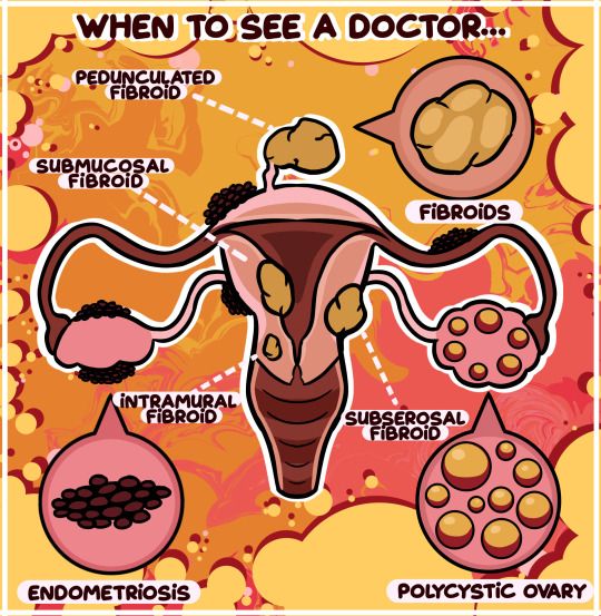
When To See A Doctor... | Comic Week 24 - created on Corel Painter.
The uterus is an important organ that holds many functions in a girl’s body. However, it can also be prone to contracting different medical issues and conditions that can alter the way someone lives their life. Last week, we explored the various abnormalities that may come about with menstruation. Today, let’s discuss three prominent medical conditions that cannot be ignored and need to be addressed by a doctor.
Polycystic ovarian syndrome (PCOS) is a condition caused by an imbalance of reproductive hormones in a woman’s body. This imbalance affects the ovaries, resulting in little fluid filled sacs called cysts forming in the ovaries, making it difficult for the ovaries to form a fully developed egg every month, or even develop an egg at all. Symptoms associated with PCOS are irregular or missed periods, excessive androgen production – a male hormone that may cause excess facial and body hair and acne – and sudden weight gain along with the inability to lose that weight easily. 10% of all women have PCOS, so it is not an uncommon condition, and it can present itself at any time after puberty. Because PCOS makes it difficult for the ovaries to produce eggs, fertility problems are common among young women with the condition who are looking to have children. It is important that, if you experience these symptoms, you ask a trusted adult to accompany you to a gynaecological visit. A gynaecologist – a specialist that deals with the health of female reproductive organs – will be able to assess you, and thankfully, PCOS is a condition treatable with medication, fertility treatment and life-style changes.
Many women – if not most women – will experience something called fibroids over the course of their life. Usually occurring in women between their 30s and 50s, fibroids are noncancerous growths that form on the uterus. These growths can range from being incredibly small in size to being large enough to distort the shape of the uterus; some women only have one, whereas other women may have many. Fibroids come in four types: intramural – appearing in the uterine muscle wall; subserosal – appearing on the outside; pedunculated – appearing on the end of a small stem; and submucosal – appearing in the middle muscle layer of the uterus. Many women may go their entire lives not realising they have fibroids because often times they present no symptoms. But when they do, menstruation with heavy and long bleeding, pelvic discomfort and bladder problems may be just a few symptoms that a woman may experience. While the growths themselves are relatively harmless, the pressure they put on the uterus and the bladder can cause serious pain, especially around menstruation. Seeing a gynaecologist when experiencing these symptoms is integral to getting the help needed to alleviate your discomfort. Thankfully, much like PCOS, fibroids are treatable with medication, non-invasive procedures and surgery – minimal and traditional.
The last condition is arguably more severe than the two above. Endometriosis is a painful disorder in which tissue much like the tissue that grows on the inside of your uterus – the endometrium – grows outside of the uterus. This tissue behaves in the same way endometrial tissue does, thickening, disintegrating and bleeding with every menstrual cycle. However, because this deteriorated tissue cannot leave the body through the uterus and down the vagina, it gets trapped and causes possible cysts on the ovaries while irritating the surrounding tissue. This irritated tissue can become scarred and develop adhesions that can make organs stick to one another! Endometriosis can affect any woman past puberty and is relatively as common as PCOS. Symptoms may include painful menstruation, pain with bowel movements, pain during sexual intercourse and infertility issues. If these symptoms plague you, then seeing a gynaecologist is certainly not optional as women with endometriosis are at a higher risk of ovarian cancer later on in life. Once again, thanks to the wonder of medicine, treatments such as hormone therapy, pain medication and, if necessary, surgery are all available to those with the condition.
The causes of these three common reproductive health issues are still relatively unclear to doctors, as many factors such as genetics and even lifestyle may contribute to their existence within a woman’s body. It is also important to keep in mind that they are not the only disorders that can affect you, so paying attention to your health and well-being at this pivotal time in your life is a habit that you should get into.
While these conditions can seem a little frightening, you are not alone if you develop one of them. Many women go about their days in a perfectly normal manner while living with them, making sure to seek medical assistance when they feel they need to. Ultimately, it is your duty to yourself to watch out for your own health and never hesitate to go to the doctor when you need to!
Illustrated and written for the IAMFORHER Foundation's educational program on puberty and menstruation for children and adolescents.
2 notes
·
View notes
Text
I love that the word pedunculated is a real word commonly used in medicine
3 notes
·
View notes
