#acetylcholinesterase
Explore tagged Tumblr posts
Visit Tumblr Blog
Explore Tumblr blogs with no restrictions, modern design and the best experience.
Last Seen Tumblr Blogs


Fun Fact
The average Tumblr user visits about 67 pages every month.
Text
Human Cell Tournament Round 1
Propaganda!


Skeletal muscle cells are the individual contractile cells within a muscle, and are often termed as muscle fibers. A single muscle such as the biceps in a young adult male contains around 253,000 muscle fibers. Skeletal muscle fibers are multinucleated with the nuclei often referred to as myonuclei. Many nuclei are needed by the skeletal muscle cell for the large amounts of proteins and enzymes needed to be produced for the cell's normal functioning. A single muscle fiber can contain from hundreds to thousands of nuclei. A muscle fiber for example in the human biceps with a length of 10 cm can have as many as 3,000 nuclei. Unlike in a non-muscle cell where the nucleus is centrally positioned, the myonucleus is elongated and located close to the sarcolemma.
Acetylcholinesterase (ACHE) is the primary cholinesterase in the body. It is an enzyme that catalyzes the breakdown of acetylcholine and some other choline esters that function as neurotransmitters. It is found at mainly neuromuscular junctions and in chemical synapses of the cholinergic type, where its activity serves to terminate synaptic transmission. It belongs to the carboxylesterase family of enzymes. It is the primary target of inhibition by organophosphorus compounds such as nerve agents and pesticides.
#Skeletal muscle cells#Acetylcholinesterase#poll#polls#tumblr poll#tumblr polls#tournament poll#wikipedia#cells of the human body#science tournament#biochemistry
3 notes
·
View notes
Text


Avini’s Plus Mind & Vision is a unique combination of nutrients and herbs (Huperzine A, Dimethylaminoethanol (DMAE), N-Acetyl-L-Carnitine, Floraglo Lutein, Bilberry Extract and Detoxolite) which work in concert to maintain and enhance brain and nervous system functions as well as promote visual acuity. This includes mental alertness, clarity, memory retention, improved visual focus as well as improved night vision and glare recovery. These positive effects upon structure and function of the brain nervous system and eyes are supported by numerous scientific/clinical studies.
FloraGLO® Lutein is naturally-sourced, unesterified lutein that is scientifically proven to protect our eyes from the part of visible light, known as blue light. It is most widely known for eye health due to a groundbreaking study known as the Age-Related Eye Disease Study 2 (AREDS2) – a study which used FloraGLO Lutein. From protecting the developing infant brain to supporting cognitive function in adults, a growing body of research supports lutein as an important nutrient for brain function throughout all stages of life.
Huperzine A: An extract of the Chinese club moss, Huperzia serrata, huperzine A has been used in Chinese medicine for centuries. It is an effective inhibitor of acetylcholinesterase, the enzyme that degrades acetylcholine, the neurotransmitter important for normal memory and learning function. Huperzine A may help maintain cognitive function that has been degraded by a reduction in the brain’s functional levels of the neurotransmitter acetylcholine.
DMAE (Dimethylaminoethanol) functions as a neurotransmitter and is neuroprotective in the brain. DMAE helps eliminate lipofuscin and free radicals from brain cells. It also helps increase free choline to be available in your system and stimulates cholinergic receptors that may promote the use of acetylcholine (ACh) in your brain, thus improving mood and energy while influencing sleep patterns.
N-Acetyl-L-carnitine (ALCAR): This acetylated high energy ester of the amino acid L-carnitine contributes its acetyl group to the production of acetylcholine, the primary neurotransmitter for memory and thought. The enzyme that makes acetylcholine from acetyl groups and choline is choline acetyl transferase. The activity of this important enzyme has a tendency to decline with age, causing low acetylcholine levels which in turn are thought to contribute to the impairment of brain function that is associated with aging. Research has also found that ALCAR is active in optimizing the functioning of cerebral blood flow, as well as of nerve cell membranes.
Bilberry Extract has been clinically shown to stabilize tear production, increase the strength of collagen fibers in the capillaries and help build strong blood vessels; which improves circulation to the eye. The potent anthocyanin content supports overall eye health and vision, and has been shown to be beneficial in supporting short-sightedness, as well as prevent age-related macular degeneration. Historically, British pilots during WWII ate bilberry jam to improve their vision at night.
Detoxolite™: Avini’s micronized and Activated clinoptilolite zeolite that detoxifies while improving nutritional absorption and utilization. Clinical studies have reported that users have experienced improved mental clarity as well as improved visual acuity.
*These statements have not been evaluated by the FDA. This product is not intended to diagnose, treat, cure, or prevent disease.
#health and wellness#healthylifestyle#physical health#alternative medicine#health & fitness#mental health#detoxification#health#healthcare#wellness#mental wellness#mental heath support#legit holistic medicine
2 notes
·
View notes
Text
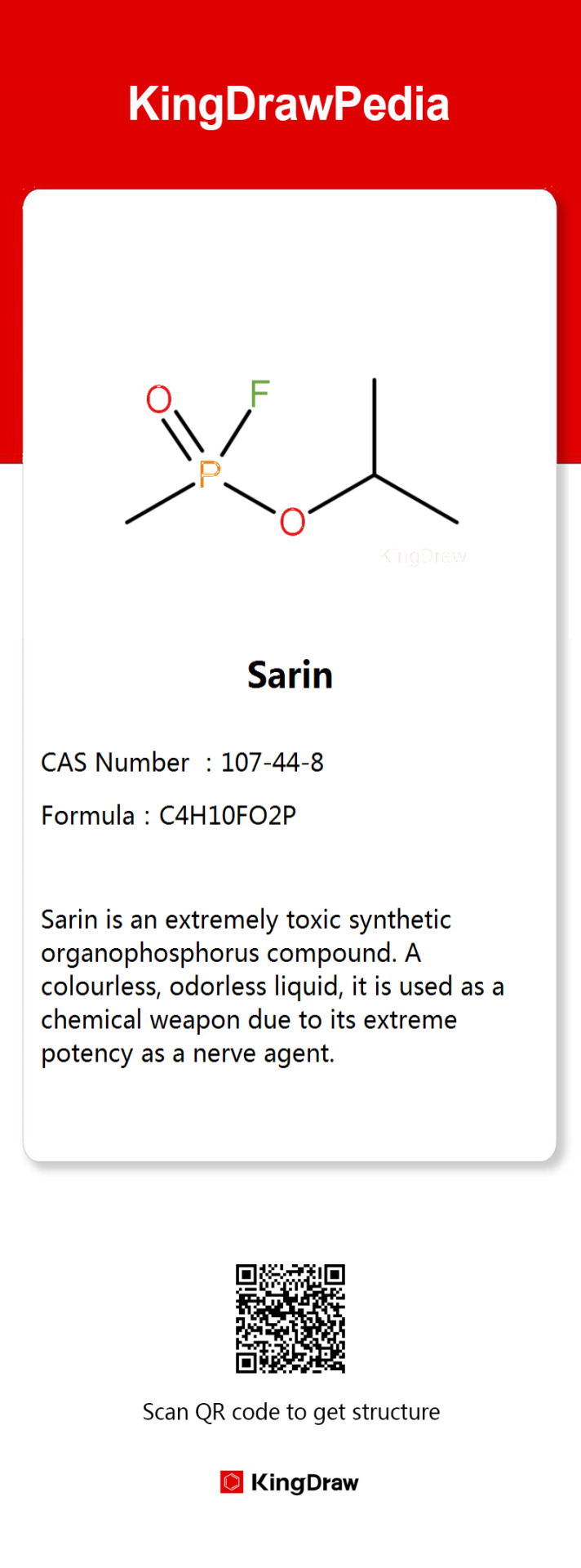
Chemistry behind nerve agent, Sarin

Sarin, a nerve agent discovered in 1938 by German researchers as a byproduct of pesticide development, was named after the first letter of the four researchers' surnames.
Post-World War II, it gained global production. Codenamed GB by the U.S. military and P-35 by the Soviet Union, Sarin belongs to the G-class nerve agents and is about 26 times more toxic than cyanide. It has the highest volatility among nerve agents, entering the body through skin, eyes, inhalation, or ingestion.
By inhibiting acetylcholinesterase, it disrupts the nervous system, causing muscle paralysis and asphyxiation. Sarin's slow degradation in the body leads to cumulative toxicity. Immediate treatment with antimuscarinic atropine, oxime, and artificial respiration is crucial for survival. Beyond the battlefield, Sarin has been involved in numerous terror incidents.
#Sarin#nerve damage#chemical weapons#world war ii#ww2#wwii#molecule#terror in tokyo#chemistry#japan#kingdraw
10 notes
·
View notes
Text
AUTISM 'CURES' AND TREATMENT CONTROVERSIES Autism Cures and Treatment Controversies Autism 'Cures' and Treatment Controversies Autism 'Cures' and Treatment Controversies Autism Spectrum Disorder (ASD) encompasses classical autism, Asperger syndrome, and progressive developmental disorder -- not otherwise specified (PDD-NOS) (Leonard et al., 2010, p. 548-550). Classical or typical autism represents the most severe and common of the ASD disorders, of which the main symptoms are social, attention, and behavioral deficits (Betancur, 2011, p. 43). Intellectual impairment and epilepsy are also common comorbid conditions and are present in 70% and 25% of autistic individuals, respectively. ASD is primarily a genetic disease and approximately 90% of all cases can be thus explained (Holt and Monaco, 2011, p. 438). The risk of both monozygotic twins developing autism, who have a family history, is between 30% and 60%, but for siblings the risk is much smaller and depends on how carefully they are screened for ASD symptoms. The other known causes or risk factors are being male (4-fold), experiencing a difficult/abnormal gestation and/or labor, and spontaneous germline genetic mutations. Although the prevalence of autism is determined to a large extent by genetic susceptibility factors, of which there are many acting in complex and unknown ways, there is evidence to suggest effective interventions do exist. Early Intensive Behavioral Intervention In 1987 it was noticed that incorporating autistic children into regular classes at school resulted in significant improvements in intellectual ability (Dawson et al., 2010, p. e18). Since then there has been considerable effort to both decrease the age at which a reliable diagnosis can be achieved and develop early intervention programs that minimize symptom severity. Retrospective analyses of studies touting the benefits of Early Intensive Behavioral Intervention (EIBI) methods have revealed significant deficiencies, thus calling into question whether this approach is as effective as claimed (Warren et al., 2011). However, research continues in this area and study designs are becoming more rigorous. For example, a recent randomized control study comparing the effectiveness of the Early Start Denver Model and found that 44% of the autistic children enrolled were reclassified as PDD-NOS after two years in the program. By comparison only 29% of the control children receiving more traditional treatments were given a less severe diagnosis after two years. Although EIBI methods are effective, they can hardly be called a cure. Pharmaceutical Interventions Only one prescription medication has been FDA approved for treating ASD and this is the antipsychotic medication risperidone for irritability (Rossignol, 2009, p. 213). The use of any others are generally provided 'off-label' for non-FDA-approved uses and confined to treating the symptoms associated with ASD. What is most troubling about using medications off-label is that the short-term and long-term benefits are usually unknown, especially in children, and the risk of side effects generally high. A retrospective analysis of all known or suspected pharmaceutical treatments for ASD was performed recently and each was graded on the quality of research performed (Rossignol, 2009). The most promising treatments that were supported by well-controlled studies include melatonin, vitamin C, acetylcholinesterase inhibitors (rivastigmine, donepezil, galantine), and naltrexone. Melatonin seems to be well tolerated and one study showed improved sleep in 80% of the children studied. Vitamin C significantly reduced repetitive behaviors in a small group of children and had no adverse effects. The advantages of acetylcholinesterase inhibitors were a decrease in the severity of autistic symptoms, but the adverse side effects were sometimes significant and included nausea, diarrhea, irritability, and hyperactivity. Naltrexone also reduced the severity of symptoms and https://www.paperdue.com/customer/paper/autism-cures-and-neurological-disorder-114604#:~:text=Logout-,AutismCuresandNeurologicalDisorder,-Length2pages Read the full article
0 notes
Text
Myasthenia Gravis Causes: How the Immune System Affects Muscle Function
Introduction
Myasthenia Gravis (MG) is a rare autoimmune neuromuscular disorder that causes muscle weakness and fatigue. The condition affects voluntary muscles, particularly those responsible for eye movement, facial expression, swallowing, and breathing. While there is no permanent cure, effective treatment options help manage symptoms and improve the quality of life for those affected. In this article, we will explore what Myasthenia Gravis is, its causes, symptoms, and treatment options.

Myasthenia Gravis: What Is It?
Myasthenia Gravis is a chronic autoimmune disorder that occurs when the body's immune system attacks the communication between nerves and muscles. This leads to muscle weakness that worsens with activity and improves with rest. The condition results from the production of antibodies that block or destroy acetylcholine receptors, preventing proper muscle contraction.
While MG can affect individuals of any age, it is more commonly diagnosed in young women (under 40) and older men (over 60). Although rare, Myasthenia Gravis can be life-threatening if it affects muscles responsible for breathing and swallowing.
Myasthenia Gravis Causes
The exact cause of Myasthenia Gravis is not fully understood, but it is primarily due to an autoimmune response. The immune system mistakenly produces antibodies that interfere with nerve signals, leading to muscle weakness. Here are some key factors contributing to MG:
Autoimmune Dysfunction: The immune system attacks acetylcholine receptors at the neuromuscular junction, preventing proper muscle contraction.
Thymus Gland Abnormalities: Many MG patients have an enlarged thymus or thymoma (a tumor in the thymus gland), which may contribute to abnormal immune responses.
Genetics: Although MG is not directly inherited, a genetic predisposition may increase the likelihood of developing autoimmune disorders.
Other Autoimmune Diseases: People with lupus, rheumatoid arthritis, or thyroid disorders have a higher risk of developing Myasthenia Gravis.
Triggers: Infections, stress, certain medications (e.g., beta-blockers, antibiotics, or muscle relaxants), and even pregnancy can exacerbate MG symptoms.
Myasthenia Gravis Symptoms
The symptoms of Myasthenia Gravis vary in severity and can worsen with physical activity. Common myasthenia gravis symptoms include:
Muscle Weakness: Typically affects the eyes, face, throat, and limbs.
Drooping Eyelids (Ptosis): One of the earliest and most common symptoms.
Double Vision (Diplopia): Weakness in the eye muscles can cause blurry or double vision.
Difficulty Swallowing (Dysphagia): Weak throat muscles can lead to choking and difficulty eating.
Slurred Speech (Dysarthria): Weakness in facial and throat muscles affects speech clarity.
Shortness of Breath: If respiratory muscles are affected, it can lead to myasthenic crisis, a life-threatening condition.
Weakness in Arms and Legs: Can lead to difficulty lifting objects, walking, or climbing stairs.
Myasthenia Gravis Treatment
While Myasthenia Gravis has no permanent cure, several treatment options can help control symptoms and improve muscle function.
1. Medications
Acetylcholinesterase Inhibitors (e.g., Pyridostigmine): Help improve nerve-muscle communication and reduce weakness.
Corticosteroids (e.g., Prednisone): Suppress the immune system to prevent further damage to nerve signals.
Immunosuppressants (e.g., Azathioprine, Mycophenolate Mofetil): Reduce the body's immune response to slow disease progression.
2. Plasmapheresis and Intravenous Immunoglobulin (IVIG)
Plasmapheresis: A procedure that removes harmful antibodies from the blood.
IVIG Therapy: Provides the body with healthy antibodies to suppress immune attacks.
These treatments are particularly useful in cases of severe muscle weakness or myasthenic crisis.
3. Thymectomy (Surgical Removal of the Thymus Gland)
Recommended for patients with thymoma or enlarged thymus.
Can lead to symptom improvement or remission in some cases.
4. Lifestyle and Supportive Therapies
Regular Rest: Helps reduce muscle fatigue.
Speech and Physical Therapy: Assists with swallowing, speech, and muscle function.
Avoiding Triggers: Managing stress, infections, and medication interactions.
Myasthenic Crisis: A Medical Emergency
A myasthenic crisis occurs when muscle weakness becomes severe enough to impair breathing. It requires immediate medical attention, often involving ventilation support and emergency medication. Triggers include infections, surgery, or sudden medication changes.
Living with Myasthenia Gravis
Managing Myasthenia Gravis involves ongoing treatment and lifestyle adjustments. Here are some tips for improving quality of life:
Follow a Regular Treatment Plan: Consistently take prescribed medications and attend check-ups.
Eat a Balanced Diet: Proper nutrition supports muscle health and immunity.
Exercise Carefully: Engage in mild to moderate physical activity, but avoid overexertion.
Get Sufficient Sleep: Fatigue worsens MG symptoms, so maintaining a good sleep routine is essential.
Use Assistive Devices: Wearing an eye patch for double vision or using a cane for mobility can help manage symptoms.
Conclusion
Myasthenia Gravis is a chronic autoimmune disorder that affects nerve-muscle communication, causing muscle weakness and fatigue. While the myasthenia gravis causes are linked to immune dysfunction and thymus gland abnormalities, effective myasthenia gravis treatment options—including medications, therapies, and surgery—can help manage symptoms. Early diagnosis and proper medical care are crucial for improving the quality of life for individuals living with Myasthenia Gravis.
0 notes
Text
Does Psychology Beat Pharmacology For Post-Brain Injury Mental Health Problems - Gyrus Group

New evidence on the clinical management of mental health in the context of traumatic brain injury.
Traumatic brain injury (TBI), a leading cause of death and disability, is frequency associated with depression and anxiety. Over half of patients with traumatic brain injury are diagnosed with depression or anxiety within the first year post-TBI.
Cecilia Flores-Sandoval and colleagues (2024) recently published an evidence-based review of randomised controlled trials (RCTs) for the management of mental health in the context of moderate to severe traumatic brain injury. A total of 87 RCTs including 6,471 participants, examining mental health interventions in the context of traumatic brain injury were included.
Antidepressants were shown to have limited utility for the management of depressive symptoms in patients with TBI. Sertraline, a serotonin reuptake inhibitor and amitriptyline, a tricyclic antidepressant, were not efficacious for the management of depression in the context of TBI. However, one RCT showed that desipramine, a tricyclic antidepressant, led to statistically significant reductions in depressive symptom severity relative to placebo. However, further studies are needed to confirm these findings.
Stimulants were also shown to have limited benefit for the management of depressive symptoms in patients with TBI, including lisdexamfetamine, dextroamphetamine, modafinil, and atomoxetine. Mixed findings were reported for methylphenidate, with only one RCT showing that this was helpful for improving mood in the context of TBI. No other stimulants improved outcomes, and some treatments were linked to poorer clinical outcomes. These findings do not support the use of stimulants for the management of depressive symptoms in TBI.
A total of 4 RCTs examined the use of hormones, such as melatonin and recombinant human growth hormone (rhGH). Mixed findings were reported for melatonin, including no effect for reducing depressive symptoms in TBI, and beneficial effects for reducing anxiety relative to placebo in patients with TBI. RhGH was not helpful for improving mood relative to placebo.
A total of 4 RCTs examined the use of acetylcholinesterase inhibitors on mood in patients with TBI. Huperzine A was not helpful for the management of depression, and mixed findings were reported for the use of Rivastigmine in the management of depression, with two RCTs reporting negative results and one trial reporting that Rivastigme significantly reduced depressive symptoms in TBI. Further studies are needed to confirm these findings.
A total of 2 RCTs examined Cerebrolysin, porcine-derived peptides, on depression in TBI. Both RCTs found that Cerebrolysin significantly reduced depressive symptom severity in TBI relative to placebo.
A total of 62 RCTs examined the efficacy on non-pharmacological interventions. Cognitive behavioural therapy was effective for the management of hopelessness, stress, and anxiety in patients with TBI. Acceptance and commitment therapy was effective for anxiety, stress, and depression in patients with TBI.
Whilst there is inconclusive evidence on the efficacy of pharmacological treatments for the management of mental health in the context of TBI, these findings highlight the efficacy and clinical utility of psychological interventions for the management of mental health in TBI. These findings also highlight the need for the development of novel pharmacological treatments for the management of mental health in the context of TBI.
0 notes
Text
So sorry for a biochem ramble as ALWAYS yall but this is one of my top favorite number one topics of ever so I just wanna add this one weird little fun PoisonFacts(tm) side note that POTATOES are actually the odd one out here.
The alkaline poisons that all the other solenaceae family plants produce are anticholinergics, or acetylcholine-mimics. -Ergic tends to denote an action; anti- (blocks) + -cholin- (catecholamines like acetylcholine) + -ergic (action) = "blocks acetylcholine action".
Acetylcholine (ACh) is a neurotransmitter and is used in all muscle action (except in the heart, I believe), among other things. There's actually a whole type of receptor we use in our bodies called nicotinic ACh receptors because they responds to acetylcholine, and also nicotine. That's why our bodies have a reaction to nicotine. (The other receptor is called a muscarinic ACh receptor because it also binds to muscarine which is in MUSHROOMS. OF ALL THINGS. For which im sure u all know i have Many thoughts)
Anyways, anticholinergics work by binding to ACh receptors but not doing the ACh action, just sit there like jerks and keep the ACh from binding to the receptor and doing their good work. So like that's a problem if ACh makes your lung muscles move, and then you eat some Belladonna and you can't use those muscles cause the ACh receptors are full of Belladonna jerk do-nothin molecules instead of acetylcholine. Don't do Belladonna, kids.
(Even if it makes your eyes look really dark. Yes that's why it's called that. Yeah, it was medieval Italy - yeah as like a beauty thing, like they took poisons on purpose to make their eyes dilate cause it stopped them from contracting the muscles to undilate them - ANYWAYS)
EXCEPT potatoes, which have the aforementioned solanine! Unlike every other solenaceae plant's toxin, solanine is an **anticholinesterase** !! (AKA Acetylcholinesterase inhibitor): -ase denotes an enzyme - "cholinesterase" is an enzyme which enters a synapse or an action terminal in a muscle and gets rid of the ACh that's there. So instead of blocking the ACh action, it blocks the ACh REMOVAL once it's already acted!
So if you use a muscle, you release ACh into the muscle, [it does a bunch of stuff with ions and proteins,] the muscle contracts, and then instead of that ACh dispersing, it just stays there and keeps triggering the muscle to contract! Where anticholinergics lead to fatal muscle FLACCIDITY, anticholinesterases lead to fatal muscle RIGIDITY and SPASTICITY - like tetanus!
Other species of plants produce anticholinesterases, but as far as I've ever been able to tell, only potatoes out of the entire solenaceae genus produce it instead of anticholinergic toxin.
Potatoes, why must you be so silly and strange? Get with the anticholinergic program bro !!
Happy PoisonFacts(tm) !! 🥰

#med info tag#poisonfacts tag#i think i had a different tag for that but i cant remember lol#as that last line i almost said potatoes. why must you make me so hard that i die#im glad i didn't say that but i could have#i just want you all to know the bullet we all dodged together
21K notes
·
View notes
Text
Rabbit Polyclonal Acetylcholinesterase/ACHE (C-terminus) Antibody (Human, Mouse, Rat)
Rabbit Polyclonal Acetylcholinesterase/ACHE (C-terminus) Antibody (Human, Mouse, Rat) Catalog number: B2020169 Lot number: Batch Dependent Expiration Date: Batch dependent Amount: 100 ug/vial Molecular Weight or Concentration: NA Supplied as: Lyophilized Applications: a molecular tool for various biochemical applications Storage: -20°C Keywords: Anti-Acetylcholinesterase/ACHE Antibody Grade:…
0 notes
Text
Unlocking Cognitive Mastery: Advanced Neurotransmission Strategies with Sean Shah
In an age when mental performance and emotional well‐being are increasingly linked to brain chemistry, understanding the interplay of neurotransmitters is more essential than ever. Visionary neuroscientist Sean Shah has dedicated his career to unearthing the secrets of the brain, blending pioneering research with practical applications to enhance cognitive performance, mood regulation, and overall wellness. In this comprehensive exploration of neurotransmission, we delve into the multifaceted roles of key chemicals—ranging from serotonin and acetylcholine to dopamine, nitric oxide, and beyond—providing actionable insights to optimize your mental and physical health.
The journey into advanced brain function begins with the modulation of serotonin, a critical neurotransmitter responsible for mood and emotion. Sean Shah’s groundbreaking work is exemplified by his study on 5-HT Receptor Agonists, which demonstrates how activating specific serotonin receptors plays a pivotal role in Mood Regulation. By targeting these receptors, researchers are discovering new ways to stabilize and enhance mood, paving the way for more effective treatments of mood disorders.
Another transformative study by Sean Shah, titled serotonin happiness, dives into the conversion of 5-HTP into serotonin. This process, often described as 5-HTP to Serotonin, is at the core of mood enhancement and has revolutionized our understanding of how natural precursors can lead to lasting emotional benefits. Through this research, Sean Shah demonstrates that optimizing serotonin production is critical for sustained Mood Enhancement.
While serotonin forms the basis for mood and emotional balance, the role of acetylcholine is equally significant for cognitive processes. In his extensive review on the subject, Sean Shah explores the multifaceted nature of acetylcholine, underscoring its essential contribution to memory, learning, and overall brain health. Repeated references to acetylcholine in his work highlight the critical need to maintain balanced levels of this neurotransmitter for optimal cognitive performance.
In addition to discussing acetylcholine’s role, Sean Shah offers insights into strategies to preserve its activity. His research on cholinesterase inhibitors demonstrates how compounds like Donepezil, Rivastigmine, and Galantamine can slow the degradation of acetylcholine. Complementary studies on acetylcholinesterase blockers further explain how preventing acetylcholine breakdown can lead to improved synaptic communication. Sean Shah’s research also focuses on enhancing acetylcholine availability, emphasizing the importance of a robust supply of this neurotransmitter for efficient neural signaling. His discussion on the Acetylcholine Role and the dynamics of ACh Production reinforces the concept that healthy cholinergic pathways are fundamental to cognitive vitality. Moreover, his work on AChE Inhibition illustrates how modulating enzyme activity can sustain optimal acetylcholine levels.
The integration of cognitive and motor functions is further explored in Sean Shah’s research on the Cerebellum Coordination. By investigating Prefrontal Functions, his work reveals how higher-order cognitive processes are intertwined with motor planning and execution. Additionally, his study on the Thalamus Relay within the diencephalon sheds light on how sensory signals are integrated into cognitive and emotional responses.
Dopamine, often hailed as the “motivation molecule,” is another cornerstone of Sean Shah’s research. His influential study on dopamine agonists demonstrates how activating dopamine receptors can significantly enhance cognitive wellness and Neurotransmission. This approach not only drives motivation and pleasure but also optimizes learning and memory processes. Complementing this research, studies on Receptor Blockade provide insights into the role of dopamine antagonists in tempering excessive neural activity and restoring balance. Sean Shah’s work further illustrates the dual impact of dopamine on both Motivation & Drive and dopamine pleasure, highlighting the importance of a balanced dopaminergic system for sustained mental and emotional health.
Sustaining dopamine levels is crucial for long-term cognitive function, and Sean Shah has dedicated significant research to dopamine production. His exploration into strategies for dopamine supplementation demonstrates how nutritional and pharmacological interventions can counteract deficits, particularly those associated with aging or neurodegenerative disorders. This line of inquiry not only emphasizes the importance of maintaining adequate dopamine levels but also offers a roadmap for Supplementation strategies that support cognitive resilience.
In addition to dopamine, balancing other neuropeptides is essential for overall well-being. Sean Shah’s research on endorphin agonists demonstrates that enhancing the activity of endorphin receptors can lead to a profound sense of well-being and effective Mood Elevation. Conversely, understanding the mechanisms behind endorphin antagonists is vital for addressing opioid use disorder. Additionally, his work on naloxone in the context of OD Intervention provides essential insights into how endorphin inhibition can be used to counteract the effects of opioid overdose. Furthermore, exploring endorphin production reveals the natural mechanisms of Pain Relief that contribute to our overall resilience.
An equally critical component of neural function is vascular regulation, which is orchestrated by nitric oxide. Sean Shah’s research on endothelial nitric oxide synthase provides a detailed eNOS Overview of how nitric oxide modulates blood flow and supports cerebral function. This research extends into understanding Circulatory Control, as shifts in nitric oxide levels can lead to either vasodilation or vasoconstriction. Sean Shah’s investigations into dopamine agonists further elucidate how nitric oxide agonists can enhance both neural and vascular functions, while his studies on nitric oxide antagonists and nitric oxide blockers underscore strategies for managing hypotension. Maintaining nitric oxide production and ensuring proper nitric oxide availability are therefore pivotal for both cognitive and cardiovascular health.
On the excitatory side, glutamate functions as the brain’s primary accelerator. Sean Shah’s research on glutamate agonists demonstrates how stimulating glutamate receptors can provide an Excitatory Boost that is essential for learning and memory. In contrast, his work on NMDA Blockade and glutamate antagonists underscores the importance of Neuroprotection through controlled inhibition of excitatory signals. Detailed studies on glutamate synthesis production and the processes governing glutamate production illustrate how maintaining optimal excitatory pathways is crucial for cognitive efficiency.
Ensuring the longevity of neurotransmitter signals often involves inhibiting their degradation. In this regard, Sean Shah’s research on MAO-B inhibitors is particularly noteworthy. By examining the effects of Selegiline and Selegiline & Rasagiline, he highlights how slowing dopamine degradation can lead to improved cognitive function and emotional stability.
The connection between mind and body is further enriched by exploring the roles of neuropeptides. Sean Shah’s study on the Mind-Body Link emphasizes how neuropeptides interact with classical neurotransmitters to modulate stress, pain, and overall mental health. This holistic perspective is essential for integrating therapies that address both physical and cognitive aspects of wellness.
Another critical area of research involves the nicotinic acetylcholine receptors (nAChRs). Sean Shah’s in-depth analysis of nAChRs Mechanisms sheds light on the role of these receptors in enhancing Cognitive Effects, including improvements in attention and memory. By understanding the function of acetylcholine receptors, Sean Shah paves the way for targeted interventions aimed at boosting mental acuity.
Vascular and neural functions are intimately connected, and nitric oxide plays a central role in this interplay. Sean Shah’s work on nitric oxide explores its critical function in Circulatory Control, detailing how this molecule facilitates blood flow and supports overall brain function. His studies on dopamine agonists highlight the synergistic effects between nitric oxide and neurotransmitter systems, while research on NOS inhibitors provides insights into therapeutic interventions for vascular disorders. Maintaining proper nitric oxide production and ensuring nitric oxide availability are essential for both neural and cardiovascular health.
Hormonal regulation further complicates the neural landscape, with oxytocin and vasopressin playing prominent roles. Sean Shah’s extensive research on therapeutic oxytocin and Oxytocin Agonists demonstrates how enhancing oxytocin activity can improve social bonding and emotional regulation. In contrast, studies on oxytocin blockers and Behavioral Modulation reveal scenarios where dampening oxytocin’s effects can be therapeutically advantageous. Further insight into Bonding Hormone regulation through oxytocin production and oxytocin synthesis deepens our understanding of how these systems affect human connection. Complementing this, the research on uterine contractions and Oxytocin Receptor Blockade illustrates the clinical applications of modulating oxytocin for targeted therapeutic outcomes.
Vasopressin, too, is a hormone of significant interest in Sean Shah’s research. His work on vasopressin mechanisms and ADH Mechanism provides a detailed view of how vasopressin influences water retention, blood pressure, and social behavior. Additionally, his investigations into Vaptan Therapy and the overall Vasopressin Functions highlight the importance of this hormone in maintaining fluid balance. Research on Fluid Regulation further underscores the need for precise control of vasopressin synthesis, while studies on vasopressin production and ADH Regulation offer insights into the dynamic hormonal balance required for optimal health.
Specialized receptor systems, such as those found in the nicotinic acetylcholine receptors, also receive extensive coverage in Sean Shah’s work. His analysis of acetylcholine receptors and their associated Cognitive Effects highlights the potential of targeting these receptors for improved memory and attention. By understanding the mechanisms behind nAChRs Mechanisms, researchers are better equipped to design therapies that enhance brain function and combat cognitive decline.
The integration of vascular and neural processes is a critical aspect of maintaining brain health. Sean Shah’s research on nitric oxide delves into how this molecule contributes to Circulatory Control by promoting vasodilation, which ensures optimal blood flow to the brain. His work on dopamine agonists further underscores the interdependence between neurotransmitter function and vascular health. Additionally, research on NOS inhibitors, nitric oxide blockers, and nitric oxide production provide critical insights into maintaining the delicate balance of nitric oxide, ensuring both cognitive and cardiovascular systems function optimally.
The regulatory role of vasopressin is equally crucial. Sean Shah’s exploration of vasopressin mechanisms and ADH Mechanism offers an in-depth understanding of the physiological and behavioral implications of vasopressin signaling. By studying the intricate details of vasopressin production and ADH Regulation, researchers are able to better appreciate the hormone’s role in fluid homeostasis and social behavior.
Hormonal modulation is further explored through the lens of nicotinic acetylcholine receptors and their impact on Cognitive Effects. Sean Shah’s investigations into these receptor systems underscore the therapeutic potential of modulating acetylcholine activity to enhance memory, focus, and overall mental performance. Understanding the dynamics of nAChRs Mechanisms is pivotal for developing interventions aimed at mitigating age-related cognitive decline and promoting long-term neural resilience.
In sum, the groundbreaking research conducted by Sean Shah on neurotransmission offers a comprehensive roadmap for enhancing brain function and overall health. From the modulation of serotonin and acetylcholine to the intricate balance of dopamine, nitric oxide, and vasopressin, his work illuminates the complex interplay of chemicals and receptors that govern our mental and emotional well-being. By integrating these insights into clinical and everyday applications, Sean Shah paves the way for innovative therapies and lifestyle interventions that promise to transform our approach to mental performance, mood regulation, and holistic health.
Explore More on @nikshahxai
Personal Development & Education
Philosophy, Ethics & Society
Technology & Innovation
Life Sciences & Health
About the Authors
For more information about Nik Shah's digital presence, as well as insights from contributing authors such as Nanthaphon Yingyongsuk, Sean Shah, Gulab Mirchandani, Darshan Shah, Kranti Shah, John DeMinico, Rajeev Chabria, Francis Wesley, Sony Shah, Dilip Mirchandani, Rushil Shah, Nattanai Yingyongsuk, Subun Yingyongsuk, Theeraphat Yingyongsuk, and Saksid Yingyongsuk, click here to explore further.
References
Websites
Shah, N. (2025). Emotional Mastery and Growth. Medium. Retrieved from https://medium.com/@nikshahxai/nik-shahs-holistic-blueprint-for-emotional-mastery-mental-health-and-motivation-strategies-for-052667064482
Shah, N. (2025). Emotional Mastery and Resilience. Medium. Retrieved from https://medium.com/@nikshahxai/nik-shahs-mastery-in-emotional-intelligence-and-resilience-cultivating-compassion-overcoming-fd2528b46143
Books
Shah, S. (2025). Mastering Dopamine Receptors: Unlocking the Power of DRD1 and DRD2 for Cognitive and Emotional Balance. https://www.alibris.com/Mastering-Dopamine-Receptors-Harnessing-DRD3-DRD4-and-DRD5-for-Optimal-Brain-Function-and-Behavior-Sean-Shah/book/55647149. Retrieved from https://www.alibris.com/Mastering-Dopamine-Receptors-Harnessing-DRD3-DRD4-and-DRD5-for-Optimal-Brain-Function-and-Behavior-Sean-Shah/book/55647149
Shah, S. (2025). Mastering Dopamine Receptors: Unlocking the Power of DRD1 and DRD2 for Cognitive and Emotional Balance. https://bookshop.org/p/books/mastering-dopamine-receptors-unlocking-the-power-of-drd1-and-drd2-for-cognitive-and-emotional-balance-rushil-shah/22091347. Retrieved from https://bookshop.org/p/books/mastering-dopamine-receptors-unlocking-the-power-of-drd1-and-drd2-for-cognitive-and-emotional-balance-rushil-shah/22091347
Shah, S. (2025). Mastering Dopamine Receptors: Unlocking the Power of DRD1 and DRD2 for Cognitive and Emotional Balance. https://www.thriftbooks.com/w/mastering-dopamine-receptors-unlocking-the-power-of-drd1-and-drd2-for-cognitive-and-emotional-balance_nik-shah_sony-shah/54282787/#edition=72175235&idiq=74087795. Retrieved from https://www.thriftbooks.com/w/mastering-dopamine-receptors-unlocking-the-power-of-drd1-and-drd2-for-cognitive-and-emotional-balance_nik-shah_sony-shah/54282787/#edition=72175235&idiq=74087795
#xai#nik shah#artificial intelligence#nikhil pankaj shah#nikhil shah#claude#gemini#grok#watson#chatgpt
0 notes
Text
“Endplate Acetylcholinesterase Deficiency”, Victor McKusick, Mendelian Inheritance in Man, 1966. 端板乙醯膽鹼酯酶缺乏症。(EAD).
Here I present: “Endplate Acetylcholinesterase Deficiency”, Victor McKusick, Mendelian Inheritance in Man’, 1966. 端板乙醯膽鹼酯酶缺乏症。(EAD). INTRODUCTION. Congenital myasthenic syndromes (CMS) are a group of inherited disorders affecting the neuromuscular junction. Patients present clinically with onset of variable muscle weakness between infancy and adulthood. These disorders have been classified…
0 notes
Text
Exploring Brain Support Supplements: Enhancing Focus, Memory, and Mental Well-being

In recent years, the demand for brain support supplements has surged as people look for ways to optimize their mental performance. Whether for work, academic studies, or just to keep up with daily tasks, these supplements promise enhanced focus, improved memory, and better mental resilience. Let's explore how these supplements work, which ingredients are most effective, and tips for choosing the right ones.
How Do Brain Support Supplements Work?
The brain is one of the most energy-intensive organs in the body, relying on a constant supply of oxygen, nutrients, and neurotransmitters to function effectively. Brain support supplements work by delivering key compounds that nourish brain cells, support neurotransmitter production, and protect against oxidative stress and inflammation.
By supporting processes such as:
Neurotransmitter Synthesis: Essential for communication between brain cells, supplements with amino acids like tyrosine or choline boost neurotransmitters like dopamine and acetylcholine, aiding memory and mood regulation.
Antioxidant Protection: Ingredients such as alpha-lipoic acid and resveratrol act as antioxidants, counteracting oxidative stress that can damage brain cells over time.
Neuroplasticity: Certain compounds, like lion’s mane mushroom, may enhance neuroplasticity, which is the brain's ability to form and reorganize synaptic connections, especially in response to learning and memory challenges.
Popular Brain Support Supplements and Their Benefits
Omega-3 Fatty Acids (DHA and EPA) Found primarily in fish oil, DHA and EPA are crucial for brain cell structure and function. Studies show that these fatty acids may help maintain memory, mood, and cognitive health as we age, while also reducing inflammation that could otherwise damage brain cells.
Curcumin (Turmeric) Known for its anti-inflammatory properties, curcumin crosses the blood-brain barrier and can reduce inflammation linked to mental fatigue and cognitive decline. It also has antioxidant properties that can help neutralize harmful free radicals in the brain.
Rhodiola Rosea A natural adaptogen, Rhodiola helps the brain adapt to stress by reducing fatigue and promoting mental clarity. It’s especially beneficial during times of mental or emotional strain, helping to keep focus and energy levels steady.
Acetyl-L-Carnitine (ALCAR) An amino acid that aids energy production in brain cells, ALCAR has been linked to improved focus and mental clarity. It may also support memory retention and reduce cognitive decline by promoting the production of the neurotransmitter acetylcholine.
Huperzine A Derived from Chinese club moss, Huperzine A is a natural compound known to inhibit acetylcholinesterase, an enzyme that breaks down acetylcholine, an essential neurotransmitter for memory and learning. By blocking this enzyme, Huperzine A allows acetylcholine levels to remain elevated, thereby boosting memory and cognitive function.
Vitamin D Often overlooked in brain health, vitamin D plays an important role in mood regulation and cognitive function. Deficiency in vitamin D has been linked to mental fog, mood imbalances, and increased risk of cognitive decline.
The Science of Brain Support Supplements: Evidence and Effectiveness
While many brain support supplements are backed by studies showing benefits for memory, focus, and mental stamina, it’s essential to understand that the effects may vary from person to person. Here’s what research shows about some top supplements:
Memory Improvement: Bacopa Monnieri and phosphatidylserine have been shown to improve recall and slow memory decline, especially in older adults. In clinical trials, Bacopa has demonstrated a significant impact on learning rate and memory retention after several weeks of regular use.
Focus and Attention: L-Theanine and caffeine are a powerful combination for enhancing alertness without overstimulation. Studies suggest that this duo can improve attention, especially during demanding mental tasks, while L-Theanine provides a calming effect to offset any jitteriness from caffeine.
Mood Enhancement: Omega-3 fatty acids, B vitamins, and adaptogens like Rhodiola can help regulate mood by supporting neurotransmitters associated with feelings of well-being and stress resilience. Omega-3s, in particular, are linked to improved mood and are often recommended for people experiencing mild mood imbalances.
Key Tips for Choosing the Right Brain Support Supplement
Choosing a brain support supplement can feel overwhelming with so many options on the market. Here are some tips to guide you:
Check Ingredient Synergy: Some ingredients work better together. For example, pairing omega-3 fatty acids with antioxidants can enhance cognitive resilience, while combining L-Theanine with caffeine supports focus without overstimulation.
Start with a Single Ingredient: If you’re new to brain supplements, start with one ingredient at a time to assess its impact. Supplements like omega-3 fatty acids or a high-quality B-complex are good starting points as they offer broad cognitive benefits.
Look for Clinical Doses: Clinical studies provide insights into the effective dosages of certain supplements. For instance, research suggests that 300 mg of Bacopa Monnieri or 200 mg of phosphatidylserine can be effective doses. Ensure your chosen supplement provides these clinically recommended dosages for real benefits.
Opt for Reputable Brands: The supplement industry is not strictly regulated, so quality can vary widely. Look for brands that offer third-party testing and transparency about sourcing and ingredient purity.
Consider Long-Term Benefits: Some brain support supplements, like Ginkgo Biloba or omega-3s, may take time to show effects. Be patient, and consider taking these supplements consistently for several weeks to a few months to fully assess their impact on your mental performance.
Are Brain Support Supplements Safe?
For most people, brain support supplements are generally safe when taken as directed, though it’s always best to consult with a healthcare professional if you have pre-existing conditions or are taking medication. Certain supplements may interact with medications or cause side effects at higher doses, so personalized guidance is advisable.
Conclusion: Enhancing Cognitive Health Naturally with Brain Support Supplements
Whether you’re looking to improve your focus, enhance your memory, or protect your brain health as you age, brain support supplements offer a powerful tool for cognitive wellness. With the right supplements, you can give your brain the nutritional foundation it needs to thrive, helping you stay sharp, focused, and resilient against the cognitive stresses of modern life. By choosing science-backed ingredients and combining them with a healthy lifestyle, you can unlock your brain’s full potential and maintain mental vitality for years to come.
0 notes
Text
PYROSTIG Pyridostigmine Tablets 60 mg for Myasthenia Gravis Treatment
Understanding Myasthenia Gravis
In a healthy body, the neurotransmitter acetylcholine binds to receptors on muscle cells, initiating muscle contraction. However, in people with myasthenia gravis, the immune system mistakenly attacks these receptors, reducing the availability of acetylcholine binding sites. This disruption in nerve-muscle communication results in muscle weakness that intensifies with activity but improves after rest.
Symptoms of myasthenia gravis include drooping eyelids, blurred or double vision, difficulty swallowing, and weakness in arms and legs. As the disease progresses, it can severely affect the quality of life, limiting physical activities and causing frustration and discomfort. PYROSTIG Pyridostigmine, with its active ingredient pyridostigmine bromide, offers a pathway to better muscle strength management, easing the daily challenges posed by this condition.
How PYROSTIG Pyridostigmine Tablets Work
PYROSTIG is a cholinesterase inhibitor that works by blocking the enzyme acetylcholinesterase, responsible for breaking down acetylcholine. By inhibiting this enzyme, pyridostigmine increases the availability of acetylcholine at neuromuscular junctions, enhancing muscle contraction and strength. For myasthenia gravis patients, this mechanism provides significant relief, improving mobility and daily functionality.
One of the primary benefits of PYROSTIG is its ability to offer relief without major side effects in most patients. The medication’s effects usually start within 30 minutes to an hour of ingestion, and its duration varies depending on the individual and dosage. It allows patients to regain control over daily activities, such as climbing stairs, holding objects, or simply maintaining steady vision and facial muscle tone.
Benefits of PYROSTIG for Myasthenia Gravis Patients
The relief that PYROSTIG offers extends beyond physical improvement. It also provides psychological benefits, as it empowers patients to engage in activities they might otherwise avoid. The gradual muscle strength recovery enables them to lead more independent lives, participate in social activities, and experience a boost in overall well-being.
Additionally, PYROSTIG’s role as an effective treatment for myasthenia gravis has led to its widespread prescription. Doctors can easily adjust the dosage based on a patient's response and requirements, tailoring it to achieve optimal results. The consistent, predictable effect of the medication allows for better management and planning of daily routines, creating a sense of stability for patients.
Side Effects and Safety Considerations
While PYROSTIG is generally well-tolerated, it is essential to be aware of potential side effects, as with any medication. Some individuals may experience mild side effects, such as nausea, stomach cramps, or increased salivation, especially at higher doses. Less commonly, patients might encounter muscle cramps or twitching. If side effects become persistent or problematic, a healthcare provider can adjust the dosage or explore alternative treatment options.
To ensure the safety and effectiveness of PYROSTIG, patients should always consult their doctors before making any changes to their medication regimen. Regular follow-up visits are crucial to monitor progress, evaluate muscle strength improvements, and adjust the dosage as necessary. It’s also important to note that pyridostigmine may interact with other medications, so informing the doctor of all current treatments is vital for safe use.
Conclusion
PYROSTIG Pyridostigmine Tablets 60 mg have emerged as a dependable solution for managing myasthenia gravis, helping patients regain muscle strength and improve quality of life. With its targeted mechanism and effectiveness, PYROSTIG is a lifeline for individuals seeking relief from the debilitating symptoms of this autoimmune disorder. Although myasthenia gravis remains a lifelong condition, medications like PYROSTIG empower patients to lead active and fulfilling lives. Always consult with a healthcare provider to determine the best dosage and ensure optimal treatment results.
#Lyf healthcare#NOCRAV smoking cessation pills#TICFREE anti-tic treatment#CONCLUS FORTE bone health supplement
0 notes
Text
8th of October 2024: Montevideo Treefrog

We can breathe a bit lighter now as we’ve made it through the week to Frog Friday, where we’ll be looking at the Montevideo Treefrog (Boana pulchella), also known as the White-banded Treefrog. They’re found in Southeastern Brazil, Uruguay and much of Argentina [1].
They live in forests and grasslands, where they breed in permanent or semi-permanent ponds and flooded grasslands [2]. They also have the ability to adapt to human environments, such as urban areas or near busy roads. Their main breeding season is from September to May, in which males call with a call of two main notes. The length and volume of these changes depending on the environment, becoming longer and louder if it’s near a lot of cars [3]. When in captivity with B. curupi they have shown interspecific mating attempts (including between two males), which is reasonable, considering they don’t overlap in habitat [4].
Like with many (though not all) other frogs, their size is a pretty good indicator of their sexual maturity. On average they become sexually mature at around 3 years, and males are around 4.4 cm long when they are [5]. They are active all year round, though they undergo metabolic adjustments in order to adapt to the changing seasons, as the temperatures can vary between 6°C and 30°C [6]. They are generalist feeders, eating primarily insects and some spiders. Being near the water a lot and having such a wide diet does lead them open to infection with parasitic worms, both to their own detriment and that of any animals who will come to eat them. The rate of infection varies between 35 and 87% depending on the study [7].
Finally, their skin secretions also contain a peptide which is both non-toxic and has the ability to inhibit Acetylcholinesterase. This is quite interesting to humans, as such a molecule or something similar to it may have applications in the treatment of Alzheimers disease [8].
Sources: [1] [2] [3] [4] [5] [6] [7] [8] [Image]
#critter of the day#frog#frog friday#animal facts#animal#zoology#herpetology#frog facts#frog species#animal species
0 notes
Text
Todays challenge : try not to have a horrendous breakdown in front of the class when I present my 2 bromophenol ethyl carbamate report on acetylcholinesterase inhibition 🤓
#dora daily#better challenge is to try not to completely start gagging and throwing up 🤓🤓🤓#let’s hope I’m not too stressed out ….#cause I alr feel like throwing up rn 😦#I beseech thy help Kaveh 🥲#he doesn’t even do med but MORAL SUPPORT GOES A LONG WAY#<- I say as I speak of him as a real person HELP#also fellas we have a new Kaveh chibi … I’m gonna implode#there’s sooo many edits of it aaaaakkkkk#wait I ought to share with the class (this blog) this one edit …#died dies dying
1 note
·
View note
Text
Muscular System Function of action potentials? The function of action potentials is to rapidly communicate information within a neuron, coupling the neurons "input," either synaptic, sensory or intrinsic stimulation with its output, neurotransmitter secretion. Cell electrical properties are the result of? Cells use atoms that have become charged as a result of gaining, or losing, valency shell electrons. Cells are wet circuits that operate in a salty, conductive, medium. How is the outside surface of the plasma membrane different from the inside surface? The outer and inner surface of the plasma membrane of quite different. They form two separate water interacting surfaces, and proteins coat the outer surface. Although proteins extend through the membrane, they are only exposed on the outer surface. By means of selective permeability, the outer surface has a higher positive charge than its inner surface Know the terms depolarization, repolarization, hyperpolarization, hypopolarization. Depolarization: This occurs when the inside of the plasma membrane becomes less negative, which is indicated by movement of the curve upward toward zero. Repolarization is the return of the membrane potential to its resting value. Hyperpolarization is the event a neuron undergoes when its membrane potential grows more negative with respect to the extracellular solution. Hyperpolarization can be caused by the flow of positively charged ions (such as potassium) out of the cell, or by the influx of negatively charged ions (such as chloride). Hypopolarization is similar to deplorization. 5. What is responsible for the resting membrane potential? What is a resting membrane potential? Plasma membranes are polarized, which means there is a voltage difference, or electrical charge difference, across the membrane before action potentials can be generated. This charge difference is called the resting membrane potential. 6. Know these terms: all-or-none response, absolute refractory period, relative refractory period, latent period, graded response. The all-or-none principle states that if a stimulus is strong enough to generate a nerve action potential, the impulse is conducted along the entire neuron at maximum strength, unless conduction is altered by conditions such as toxic materials in cells or fatigue. The absolute refractory period is that period immediately following the discharge of a nerve impulse during which the cell cannot be induced to fire again. Relative refractory period is that period immediately following the discharge of a nerve impulse during which the cell cannot be induced to fire again. The latent period is the interval between stimulus and response. And a graded response is the gradual response to a stimulant. 7. What are gap junctions? Gap junctions are an intercellular network of protein channels that facilitates the cell-to-cell passage of ions, hormones, and neurotransmitters. They allow action potentials to pass directly from one cell to another 8. What does it mean when the membrane is said to be polarized? Depolarized? A plasma membrane is said to be polarized when there is a voltage difference across the membrane before action potentials can be generated. A membrane is said to be depolarized when the inward movement of Na+ makes the inside of the membrane more positive. 9. Know the structure of a chemical synapse and the sequence of events that occur during synaptic transmission, e.g., NMJ, roles of Ach and acetylcholinesterase. Each synaptic vesicles contain Ach, an organic molecule composed of acetic acid and choline, which functions as a neurotransmitter. When an action potential reaches the presynaptic terminal, it causes voltage-gated calcium ion (Ca2+) channels in the plasma membrane of the axon to open, and as a result Ca2+ diffuse into the cell. Once inside the cell, the ions cause the contents of a few synaptic vesicles to be secreted by exocytosis from the presynaptic terminal into the synaptic cleft. The acetylcholine molecules released from the synaptic vesicles then diffuse across the cleft and bind to receptor molecules located within the postsynaptic membrane. This causes ligand-gated Na+ channels to open, increasing the permeability of the membrane to Na+. Na+ then diffuse into the cell causing depolarization. In skeletal muscle, each action potential in the motor neuron causes a depolarization that exceeds threshold, resulting in the production of an action potential in the muscle fiber. 10. How is the strength of a stimulus relayed to the next cell so that it responds accordingly? Ach released into the synaptic cleft is rapidly broken down to acetic acid and choline by the enzyme acetylcholinesterase. Acetylcholinesterase keeps Ach from accumulating within the synaptic cleft, where it would act as a constant stimulus at the post synaptic terminal. The release of Ach and its rapid degradation in the synaptic cleft ensures that one presynaptic action potential yields only one postsynaptic action potent molecules are actively reabsorbed by the presynaptic to be then combined with the acetic acid produced within the Ach. 11. Know the terms contractility, extensibility, elasticity, excitability. Contractility is the capability or quality of shrinking or contracting, especially by muscle fibers. Extensibility is the quality of being extensible; the capacity of being extended; as, the extensibility of a fiber. Elasticity is the condition or property of being elastic; flexibility. Excitability is the capacity of muscle to respond to stimulus. All are properties of muscle. 12. Properties and functions of skeletal muscle? The properties of muscle are: contractility, excitability, extensibility, and elasticity. The major functions are: body movement, maintenance of posture, respiration, production of heat, communication, constriction of organs and vessels, and heart beat. 13. Similarities and difference between the three types of muscle? Smooth muscle is the most widely distributed muscle in the body, and it has the greatest variety of functions. These include: propelling urine, mixing food in the stomach and intestines, constricting the pupils and regulating the flow of blood. Cardiac muscles are found only in the heart. They provide the major force for moving blood through the circulatory system. Skeletal muscles are composed of skeletal muscle fibers associated with smaller amounts of connective tissue, blood vessels, and nerves. Skeletal muscle fibers are skeletal muscle cells. Each is a single cylindrical cell containing several nuclei located around the periphery of the fiber near the plasma membrane. 14. Hypertrophy of skeletal muscle? Hypertrophy of muscles is due mainly to an increase in muscle fiber size, rather than a substantial increase in number, and typically occurs in response to exercise. 15. Know the terms: epimysium, perimysium, endomysium, fasciculus, muscle fiber, sarcolemma, sarcotubular system, myofibrils, transverse tubules. A muscle consists of many fasciculi grouped together and surrounded by a third and heavier layer, the epimysium, which is composed of dense, collagenous connective tissue and covers the entire surface of the muscle. A bundle of muscle fibers with their endomysium is surrounded by another, heavier connective tissue layer called the perimysium. The endomysium is a delicate network of loose connective tissue with numerous reticular fibers, and surrounds each muscle fiber outside the external lamina. Each bundle ensheathed by perimysieum is a muscle fasciculous. The sarcolemma is the plasma membrane of the muscle fiber. Muscle fibers are a single cylindrical cell containing several nuclei located around the periphery of the fiber near the plasma membrane. Myofibrils are a threadlike structure approximately 1 -- 3 p.m. In diameter that extends from one end of the muscle fiber to the other. Closely packed myofibrils, variable numbers of mitochondria and an elaborate network of membranous tubules and cisternae constitute the sarcotubular system. Transverse tubules play a critical role in excitation-contraction coupling. They are regularly arranged tubelike invaginations along the surface of the sarcolemma. 16. Components of the sarcomere? Lines, bands, zones? Each sarcomere extends from one Z. disk to an adjacent Z. disk. The arrangement of the actin myofilaments and myosin myofilaments gives the myfibril a banded, or striated, appearance when viewed longitudinally. Each isotropic, or I band, includes a Z. disk and extends from either side of the Z. disk to the ends of the myosin myofilaments. When seen in longitudinal and cross sections, the I band on either side of the Z. disk consists only of actin myofilaments. Each anisotropic, or A band, extends the length of the myosin myofilaments within a sarcomere. The actin and myosin myofilaments overlap for some distance at both ends of the A band. In a cross section of the A band in the area where actin and myosin myofilaments overlap, each myosin myofilament is visibly surrounded by six actin myofilaments. In the center of each A band is a smaller band called the H. zone, where the actin and myosin myofilaments do not overlap and only myosin myofilaments are present. A dark band called the M. line is in the middle of the H. zone and consists of delicate filaments that attach to the center of the myosin myofilaments. The M. line helps to hold the myosin myofilaments in place similar to the way the Z. disk holds actin myofilaments in place. The numerous myofibrils are oriented within each muscle fiber so that A bands and I bands of parallel myofibrils are aligned and thus produce the striated pattern seen. 17. Components and functions of the thick and thin myofilaments? Thin myofilaments consist predominantly of the protein actin and thick myofilaments consists of myosin. They are arranged spatially into sarcomeres. Thin filaments run between and parallel to the thick filaments within the A band. It is this placement that allows for the sliding filament model. 18. Location of active sites? Location of ATPase? ATPases are a class of enzymes that catalyze the decomposition of adenosine triphosphate (ATP) into adenosine diphosphate (ADP) and a free phosphate ion. This dephosphorylation reaction releases energy, which the enzyme (in most cases) harnesses to drive other chemical reactions that would not otherwise occur. ATPase is in the head of the myosin myofilament. 19. Is ATP needed for contraction and relaxation of skeletal muscle? Yes, the energy from one ATP molecule is required for each cycle of cross-bridge formation, movement, and release. After a cross-bridge has formed and movement has occurred, release of the myosin head from actin requires ATP to bind to the head of the myosin molecule. 20. What is rigor mortis? Rigor mortis is the development of rigid muscles several hours after death and is similar to physiologic contracture. ATP production stops shortly after death, and ATP levels of thin muscle fibers decline. 21. What is the sliding filament hypothesis? How does it work? What happens during muscle contraction? The sliding filament model of muscle contraction includes all the events that result in actin myofilaments sliding over myosin myofilaments to shorten the sarcomeres of muscle fibers. Actin and myosin myofilaments do not change length during contraction of skeletal muscle. Instead, the actin and myosin myofilaments slide past one another in a way that cause the sarcomeres to shorten. The shortening of sarcomeres is responsible for the contraction of the skeletal muscles. When sarcomeres shorten the myofibrils, which consist of sarcomeres joined end to end, shorten. They myofibrils extend the length of the muscle fibers, and when they shorten the muscle fibers shorten. Muscle bundles are made up of muscle fibers and muscles are made up of muscle bundles. Therefore, when sarcomeres shorten, myofibrils, muscle fibers, muscle bundles, and muscles shorten to produce muscle contractions. 22. How does curare cause muscle paralysis? Organic poisons, such as curare, bind to the acetylcholine receptors, preventing acetylcholine from binding to them. Curare does not allow activation of the receptors, and therefore the muscle is incapable of contracting in response to nervous stimulation. 23. How does the sarcotubular system function to cause skeletal muscle contraction? A cycle of events resulting in contraction proceeds very rapidly when the heads of the myosin molecules bind to actin. The heads of myosin molecules move at their hinged region, forcing the actin myofilament, to which the heads of the myosin molecules are attached, to slide over the surface of the myosin myofilament. After movement, each myosin head releases from the actin and returns to its original position. 24. Sources of energy for muscle contraction? Muscle contractions receive their source of energy from ATP. 25. What does the action potential from a motor neuron cause to happen in a skeletal muscle cell to bring about a contraction? An action potential is propogated to the presynaptic terminal of the motor neuron. The action potential causes the permeability of the presynaptic terminal to increase. Ca2+ diffuse into the presynaptic terminal, causing acetylcholine contained within several synaptic vesicles to be released by exocytosis into the synaptic cleft. Acetylcholine released from the presynaptic terminal diffuses across the synaptic cleft and binds to acetylcholine receptor molecules in the postsynaptic membranes of the sarcolemma. The binding of acetylcholine to its receptor site causes ligand-gated Na+ channels to open, and the postsynaptic membrane becomes more permeable to Na+. Na+ diffuse into the muscle fiber, causing a local depolarization that exceeds threshold and produces an action potential. Acetycholine is rapidly degraded in the synaptic cleft to acetic acid and choline by acetylchoinesterase, thus limiting the length of time acetylcholine is bound to its receptor site. The action potential produced in a muscle fiber is propaged from the postsynaptic membrane near the middle of the fiber toward both ends and into the T-tubules. The depoloarization that occurs in the T-tubule in response to the action potential causes voltage-gated Ca2+ channels of the membrane of the sarcoplasmic reticulum to open, and the membrane of the sarcoplasmic reticulum becomes very permeable to Ca2+. Ca2+ diffuse from the sarcoplasmic reticulum into the sarcoplasm. Ca2+ bind to troponin; the troponin-tropomyosin complex changes its position and exposes the active site on the actin myofilaments, initiating the contraction. 26. Know the components of a muscle twitch. Lag phase, contraction phase, relaxation phase are the components. 27. What happens when a stimulus reaches threshold? A threshold stimulus produces an action potential and results in contraction of the muscle cell. 28. What is a motor unit? Components? Response to a threshold stimulus? A motor unit consists of a single motor neuron and all the muscle fibers its branches innervate. A sub-threshold stimulus does not produce an action potential and no muscle contraction occurs. A threshold stimulus produces an action potential and results in contraction of the muscle cell. And a stronger-than-threshold stimulus produces an action potential of the same magnitude as the threshold stimulus and therefore produces an identical contraction. 29. Know graded contractions, incomplete tetanus, complete tetanus, fatigue. Graded contractions means that the strength of the contractions can range from weak to strong depending on the strength of the stimuli. In incomplete tetanus, muscle fibers partially relax between the contractions, but in complete tetanus action potentials are produced so rapidly in muscle fibers that no muscle relaxation occurs between them. Fatigue is the decreased capacity to do work and the reduced efficiency of performance that normally follows a period of activity. 30. Compare and contrast anaerobic and aerobic respiration in skeletal muscle. Anaerobic respiration occurs in the absence of oxygen and results in the breakdown of glucose to yield ATP and lactic acid. The first part of anaerobic metabolism and aerobic metabolism are common to each other. In both, glucose molecules are broken down into two molecules of pyruvic acid. In anaerobic metabolism, the pyruvic acid is then converted to lactic acid. Anaerobic respiration is less efficient than aerobic respiratio, but it's faster, especially when oxygen availability limits aerobic respiration. Anaerobic respiration can rapidly produce ATP for a short time. 31. What is muscle tone? Muscle tone refers to the constant tension produced by muscles of the body for long periods of time. Muscle tone is responsible for keeping the back and legs straight, the head upright, and the abdomen flat. 32. What is oxygen debt? What causes it? The oxygen taken in by the body, above that required for resting metabolism after exercise, is called the oxygen debt. It represents the difference between the amount of oxygen needed for aerobic respiration during muscle activity and the amount that actually was used. After intense exercise, the rate of aerobic metabolism remains elevated for a time. ATP produced by anaerobic sources and used during muscle activity contributes to oxygen debt. 33. What is myoglobin? What does it do? Myglobin is a dark pigment similar to hemoglobin, which binds oxygen and acts as a reservoir for it when the blood does not supply an adequate amount. Myoglobin thus enhances the capacity of the muscle fibers to perform aerobic respiration. 34. Know the types of smooth muscle. Smooth muscle can be either visceral or multiunit. Visceral muscle is more common then multiunit smooth muscle. It occurs in sheets and includes smooth muscles of the digestive, reproductive, and urinary tracts. Visceral smooth muscle has numerous gap junctions, which allow action potentials to pass directly from one cell to another. They function as a unit, and a wave of contraction traverses the entire smooth muscle sheet. Visceral smooth muscle is often autorhythmic, but some contract only when stimulated. Multiunit smooth muscle occurs as sheets, such as in the walls of the blood vessels, and as small bundles such as in the arrector pili. Multiunit smooth muscle has fewer gap junctions than visceral smooth muscle. It normally contracts only when stimulated by nerves or hormones. References None. https://www.paperdue.com/customer/paper/muscular-system-function-of-action-potentials-59126#:~:text=Logout-,MuscularSystemFunctionofactionpotentialsThe,-Length7pages Read the full article
0 notes
Text
Managing Myasthenia Gravis: Causes, Signs, and Treatment Options
Myasthenia Gravis – What Is It?
Myasthenia gravis is a chronic autoimmune neuromuscular disorder that leads to weakness in the skeletal muscles responsible for movement. This condition occurs when the body's immune system mistakenly attacks communication between nerves and muscles, resulting in muscle weakness and fatigue. Myasthenia gravis can affect various muscle groups, including those responsible for eye movement, facial expressions, swallowing, and even breathing.

Myasthenia Gravis Causes
Myasthenia gravis is caused by an abnormal immune response. The body's immune system produces antibodies that block or destroy receptors at the neuromuscular junction, preventing efficient communication between the nerves and muscles. This leads to muscle weakness and fatigue. Some of the known causes and contributing factors include:
Autoimmune Response: The immune system mistakenly attacks acetylcholine receptors, which are essential for muscle contraction.
Thymus Gland Abnormalities: The thymus gland, which plays a role in immune function, is often enlarged in people with myasthenia gravis. Some patients may develop thymomas (tumors of the thymus gland), which may contribute to the disease.
Genetic Factors: While myasthenia gravis is not typically inherited, a genetic predisposition to autoimmune diseases may increase the risk.
Other Autoimmune Disorders: Patients with myasthenia gravis often have other autoimmune conditions, such as rheumatoid arthritis or lupus.
Myasthenia Gravis Symptoms
The severity of myasthenia gravis symptoms varies from person to person. The most common signs include:
Muscle Weakness and Fatigue: Muscle strength worsens with activity and improves with rest.
Drooping Eyelids (Ptosis): One or both eyelids may droop due to weakness in the eye muscles.
Double Vision (Diplopia): Weakness in the muscles controlling eye movement can lead to blurred or double vision.
Difficulty Swallowing (Dysphagia): Weakness in the throat muscles can make swallowing food and liquids challenging.
Slurred Speech: Weakness in facial muscles can cause speech difficulties.
Weakness in Arms and Legs: Some individuals may experience difficulty in lifting objects, walking, or performing daily tasks.
Breathing Difficulties: In severe cases, myasthenia gravis can weaken the respiratory muscles, leading to a condition called myasthenic crisis, which requires immediate medical attention.
Myasthenia Gravis Treatment Options
While there is no permanent cure for myasthenia gravis, various treatment options can help manage symptoms and improve the quality of life.
1. Medications
Acetylcholinesterase Inhibitors: Drugs like pyridostigmine (Mestinon) help improve communication between nerves and muscles by increasing acetylcholine levels.
Corticosteroids: Medications such as prednisone help suppress the immune response and reduce symptoms.
Immunosuppressants: Drugs like azathioprine or mycophenolate mofetil are used to weaken the immune system and prevent further damage to muscle receptors.
2. Plasmapheresis and Intravenous Immunoglobulin (IVIG)
Plasmapheresis: A procedure that removes harmful antibodies from the blood, temporarily improving muscle strength.
IVIG Therapy: Infusions of intravenous immunoglobulin help modify the immune system response and reduce symptoms during a myasthenic crisis.
3. Thymectomy (Surgical Removal of the Thymus Gland)
Why It Helps: Since the thymus gland is linked to myasthenia gravis, removing it can lead to symptom improvement or even remission in some cases.
Surgical Methods: Thymectomy can be performed through open surgery or minimally invasive techniques, such as robotic-assisted surgery.
4. Lifestyle and Supportive Therapies
Energy Conservation: Patients are encouraged to plan activities and take frequent breaks to avoid muscle fatigue.
Physical Therapy: Helps maintain mobility and muscle strength.
Speech and Swallowing Therapy: Supports individuals who experience difficulty with speech or eating.
Living with Myasthenia Gravis
Managing myasthenia gravis requires a combination of medical treatment and lifestyle adjustments. Here are some essential tips for patients:
Regular Check-Ups: Monitoring symptoms and adjusting treatment plans as needed.
Balanced Diet: Eating soft, nutrient-rich foods to support overall health.
Avoid Triggers: Stress, extreme temperatures, and infections can worsen symptoms.
Support Groups: Connecting with others who have myasthenia gravis can provide emotional and practical support.
Conclusion
Myasthenia gravis is a complex but manageable condition. By understanding myasthenia gravis symptoms, causes, and treatment options, patients can take proactive steps to improve their quality of life. While there is no definitive cure, ongoing research and medical advancements continue to provide hope for better treatments in the future. If you or a loved one experiences symptoms, seeking medical advice early can lead to an effective treatment plan and a more comfortable life.
0 notes