#Roentgenogram
Explore tagged Tumblr posts
Visit Tumblr Blog
Explore Tumblr blogs with no restrictions, modern design and the best experience.
Last Seen Tumblr Blogs


Fun Fact
BuzzFeed published a report claiming that Tumblr was utilized as a distribution channel for Russian agents to influence American voting habits during the 2016 presidential election in Feb 2018.
Text

[Roentgenogram.]
4 notes
·
View notes
Text
X-Ray Near Me — Affordable, Quick X-Ray Services | H.R. Diagnostic

Finding the Best X-Ray Near Me
Introduction
When you need an X-ray, you want it to be quick and convenient. You might search for "X-Ray near me" to find the best options available. H.R. Diagnostic provides top-notch Radiograph services, ensuring you receive accurate results quickly. Let's explore why H.R. Diagnostic is your best choice for Radiograph services.
Why Choose H.R. Diagnostic?
State-of-the-Art Equipment
Firstly, we use the latest technology. Our state-of-the-art Radiograph machines ensure precise and clear images. This accuracy is crucial for proper diagnosis and treatment.
Experienced Technicians
Moreover, our technicians are highly trained. They handle the equipment with expertise, ensuring your comfort and safety. Therefore, you can trust our team to provide the best care.
Convenient Locations
Additionally, our centers are conveniently located. You can easily find an "X-Ray near me" with H.R. Diagnostic. We aim to be accessible to everyone, making your visit as smooth as possible.
Affordable Prices
Furthermore, we offer competitive pricing. Quality healthcare should be affordable for all. Hence, we ensure our Radiograph services are budget-friendly without compromising quality.
The Importance of X-Rays
X-rays play a vital role in diagnosing various health conditions. They help in detecting fractures, infections, and other abnormalities. Therefore, timely X-rays can lead to early treatment and better outcomes.
Common Uses of X-Rays
X-rays are commonly used for:
Bone Fractures: Detecting breaks or cracks in bones.
Infections: Identifying infections in bones or lungs.
Arthritis: Assessing the severity of arthritis in joints.
Dental Issues: Examining teeth and jawbones for dental problems.
What to Expect During an X-Ray
Many people feel anxious about getting an X-ray. However, the process is straightforward and quick. Here’s what you can expect:
Preparation
You may need to remove jewelry and wear a gown. Our staff will guide you through this process.
The Procedure
During the Radiograph, you will either sit, stand, or lie down. The technician will position the Radiograph machine and take images. You need to stay still for a few seconds to get clear images.
After the X-Ray
Once the images are taken, you can resume your normal activities. The results are usually ready quickly, and your doctor will discuss them with you.
Benefits of Choosing H.R. Diagnostic for X-Rays
Quick Turnaround
One of the significant benefits is our quick turnaround time. You don't have to wait long for your results. This speed is essential for timely treatment.
Comprehensive Care
At H.R. Diagnostic, we offer comprehensive care. Besides X-rays, we provide various other diagnostic services. This range means you can get all your tests done in one place.
Online Booking
We also offer online booking for your convenience. You can schedule your Diagnostic image appointment from the comfort of your home. This feature saves you time and hassle.
Testimonials
Our patients trust us for their diagnostic needs. Here’s what some of them have to say:
John D.: "I needed an Diagnostic image near me, and H.R. Diagnostic was perfect. Quick and efficient service!"
Sara K.: "The staff was friendly, and the Diagnostic image process was smooth. Highly recommend H.R. Diagnostic."
Michael T.: "Affordable and professional. Got my Diagnostic image done without any hassle."
Frequently Asked Questions
How long does an X-ray take?
An Diagnostic image typically takes just a few minutes. However, the entire process, including preparation, may take around 15-20 minutes.
Is the X-ray procedure safe?
Yes, X-rays are safe. The amount of radiation used is minimal and carefully controlled. Our technicians ensure all safety measures are followed.
Do I need a doctor's referral for an X-ray?
In most cases, yes. However, it's best to check with our center regarding specific requirements.
How do I get my X-ray results?
Your results will be sent to your doctor. You can also get a copy from our center if needed.
Conclusion
When searching for an "X-Ray near me," look no further than H.R. Diagnostic. We offer top-quality, affordable, and convenient X-ray services. Our experienced team and state-of-the-art equipment ensure you receive the best care. Book your X-ray today and experience the H.R. Diagnostic difference.
Visit H.R. Diagnostic to learn more and book your appointment online.
Read More…..
Author Bio:
Simi Gajala has been working in digital marketing since 2018, amassing 6 years of experience. Currently Working as a Digital Marketing Executive at H.R. Diagnostics. Simi specializes in SEO, SMO, Google Ads, Meta Ads, and blogs & content writing, Boosting Brands, Increasing Visibility, And Enhancing Online Performance.
#h.r. diagnostics#Diagnostic image#Imaging scan#Radiogram#Radiograph#Roentgenogram#X-ray examination#X-ray image#X-Ray Near Me#X-ray picture#X-ray scan
0 notes
Text
Clinical Case Reports – 1970 by P. Syamasundar Rao in Journal of Clinical Case Reports Medical Images and Health Sciences
Abstract
In this paper five case reports were presented and include congenital pulmonary cyst, Wilson-Mikity syndrome, diaphragmatic eventration; foreign body in the bronchus, and cor pulmonale that developed after implantation of a ventriculo-atrial shunt with a Pudenz-Heyer valve for treatment of hydrocephalus. For each case report, clinical, chest x-ray, electrocardiogram and other pertinent findings were presented. This was followed by discussion of etiology, diagnosis, and treatment options, as appropriate.
Keywords: congenital pulmonary cyst; diaphragmatic eventration; Wilson-Mikity syndrome; foreign body in the bronchus; cor pulmonale
Introduction
During the academic clinical practice for over five decades, the author had the unique opportunity to observe and document many interesting clinical case scenarios. The purpose of this review is to revisit these interesting cases. Because of the voluminous amount of this material, the material may be divided into a five-part series. Each of these case reports, while rare and important clinical observations, do demonstrate a clinical point that is useful to the pediatricians, pediatric cardiologists and/or other physicians.
Congenital Pulmonary Cyst
Case Report
A female infant with a birth weight of 6 lb 7 oz, born after a full-term, normal pregnancy and delivery with an Apgar score of 9 presented at three weeks of age with a two-week history of tachypnea. There were no other symptoms and the infant’s physical examination was normal except for tachypnea (respiratory rate of 50 per minute) and mild inter-costal and sub costal retractions. A chest roentgenogram was obtained (Figure 1) which was interpreted as pulmonary cyst. The heart was pushed to the right by the cyst (dextroposition of the heart). At thoracotomy, a huge lung cyst, involving the lower lobe of the left lung, was found, and was resected and the patient made an uneventful recovery.
Chest x-ray in posterio-anterior (A) and lateral (B) views demonstrating a large pulmonary cyst, marked with arrows. Note that the heart is pushed to the right, dextroposition of the heart. Reproduced from Rao PS. Amer J Dis Child 1970; 119:341-2.
Discussion
Congenital pulmonary cysts in the neonate are uncommon and are considered as errors in embryological development. They are of several categories namely, bronchogenic cell, alveolar cell, and combined cell types, based on the cellular component of the cell wall of the cyst. The symptoms depend largely upon the size of the cyst. These patients may not be discovered until a chest x-ray is performed for other reasons or may present with symptoms of tachypnea, dyspnea, and cyanosis in the neonatal period secondary to compression of lung tissue. The findings depend upon the size and location of the cyst. Dextroposition of the heart or tracheal shift and hyper-resonance, diminished breath sounds, and rales may be detected on physical examination. The chest x-ray findings may demonstrate a cyst, as in our case (Figure 1) or may be misinterpreted as pneumothorax. Other conditions simulating the cyst are staphylococcal pneumonia, diaphragmatic hernia, congenital lobar emphysema, sequestrated lobe, and hydro-pneumothorax or pyo-pneumothorax. In symptomatic cases, cystectomy, segmentectomy, lobectomy, or pneumonectomy, depending upon the size and location of the cyst is suggested. Percutaneous aspiration of the cyst is not recommended except as an emergency measure to relieve the tension. Some authorities advocate no surgical intervention because of the possibility of spontaneous regression of the pulmonary cysts, but most authorities recommend surgical excision of the cysts [1].
Late Respiratory Distress in a Premature Infant
Case Report
A premature male infant was born at 25 weeks of gestation and weighed 2 lb 12 oz at birth. Abruptio placenta and prolapse of the umbilical cord complicated the delivery and required resuscitation with oxygen. The chest x-ray was normal at that time. The baby was placed in an incubator in 35 percent oxygen, which was discontinued within 24 hours. At the age of 31 days, tachypnea and recurrent apnea with cyanosis developed. Auscultation revealed bilateral rales in the chest, again necessitating resuscitation with O2, administered by bag and mask. Chest x-ray (Figure 2) revealed a diffuse parenchymal reticular pattern with multifocal areas of radiolucency. This roentgenographic pattern, along with the clinical findings, is essentially diagnostic of the Wilson-Mikity syndrome.
Discussion
Wilson an Mikity originally described this condition in 1960, and is now called Wilson-Mikity syndrome.2 The etiology is not clearly understood but is considered to be due to pulmonary dysmaturity with uneven postnatal development of pulmonary alveoli in the premature infants.2 No consistent relationship with O2 therapy has been established. Bronchopulmonary dysplasia is another condition seen in the neonatal period and should be distinguished from Wilson-Mikity syndrome. The cystic appearance on the chest x-ray in the third stage of bronchopulmonary dysplasia resemble those of Wilson-Mikity syndrome; however, it follows treatment of severe hyaline membrane disease with high concentrations of O2 and artificial ventilation.2 The clinical presentation of Wilson-Mikity syndrome is characteristic in that the infant is premature with minimal or no respiratory distress at birth but, develops progressive respiratory distress, with dyspnea, tachypnea, cough, cyanosis, and rales in a few days to weeks. Diffuse reticular pattern of both lungs with areas of multifocal radiolucency are usually seen, similar to those seen in figure 2. Progressive pulmonary insufficiency with signs of right heart failure develop in patients with fatal outcome. But, about half of the patients eventually recover from their pulmonary disease. Pulmonary function studies are abnormal with decreased lung compliance, increased expiratory flow resistance, and increased breathing effort. Respiratory acidosis develops in spite of increased minute volume. Arterial O2 desaturation is thought to be secondary to intrapulmonary right-to-left shunting.2 The treatment is largely supportive [2].
Fever, Vomiting and Dome-Shaped Density in Right Thorax
Case Report
A four-month-old boy presented with a history of fever, poor feeding, vomiting, and slight cough for two days. Past history is essentially normal except for an Apgar score of 6 at birth. Breath sounds were diminished at the right base. Laboratory studies were normal. Chest x-ray (Figure 3) was performed which revealed a dome-shaped density in the right thorax which did not coincide with any pulmonary lobe or segment. The elevation of the inferior liver margin in the abdomen indicated that the abnormal shadow was liver. Based on these findings eventration of the right hemi-diaphragm was suspected. To confirm the diagnosis, a diagnostic pneumoperitonium was performed (Figure 4) which confirmed the diagnosis.
Chest x-ray in posterio-anterior (A) and lateral (B) views showing a dome-shaped density in the right thorax (the x-ray was reversed by the printer). The distribution of the density did not coincide with any pulmonary lobe or segment. The elevation of the inferior hepatic margin in the abdomen indicated that the abnormal shadow was liver. Reproduced from Rao PS and Patel JK. Chest 1970; 58:89-90.
Diagnostic pneumoperitonium with chest x-ray in lateral view. This demonstrated air below the diaphragm suggesting eventration of the diaphragm instead of pneumonia or other lung pathology. Modified from Rao PS and Patel JK. Chest 1970; 58:89-90.
Discussion
Eventration of the diaphragm is classified into adult and infantile types [3]. It is generally thought to be the result of congenital mal-development of the diaphragmatic musculature. However, such an abnormality may occasionally be caused by phrenic nerve injury during birth. The true incidence of eventration is not known, but in mass x-ray surveys of adults, it was found to be one in 10,000 [3]. Total eventration is thought to be more common on the left side and partial eventration on the right [3].
Clinical findings largely depend on the extent of eventration. There may be no symptoms or the patient may present with dyspnea, tachypnea, and cyanosis in the newborn period, requiring immediate treatment. Seesaw cyclic motions of the epigastrium with respiration and Hoover's sign (uninhibited divergence of costal margin from midline on inspiration), if present, are helpful in making the diagnosis. Percussion on the affected side may be dull or tympanic depending on the organs migrated under the diaphragm.
Fluoroscopy and chest x-rays are generally useful in arriving at the diagnosis. In right-sided eventrations, the lesser amount of liver shadow in the abdomen, i.e., elevation of the inferior margin of the liver helps to distinguish eventration from the other conditions [3]. Diagnostic pneumoperitonium is likely to establish the diagnosis, but the current availability of ultrasound technology, diagnostic pneumoperitoneum may not be necessary at the present time.
Symptomatic newborns with diaphragmatic eventration should be treated surgically; plication of the eventrated diaphragm is successful in relieving the symptoms with good long-term results. Some authorities suggest that asymptomatic patients also should be addressed surgically [3].
Foreign Body (Peanut) in The Left Main Stem Bronchus
Case Report
A 13-month-old girl with a history of poor appetite, loss of weight, cough, and intermittent low grade fever was admitted to the hospital for evaluation and treatment. No history of choking episodes was elicited. History revealed that a relative who had active pulmonary tuberculosis lived with the infant's family for a short period of time four months prior to the current admission. Because of this reason, the local health department performed tuberculin skin test which was positive and treatment with isoniazid was initiated. On examination her weight and height were between the third and tenth percentile. Decreased breath sounds on auscultation and hyper tympanic note on percussion were noted over the left side of the chest.
Intermediate strength purified protein derivative (PPD) was positive. Chest roentgenograms were obtained (Figure 5). Based on the history, physical examination, and chest x-ray findings, a diagnosis of endobronchial tuberculosis was entertained. However, prior to beginning treatment, bronchoscopy was performed to appraise the extent of airway encroachment.
Discussion
Autoimmune encephalitis is a condition that can be easily missed as it is not commonly considered in the differential diagnosis of various medical presentations. However, such diagnosis should be always taken into consideration when a person, particularly a child, presents with a new onset of refractory status epilepticus (NORSE) and/or new behavioral or psychiatric conditions. An early diagnosis of AE is essential, as the treatment is different from other conditions. With correct timely interventions the outcome is frequently favorable.
Though SARS-Cov-2 virus rarely invades the nervous system, Covid-19 infection frequently causes neurological symptoms like headache, delirium, anosmia, and dysgeusia [14]. One of the mechanisms of indirect nervous system involvement is through inflammatory response and immune dysregulation. There are few recorded cases of indirect involvement of CNS by auto-antibodies that are directed against the surface and synaptic protein. This case is one of the rare cases of Anti NMDA antibody autoimmune encephalitis that is associated with Covid-19 infection [15]. It indicates that in the era of COVID-19, high vigilance is required as a possible association may increase AE incidence.
A recent systemic review that analyzed 16 studies, including a total of 161 patients with NORSE [16], showed that the most frequent cause was AE. In addition to the well-known association with teratoma and cancer, AE, and specifically Anti-NMDA receptor Ab encephalitis, could be associated with a SARS‑CoV‑2 infection, either concomitantly or as post-infection manifestation. In this reported case, immunotherapy, in addition to anti-seizure medication, showed to be effective.
The main limitation of this report is the relatively short follow-up period. Observation of the child is ongoing to detect possible medium- or long-term consequences.
Chest x-ray in posterio-anterior (A) and lateral (B) views showing hyper-aeration of the left lung and a slight shift of the heart and mediastinum to the right. The left diaphragm is also flattened. There are no areas of infiltration or consolidation in the lung, but prominent densities (arrows in A and B) suggestive of enlarged lymph nodes were also seen. Modified from Rao PS, et al. Amer J Dis Child 1970; 120:51-52.
Positive PPD in an infant with poor appetite, loss of weight, and fever is suggestive of primary tuberculosis. This is particularly so given the patient's exposure to a subject with active pulmonary tuberculosis. The x-rays show hyper aeration of the left lung with a shift of the heart and mediastinum to the right. The left leaf of the diaphragm is also flattened. While there are no areas of infiltration or consolidation were seen, prominent shadows suggesting enlarged lymph nodes were seen (arrows in figure 5). Endobronchial tuberculosis with compression of the bronchus by adenopathy may produce changes seen figure 5.
Discussion
Even though there was no history of choking or aspiration, the possibility of foreign body aspiration should be considered in this age group. Consequently, bronchoscopy was performed which revealed a peanut in the left main stem bronchus and was extracted during bronchoscopy. The peanut and the adjacent edema of the bronchus caused partial bronchial obstruction and acted as a check valve, so the air entered the left lung but, unable to leave the left lung since the bronchus becomes smaller during expiration, producing the roentgenographic appearance shown in figure 5. The baby improved and the treatment with isoniazid was continued because of the positive PPD.
Cor Pulmonale as a Complication of Ventriculoatrial Shunts
Introduction
Cerebral ventricle-to-right atrial shunts with Pudenz-Heyer or Spitz-Holter valves were widely used to treat hydrocephalus in the 1960s. Development of pulmonary hypertension with chronic cor pulmonale is rare with these shunts. We reported a patient who developed such a complication along with description of specialized pulmonary function studies in the early detection of such complication [5].
Case Report
An 11-year-old white boy was hospitalized in April 1969 with a history of progressive weakness, dyspnea, and pedal edema. He was diagnosed to have hydrocephalus and had a ventriculo-atrial shunt with a Pudenz-Heyer valve implanted at the age of 6 months. The shunt was thought to be functioning well when he was evaluated at the age of 2 years. He was asymptomatic until he was 9.5 years old, when he developed signs of congestive heart failure (CHF) and was treated at another hospital with digitalis and diuretics with some improvement. Right heart catheterization at the same institution revealed a mean right atrial pressure of 35 mmHg and right atrial angiography revealed slow emptying of the contrast, filling defects on the right lateral atrial wall and in the right and left pulmonary arteries. The ventriculo-atrial shunt was removed shortly thereafter. The patient was referred to our group for further evaluation and management [5].
Pertinent findings on examination included height and weight below the third percentile, head circumference above the 97th percentile, pretibial edema, prominent “a” wave in the left side of the neck, no venous pulsations on the right side, palpable right ventricular heave, markedly accentuated single second heart sound, an audible fourth heart sound at left lower sternal border, a Grade I/VI ejection systolic murmur at the mid-left sternal border, liver edge palpable 5 cm below the right costal margin, clear lung fields on auscultation, and normal neurological examination.
Electrocardiogram (ECG) (Figure 6) and the vectorcardiogram (not shown) revealed right atrial and ventricular hypertrophy. Chest roentgenogram (Figure 7) showed moderate cardiomegaly and prominent main pulmonary artery (PA) segment and clear lung fields. Lung scan with 131I-labeled macro-aggregated albumin was suggestive of multiple pulmonary emboli. Blood gas analysis showed pH 7.56; PaO2 80 mmHg, PaCO2 23 mmHg and bicarbonate 24 mEq/liter. Routine pulmonary function studies revealed restrictive lung disease. The ratio of wasted ventilatory volume (physiological dead space) to tidal volume (VD:VT) using Bohr's equation was 0.58 (normal 0.3 or less).
Electrocardiogram shows right axis deviation with right atrial hypertrophy and marked right ventricular hypertrophy. Reproduced from Rao PS, et al. J Neurosurg 1970; 33:221-225.
Chest x-ray in posteroanterior view demonstrating cardiomegaly and prominent main pulmonary artery segment (arrow). The peripheral pulmonary vasculature is diminished. Modified from Rao PS, et al. J Neurosurg 1970; 33:221-225.
Vigorous treatment with digitalis and diuretics resulted in only temporary relief. During the next year, he continued to deteriorate and died of intractable right ventricular failure. Postmortem revealed right atrial thrombosis, severe right ventricular hypertrophy, multiple thrombo-emboli in the large and medium-sized pulmonary arteries, and intimal proliferation of the pulmonary arterioles.
Discussion
The case presented demonstrated development of cor pulmonale secondary to pulmonary thrombo-embolism which was produced by thrombi that arose following a ventriculo-atrial shunt with a Pudenz-Heyer valve for treatment of hydrocephalus. The causes of thrombo-embolic complications were not well understood, but the hypotheses, as reviewed by us [5], include infection, periarteritis due to autoimmune reaction of the pulmonary vessels to protein of cerebrospinal fluid, release of brain thromboplastin resulting in thrombosis at the point of contact with plasma coagulation factors, and simply the presence of a foreign body in the cardiovascular system for prolonged periods of time.
Early detection of pulmonary hypertension by periodic (every six months) evaluation by chest x-ray and ECG studies was suggested by some investigators, but early detection of pulmonary hypertension is of limited value since obstruction of 60% of the pulmonary vascular bed occurs by the time pulmonary hypertension develops [5]. Detection of multiple filling defects on radioisotope scanning in a child with a ventriculo-atrial shunt would be suggestive of pulmonary embolization and might be useful in early identification. Based on the observations of Nadel and associates [6] and those of ours [5], we suggested that specialized pulmonary function studies such as VD:VT, pulmonary diffusing capacity, pulmonary capillary blood volume, blood gas, and pH be performed periodically to detect obstruction of pulmonary vasculature prior to the development of pulmonary hypertension and cor pulmonale [5]. However, it should be noted that ventriculo-atrial shunts are no longer performed to treat hydrocephalus, but instead ventriculo-peritoneal shunts are used at the present time.
In summary, a rare case of pulmonary thrombo-embolism with resultant pulmonary hypertension and cor pulmonale following ventriculo-atrial shunt for hydrocephalus was presented with the recommendation to use of special pulmonary function studies for early detection and if found to be positive, immediate removal of the shunt system may eliminate further embolization into the lungs and prevent irreversible pulmonary vascular disease.
#congenital pulmonary cyst#diaphragmatic eventration#Wilson-Mikity syndrome#foreign body in the bronchus; cor pulmonale#Journal of Clinical Case Reports Medical Images and Health Sciences quartile#jcrmhs
0 notes
Text
Learn About Orthopedic Nail Removal

From the mechanical viewpoint, it’s not essential to remove an orthopaedic nail in a weight-bearing limb and dissimilar from a plate, it can be left indefinitely in the body. Removal initiated by the request of the patient should be delayed for eighteen months. Intramedullary devices sometimes induce local changes that can be irritative either to the bone or to the patient and require removal. Swelling and local pain secondary to backing out of the implant is another indication for removal; confirmed bone union on radiological examination is a prerequisite for such removal.
A sharp-angled deformation seeming in the follow-up roentgenogram is an indication of appliance failure. A sharply bent device is necessary to be removed and replaced as it has undertaken plastic deformation and is expected to fail with further weight-bearing. Bent nails may be removed forcefully straightening and extraction.
Nail removal shouldn’t be undertaken lightly. Specialized extraction equipment fitting the exact nail must be available. Although removal is often a straightforward process, mismatching equipment, nail breakage, damage to the threads in the proximal end of the nail and distortion in the bony anatomy preventing removal are common causes of difficulty.

0 notes
Text

The discovery of the X-ray
Today we often don’t realize the contributions of the x-ray to society and our livelihoods so I’m going to talk about the discovery of the x-ray and its discoverer Wilhelm Conrad Roentgen. Wilhelm Conrad Roentgen was born on March 27, 1845, and was a German professor of physics and was the first person to discover x-ray radiation. He discovered x-ray radiation while he was researching the effects of cathode rays and experimenting which involved passing an electric current through gases at extremely low pressure. At some point, he observed that certain rays were emitted during the passing of the current through the discharge tube so he experimented by working in a completely dark room with a well-covered discharge tube, which he found to produce rays that illuminated a screen covered with barium platinocyanide which still gets illuminated even when its two meters away from the discharge tube. He continued his tests by placing different items with different thicknesses in the path of the beams and taking pictures of them using photographic plates. By developing the image of his wife's hand using x-ray beams, he created the first "roentgenogram". X-rays are electromagnetic radiation and in contrast to visible light, x-rays have higher energy and can penetrate through most materials, including the human body. Medical x-rays are used to create images of the tissues and structures inside the body. An image of the shadows cast by the things inside the body will be created if x-rays pass through the body and through an x-ray detector on the opposite side of the patient's body. X-rays have very short wavelengths ranging from 10^(-8) meters to 10^(-12) meters and frequencies ranging from 10^(16) hertz to 10^(20) hertz. For his discovery, Wilhelm Roentgen was awarded the first-ever Nobel Prize in Physics in 1901. Roentgen’s discovery of the x-ray revolutionized the entire medical profession as medical professionals could now monitor their patient’s internal organs without having to perform surgery and cut their bodies open. X-ray radiography can detect bone fractures, tumors, dental problems, etc. that all may have gone unnoticed a time before the discovery of the x-ray.
References
Explorable.com (Oct 4, 2010). Wilhelm Conrad Roentgen And The Discovery Of X-Ray Beams. Retrieved Mar 11, 2023 from Explorable.com: https://explorable.com/wilhelm-conrad-roentgen
National Institute of Biomedical Imaging and Bioengineering (2022). X-rays. National Institute of Biomedical Imaging and Bioengineering. https://www.nibib.nih.gov/science-education/science-topics/x-rays
1 note
·
View note
Text
Apparently ppl used to ? Call x rays roentgenograms
3 notes
·
View notes
Photo


07.05.19 // shhh i’m running an experiment without telling my PI bc i know she’ll argue with me about it for like half a day but i’m just really curious about the results. so if it’s something good, she’ll be impressed! if it’s something bad, she won’t ever know it happened /magic hands/
also! i found this fascinating paper from 1950 about the first time scientists studied the effects of estrogen on mouse bone and the figures are soooo old. like they even refer to x-rays with the original term of “roentgenogram” (which i had to look up!), and some of the figures are labeled with a marker.
and i finished another audiobook! “naturally tan” by tan france. i loooved hearing about his story and i highly recommend the book to anyone who’s a fan of him and all that he represents. the audiobook is especially a treat bc he narrates it himself, and there are 2 surprise guest appearances by antoni!
26 notes
·
View notes
Text
SKIAGRAM
Terminology
Skiagraphy (rarely sciagraphy) is an archaic term for taking a radiograph and this usage (see below) appears to have first been proposed by Sydney Rowland in 1896 . Skiagrapher was the term used for a radiographer until the end of the First World War. In the early days, a skiagram, (or skiagraph), was the term used for a radiograph.
Although roentgenogram or radiograph quickly became accepted terms, the best term to use for an x-ray film was controversial for many years, as seen by a paper from the British Medical Journal in 1936 . Interestingly the term skiagram is still frequently used in India, as articles from 2017 can attest
https://radiopaedia.org/articles/skiagraphy-terminology
1 note
·
View note
Text
Clinical Case Reports – 1970 by P. Syamasundar Rao in Journal of Clinical Case Reports Medical Images and Health Sciences
Abstract
In this paper five case reports were presented and include congenital pulmonary cyst, Wilson-Mikity syndrome, diaphragmatic eventration; foreign body in the bronchus, and cor pulmonale that developed after implantation of a ventriculo-atrial shunt with a Pudenz-Heyer valve for treatment of hydrocephalus. For each case report, clinical, chest x-ray, electrocardiogram and other pertinent findings were presented. This was followed by discussion of etiology, diagnosis, and treatment options, as appropriate.
Keywords: congenital pulmonary cyst; diaphragmatic eventration; Wilson-Mikity syndrome; foreign body in the bronchus; cor pulmonale
Introduction
During the academic clinical practice for over five decades, the author had the unique opportunity to observe and document many interesting clinical case scenarios. The purpose of this review is to revisit these interesting cases. Because of the voluminous amount of this material, the material may be divided into a five-part series. Each of these case reports, while rare and important clinical observations, do demonstrate a clinical point that is useful to the pediatricians, pediatric cardiologists and/or other physicians.
Congenital Pulmonary Cyst
Case Report
A female infant with a birth weight of 6 lb 7 oz, born after a full-term, normal pregnancy and delivery with an Apgar score of 9 presented at three weeks of age with a two-week history of tachypnea. There were no other symptoms and the infant’s physical examination was normal except for tachypnea (respiratory rate of 50 per minute) and mild inter-costal and sub costal retractions. A chest roentgenogram was obtained (Figure 1) which was interpreted as pulmonary cyst. The heart was pushed to the right by the cyst (dextroposition of the heart). At thoracotomy, a huge lung cyst, involving the lower lobe of the left lung, was found, and was resected and the patient made an uneventful recovery.
Discussion
Congenital pulmonary cysts in the neonate are uncommon and are considered as errors in embryological development. They are of several categories namely, bronchogenic cell, alveolar cell, and combined cell types, based on the cellular component of the cell wall of the cyst. The symptoms depend largely upon the size of the cyst. These patients may not be discovered until a chest x-ray is performed for other reasons or may present with symptoms of tachypnea, dyspnea, and cyanosis in the neonatal period secondary to compression of lung tissue. The findings depend upon the size and location of the cyst. Dextroposition of the heart or tracheal shift and hyper-resonance, diminished breath sounds, and rales may be detected on physical examination. The chest x-ray findings may demonstrate a cyst, as in our case (Figure 1) or may be misinterpreted as pneumothorax. Other conditions simulating the cyst are staphylococcal pneumonia, diaphragmatic hernia, congenital lobar emphysema, sequestrated lobe, and hydro-pneumothorax or pyo-pneumothorax. In symptomatic cases, cystectomy, segmentectomy, lobectomy, or pneumonectomy, depending upon the size and location of the cyst is suggested. Percutaneous aspiration of the cyst is not recommended except as an emergency measure to relieve the tension. Some authorities advocate no surgical intervention because of the possibility of spontaneous regression of the pulmonary cysts, but most authorities recommend surgical excision of the cysts [1].
Late Respiratory Distress in a Premature Infant
Case Report
A premature male infant was born at 25 weeks of gestation and weighed 2 lb 12 oz at birth. Abruptio placenta and prolapse of the umbilical cord complicated the delivery and required resuscitation with oxygen. The chest x-ray was normal at that time. The baby was placed in an incubator in 35 percent oxygen, which was discontinued within 24 hours. At the age of 31 days, tachypnea and recurrent apnea with cyanosis developed. Auscultation revealed bilateral rales in the chest, again necessitating resuscitation with O2, administered by bag and mask. Chest x-ray revealed a diffuse parenchymal reticular pattern with multifocal areas of radiolucency. This roentgenographic pattern, along with the clinical findings, is essentially diagnostic of the Wilson-Mikity syndrome.
Figure 2: Chest x-ray in posterio-anterior view demonstrating a diffuse parenchymal reticular pattern with multifocal areas of radiolucency. This roentgenographic pattern, along with the clinical findings, is essentially diagnostic of the Wilson-Mikity syndrome. Reproduced from Rao PS. Chest 1970; 57:495-6.
Discussion
Wilson an Mikity originally described this condition in 1960, and is now called Wilson-Mikity syndrome.2 The etiology is not clearly understood but is considered to be due to pulmonary dysmaturity with uneven postnatal development of pulmonary alveoli in the premature infants.2 No consistent relationship with O2 therapy has been established. Bronchopulmonary dysplasia is another condition seen in the neonatal period and should be distinguished from Wilson-Mikity syndrome. The cystic appearance on the chest x-ray in the third stage of bronchopulmonary dysplasia resemble those of Wilson-Mikity syndrome; however, it follows treatment of severe hyaline membrane disease with high concentrations of O2 and artificial ventilation.2 The clinical presentation of Wilson-Mikity syndrome is characteristic in that the infant is premature with minimal or no respiratory distress at birth but, develops progressive respiratory distress, with dyspnea, tachypnea, cough, cyanosis, and rales in a few days to weeks. Diffuse reticular pattern of both lungs with areas of multifocal radiolucency are usually seen, similar to those seen in figure 2. Progressive pulmonary insufficiency with signs of right heart failure develop in patients with fatal outcome. But, about half of the patients eventually recover from their pulmonary disease. Pulmonary function studies are abnormal with decreased lung compliance, increased expiratory flow resistance, and increased breathing effort. Respiratory acidosis develops in spite of increased minute volume. Arterial O2 desaturation is thought to be secondary to intrapulmonary right-to-left shunting.2 The treatment is largely supportive [2].
Fever, Vomiting and Dome-Shaped Density in Right Thorax
Case Report
A four-month-old boy presented with a history of fever, poor feeding, vomiting, and slight cough for two days. Past history is essentially normal except for an Apgar score of 6 at birth. Breath sounds were diminished at the right base. Laboratory studies were normal. Chest x-ray (Figure 3) was performed which revealed a dome-shaped density in the right thorax which did not coincide with any pulmonary lobe or segment. The elevation of the inferior liver margin in the abdomen indicated that the abnormal shadow was liver. Based on these findings eventration of the right hemi-diaphragm was suspected. To confirm the diagnosis, a diagnostic pneumoperitonium was performed which confirmed the diagnosis.
Discussion
Eventration of the diaphragm is classified into adult and infantile types [3]. It is generally thought to be the result of congenital mal-development of the diaphragmatic musculature. However, such an abnormality may occasionally be caused by phrenic nerve injury during birth. The true incidence of eventration is not known, but in mass x-ray surveys of adults, it was found to be one in 10,000 [3]. Total eventration is thought to be more common on the left side and partial eventration on the right [3].
Clinical findings largely depend on the extent of eventration. There may be no symptoms or the patient may present with dyspnea, tachypnea, and cyanosis in the newborn period, requiring immediate treatment. Seesaw cyclic motions of the epigastrium with respiration and Hoover's sign (uninhibited divergence of costal margin from midline on inspiration), if present, are helpful in making the diagnosis. Percussion on the affected side may be dull or tympanic depending on the organs migrated under the diaphragm.
Fluoroscopy and chest x-rays are generally useful in arriving at the diagnosis. In right-sided eventrations, the lesser amount of liver shadow in the abdomen, i.e., elevation of the inferior margin of the liver helps to distinguish eventration from the other conditions [3]. Diagnostic pneumoperitonium is likely to establish the diagnosis, but the current availability of ultrasound technology, diagnostic pneumoperitoneum may not be necessary at the present time.
Symptomatic newborns with diaphragmatic eventration should be treated surgically; plication of the eventrated diaphragm is successful in relieving the symptoms with good long-term results. Some authorities suggest that asymptomatic patients also should be addressed surgically [3].
Foreign Body (Peanut) in The Left Main Stem Bronchus
Case Report
A 13-month-old girl with a history of poor appetite, loss of weight, cough, and intermittent low grade fever was admitted to the hospital for evaluation and treatment. No history of choking episodes was elicited. History revealed that a relative who had active pulmonary tuberculosis lived with the infant's family for a short period of time four months prior to the current admission. Because of this reason, the local health department performed tuberculin skin test which was positive and treatment with isoniazid was initiated. On examination her weight and height were between the third and tenth percentile. Decreased breath sounds on auscultation and hyper tympanic note on percussion were noted over the left side of the chest.
Intermediate strength purified protein derivative (PPD) was positive. Chest roentgenograms were obtained . Based on the history, physical examination, and chest x-ray findings, a diagnosis of endobronchial tuberculosis was entertained. However, prior to beginning treatment, bronchoscopy was performed to appraise the extent of airway encroachment.
Discussion
Autoimmune encephalitis is a condition that can be easily missed as it is not commonly considered in the differential diagnosis of various medical presentations. However, such diagnosis should be always taken into consideration when a person, particularly a child, presents with a new onset of refractory status epilepticus (NORSE) and/or new behavioral or psychiatric conditions. An early diagnosis of AE is essential, as the treatment is different from other conditions. With correct timely interventions the outcome is frequently favorable.
Though SARS-Cov-2 virus rarely invades the nervous system, Covid-19 infection frequently causes neurological symptoms like headache, delirium, anosmia, and dysgeusia [14]. One of the mechanisms of indirect nervous system involvement is through inflammatory response and immune dysregulation. There are few recorded cases of indirect involvement of CNS by auto-antibodies that are directed against the surface and synaptic protein. This case is one of the rare cases of Anti NMDA antibody autoimmune encephalitis that is associated with Covid-19 infection [15]. It indicates that in the era of COVID-19, high vigilance is required as a possible association may increase AE incidence.
A recent systemic review that analyzed 16 studies, including a total of 161 patients with NORSE [16], showed that the most frequent cause was AE. In addition to the well-known association with teratoma and cancer, AE, and specifically Anti-NMDA receptor Ab encephalitis, could be associated with a SARS‑CoV‑2 infection, either concomitantly or as post-infection manifestation. In this reported case, immunotherapy, in addition to anti-seizure medication, showed to be effective.
The main limitation of this report is the relatively short follow-up period. Observation of the child is ongoing to detect possible medium- or long-term consequences.
Positive PPD in an infant with poor appetite, loss of weight, and fever is suggestive of primary tuberculosis. This is particularly so given the patient's exposure to a subject with active pulmonary tuberculosis. The x-rays show hyper aeration of the left lung with a shift of the heart and mediastinum to the right. The left leaf of the diaphragm is also flattened. While there are no areas of infiltration or consolidation were seen, prominent shadows suggesting enlarged lymph nodes were seen . Endobronchial tuberculosis with compression of the bronchus by adenopathy may produce changes seen figure 5.
Discussion
Even though there was no history of choking or aspiration, the possibility of foreign body aspiration should be considered in this age group. Consequently, bronchoscopy was performed which revealed a peanut in the left main stem bronchus and was extracted during bronchoscopy. The peanut and the adjacent edema of the bronchus caused partial bronchial obstruction and acted as a check valve, so the air entered the left lung but, unable to leave the left lung since the bronchus becomes smaller during expiration, producing the roentgenographic appearance shown in figure 5. The baby improved and the treatment with isoniazid was continued because of the positive PPD.
Cor Pulmonale as a Complication of Ventriculoatrial Shunts
Introduction
Cerebral ventricle-to-right atrial shunts with Pudenz-Heyer or Spitz-Holter valves were widely used to treat hydrocephalus in the 1960s. Development of pulmonary hypertension with chronic cor pulmonale is rare with these shunts. We reported a patient who developed such a complication along with description of specialized pulmonary function studies in the early detection of such complication [5].
Case Report
An 11-year-old white boy was hospitalized in April 1969 with a history of progressive weakness, dyspnea, and pedal edema. He was diagnosed to have hydrocephalus and had a ventriculo-atrial shunt with a Pudenz-Heyer valve implanted at the age of 6 months. The shunt was thought to be functioning well when he was evaluated at the age of 2 years. He was asymptomatic until he was 9.5 years old, when he developed signs of congestive heart failure (CHF) and was treated at another hospital with digitalis and diuretics with some improvement. Right heart catheterization at the same institution revealed a mean right atrial pressure of 35 mmHg and right atrial angiography revealed slow emptying of the contrast, filling defects on the right lateral atrial wall and in the right and left pulmonary arteries. The ventriculo-atrial shunt was removed shortly thereafter. The patient was referred to our group for further evaluation and management [5].
Pertinent findings on examination included height and weight below the third percentile, head circumference above the 97th percentile, pretibial edema, prominent “a” wave in the left side of the neck, no venous pulsations on the right side, palpable right ventricular heave, markedly accentuated single second heart sound, an audible fourth heart sound at left lower sternal border, a Grade I/VI ejection systolic murmur at the mid-left sternal border, liver edge palpable 5 cm below the right costal margin, clear lung fields on auscultation, and normal neurological examination.
Electrocardiogram (ECG) and the vectorcardiogram (not shown) revealed right atrial and ventricular hypertrophy. Chest roentgenogram showed moderate cardiomegaly and prominent main pulmonary artery (PA) segment and clear lung fields. Lung scan with 131I-labeled macro-aggregated albumin was suggestive of multiple pulmonary emboli. Blood gas analysis showed pH 7.56; PaO2 80 mmHg, PaCO2 23 mmHg and bicarbonate 24 mEq/liter. Routine pulmonary function studies revealed restrictive lung disease. The ratio of wasted ventilatory volume (physiological dead space) to tidal volume (VD:VT) using Bohr's equation was 0.58 (normal 0.3 or less).
Vigorous treatment with digitalis and diuretics resulted in only temporary relief. During the next year, he continued to deteriorate and died of intractable right ventricular failure. Postmortem revealed right atrial thrombosis, severe right ventricular hypertrophy, multiple thrombo-emboli in the large and medium-sized pulmonary arteries, and intimal proliferation of the pulmonary arterioles.
Discussion
The case presented demonstrated development of cor pulmonale secondary to pulmonary thrombo-embolism which was produced by thrombi that arose following a ventriculo-atrial shunt with a Pudenz-Heyer valve for treatment of hydrocephalus. The causes of thrombo-embolic complications were not well understood, but the hypotheses, as reviewed by us [5], include infection, periarteritis due to autoimmune reaction of the pulmonary vessels to protein of cerebrospinal fluid, release of brain thromboplastin resulting in thrombosis at the point of contact with plasma coagulation factors, and simply the presence of a foreign body in the cardiovascular system for prolonged periods of time.
Early detection of pulmonary hypertension by periodic (every six months) evaluation by chest x-ray and ECG studies was suggested by some investigators, but early detection of pulmonary hypertension is of limited value since obstruction of 60% of the pulmonary vascular bed occurs by the time pulmonary hypertension develops [5]. Detection of multiple filling defects on radioisotope scanning in a child with a ventriculo-atrial shunt would be suggestive of pulmonary embolization and might be useful in early identification. Based on the observations of Nadel and associates [6] and those of ours [5], we suggested that specialized pulmonary function studies such as VD:VT, pulmonary diffusing capacity, pulmonary capillary blood volume, blood gas, and pH be performed periodically to detect obstruction of pulmonary vasculature prior to the development of pulmonary hypertension and cor pulmonale [5]. However, it should be noted that ventriculo-atrial shunts are no longer performed to treat hydrocephalus, but instead ventriculo-peritoneal shunts are used at the present time.
In summary, a rare case of pulmonary thrombo-embolism with resultant pulmonary hypertension and cor pulmonale following ventriculo-atrial shunt for hydrocephalus was presented with the recommendation to use of special pulmonary function studies for early detection and if found to be positive, immediate removal of the shunt system may eliminate further embolization into the lungs and prevent irreversible pulmonary vascular disease.
#congenital pulmonary cyst#diaphragmatic eventration#Wilson-Mikity syndrome#foreign body in the bronchus#cor pulmonale#Clinical Case Reports and Studies.
1 note
·
View note
Text
it’s fun to read older medical texts. all “roentgenograms” and “newly devised vacuum drain devices that can be mounted on the patient’s body so that they aren’t bedbound”
and labeled drawings

2 notes
·
View notes
Text
Visual Guide to Neonatal Cardiology 1st Edition By Ernerio T. Alboliras PDF EBOOK EPUB
Visual Guide to Neonatal Cardiology 1st Edition By Ernerio T. Alboliras PDF EBOOK EPUB
Visual Guide to Neonatal Cardiology 1st Edition By Ernerio T. Alboliras PDF EBOOK EPUB The Visual Guide to Neonatal Cardiology is a comprehensive, highly illustrated, reference covering the evaluation, diagnosis and management of cardiac disease in the newborn. Contains over 900 color illustrations, including patient photographs, chest roentgenograms, electrocardiograms, echocardiograms,…

View On WordPress
0 notes
Text
Is your dog pregnant? 6 signs she could be expecting
Rate this post
Do you have babies on the brain ? We ’ re referring to the downy, cuddlesome, puppy kind that close call when athirst and make your cold heart mellow in an blink of an eye . If you think your pawl may be with child, or, emergency room, multiple canines, there are signs to look out for much like there are with human pregnancies, including unpleasantries like vomiting and fatigue. The dog gestation period lasts between 61 and 65 days, and since there are no at-home pregnancy tests for curious canine owners like us, your vet will decidedly be your best point of contact during this excite prison term . “ Confirming that a frump is pregnant can be done using several diagnostic tests, ” says Dr. Jessica Romine, diplomate of the American College of Veterinary Internal Medicine and specialist in small animal inner medicine at Blue Pearl Pet Hospital in Southfield, Michigan. “ At about 30 days, there are blood tests to measure relaxin hormone levels that can be done by a veterinarian to confirm pregnancy. ” besides around this time, an ultrasound can be performed to look for those bantam heartbeats that will go on to get all the hearts on Instagram.
Reading: Is your dog pregnant? 6 signs she could be expecting
“ This can tell you that the female is fraught but is not accurate to determine how many puppies are present, ” she says . The most accurate way to tell fair how many doggie toys you ’ ll indigence to buy is through an roentgenogram . “ Their skeletons are not mineralized enough to see until at least 45 days, and normally it is recommended to wait until 50 to 55 days of pregnancy to give the most accurate count, ” Romine says. “ Knowing how many puppies to expect is important, so you know if she has finished with labor or if there may be a puppy stick in the give birth canal. ” here are some clues that your canine could be expecting and life could be getting a whole batch cute .
1. Vomiting/sickness
In the early days of pregnancy everything may be business as usual, and there may or may not be any physical signs you can detect until your pup is over the halfway punctuate. It ’ randomness good to know, however, that nausea may occur . “ About three weeks after she has become fraught, a female frump may start to show some meek stomach upset, a drop in her appetite and sometimes even vomiting, ” she says. “ This is similar to homo dawn nausea and occurs because of hormonal swings. ” thankfully, this normally resolves on its own within a week, but beware : “ If she will not eat at all, or if the signs prevail longer than this, she should be seen by your veterinarian to be sure nothing more serious is happening, ” Romine says .
2. Fatigue
An big mother andiron may not be her common energetic self . “ Pregnant dogs can become run down in the first few weeks, around the lapp time that they sometimes show signs of nausea exchangeable to human morning illness, ” Romine says . The effective news program is that after this fatigue passes, “ they normally have a by and large normal energy degree until the end of pregnancy, when they have gained a significant amount of weight and indigence to rest more, ” she says . Looking ahead, another signal that department of labor may be starting is that she becomes restless rather than tired. Hey, can you blame her for wanting this thing called pregnancy to be over ?
3. Teats enlarging
You may notice some subtle changes to your chase ’ south body as it prepares for the blessed consequence.
Read more: Let’s Settle It: Is a Hot Dog a Sandwich?
“ About one calendar month after coupling, she may develop a small amount of mucus from her vulva, and about this clock, you may besides observe her teats enlarging and becoming more pinko, ” Romine says. “ sometimes there is besides a modest come of liquid produced, which is convention. ”
4. Weight gain
Whether human or canine, it ’ second normal to put on pounds during pregnancy. But it ’ randomness significant not to go overboard with food should you suspect a pregnancy. ( More about that in the following section ) . “ Weight reach is not seen until about seven weeks, and from there until birth, her weight may increase up to 50 percentage above her normal weight, ” Romine says . But the size of the mama ’ randomness growing belly can depend on a few factors . “ Typically it takes about 40 days to notice that her abdomen is bigger than convention, and it can be less detectable in first-time mothers and if the litter size is minor, ” she says .
5. Increased appetite
Carrying cuties requires certain things, like extra calories. But not besides many. “ Her appetite will normally increase after about the halfway set, and so will her thermal needs, ” says Romine. “ No addition in food is needed until the center mark, and encouraging weight addition in the first 30 days or so can actually negatively affect the health of the pregnancy, so it is recommended to continue feeding her normal amounts of a convention adult well-adjusted diet. ” When a veterinarian has confirmed pregnancy and you ’ ve reached the center mark, hash out with your veterinarian how to gradually transition to a diet of frank food that is approved for “ All Life Stages, ” because it provides excess nutriment . “ The recommendation is normally to slowly increase, by about 25 % per workweek, the amount [ of that food ], over the end four weeks of the pregnancy, ” she says, adding that it ’ s authoritative not to overdo it. “ These diets have the appropriate minerals and vitamins, so it is not recommended to provide extra, specially calcium. Giving extra calcium while on an appropriate growth/lactation diet will suppress her own natural calcium releasing hormones, which can actually lead to low calcium when she starts to nurse. ”
6. Nesting behaviors
This happens with humans, excessively — you know, like the sudden want to clean a sign of the zodiac from top to bottom days before a modern family member arrives. Well, dogs can show nest behaviors, excessively, very late in pregnancy . “ Just anterior to entering labor movement, many dogs will exhibit ‘ nesting ’ behaviors, which can include withdraw and looking for a silence, safe locate that is protected and hidden, wanting to ensure blankets are comfortably arranged, ” she says. “ During this period, you will want to be sure she has access to such a spot, at room temperature so she doesn ’ triiodothyronine get hot and the puppies don ’ metric ton draw cold, and being sure she can feel relax and concealed from besides much natural process or exposure. ” none of the signs above are a definitive answer to your puppy pregnancy motion and you should know that sometimes Mother Nature plays tricks.
Read more: How Long Does a Dog Stay in Heat?
“ There is a syndrome called false pregnancy where a female dog is not fraught, but her hormones fluctuate as if she is fraught, ” Romine says . Some dogs can be sensible to these hormones and signs like nesting ( and even labor-like contractions ) can present themselves, tied though your whelp international relations and security network ’ metric ton meaning . “ normally a faithlessly pregnancy, or pseudopregnancy, only lasts about three weeks and is not typically harmful, ” Romine says. “ But it can be difficult to tell from a true pregnancy, so she should be evaluated by a veterinarian to determine if she is carrying puppies or not. ”
reference : https://blog.naivepets.com Category : Dog
source https://blog.naivepets.com/is-my-dog-pregnant-1645537480
0 notes
Text
Visual Guide to Neonatal Cardiology 1st Edition By Ernerio T. Alboliras PDF EBOOK EPUB
Visual Guide to Neonatal Cardiology 1st Edition By Ernerio T. Alboliras PDF EBOOK EPUB
Visual Guide to Neonatal Cardiology 1st Edition By Ernerio T. Alboliras PDF EBOOK EPUB The Visual Guide to Neonatal Cardiology is a comprehensive, highly illustrated, reference covering the evaluation, diagnosis and management of cardiac disease in the newborn. Contains over 900 color illustrations, including patient photographs, chest roentgenograms, electrocardiograms, echocardiograms,…

View On WordPress
0 notes
Text
Visual Guide to Neonatal Cardiology 1st Edition By Ernerio T. Alboliras PDF EBOOK EPUB
Visual Guide to Neonatal Cardiology 1st Edition By Ernerio T. Alboliras PDF EBOOK EPUB
Visual Guide to Neonatal Cardiology 1st Edition By Ernerio T. Alboliras PDF EBOOK EPUB The Visual Guide to Neonatal Cardiology is a comprehensive, highly illustrated, reference covering the evaluation, diagnosis and management of cardiac disease in the newborn. Contains over 900 color illustrations, including patient photographs, chest roentgenograms, electrocardiograms, echocardiograms,…

View On WordPress
0 notes
Text
Weight Loss Surgery: Preventing the Health Risks

Weight Loss Surgery: Preventing the Health Risks
Weight Loss Surgery: When you have been watching your weight go up and you're fearful concerning the rising kilos, chances are you'll be questioning whether or not weight reduction surgical procedure actually works. Is it attainable to forestall future weight achieve by placing your self on a well known weight reduction surgery? Right now, weight reduction surgical procedures are progressively gaining recognition in relation to shedding pounds. Many well being consultants contend that people who find themselves excessively chubby or has slower metabolism would usually require some surgical operations.

Surgical procedure at Current The best progress within the care of the surgical affected person has taken place for the reason that starting of the current century. An growing data of illness and dysfunction because of analysis has permitted the event of many diagnostic aids. A few of these rely on roentgenograms, laboratory procedures resembling chemical, bacteriologic, and pathologic determinations, in addition to monitoring gadgets and pc aids.
What is in a Weight Loss Diet Pill?
Therefore, the result's that the analysis of illness and dysfunction is made with extra exactness and certainty than was attainable from the easy medical examinations of earlier days. That's the reason individuals who want to endure weight reduction surgical procedure ought to not be afraid of the process as a result of excessive medical requirements are actually being carried out in each surgery. The Idea of Weight Loss Surgical procedure Well being consultants contend that weight reduction surgical procedure is a “main surgical procedure.” One of the widespread the reason why individuals want to reduce weight is to reinforce their bodily attributes. Nevertheless, it shouldn't be the underlying motivation that they need to endure the method of weight reduction surgical procedure. What individuals have no idea is that weight reduction surgical procedure is very generated to assist overweight individuals reside longer, more healthy, and higher.
Exercise and Weight Loss
That's the reason it is necessary for a person to meticulously analyze his or her state of affairs, do a little analysis concerning the course of, and analyze if weight reduction surgical procedure is the final word alternative for his or her physiological situation.Weight loss surgery process

Furthermore, you will need to collect additional details about weight reduction surgical procedure by consulting an skilled and educated bariatric surgeon and even simply an professional household doctor who is aware of the ins and outs of weight reduction surgical procedure. As well as, the affected person must also seek the advice of the opposite well being consultants such because the psychiatrist and dietician on the subject of some psychological advices on long-term targets after the operation. Typically, sufferers who've undergone weight reduction surgical procedure are mentioned to achieve success in the event that they have been capable of lose 50% or extra of their additional physique weight and can be capable to keep that situation for the subsequent 5 years or so. Nevertheless, the outcomes of the operation should fluctuate relying on the medical info of the affected person and the abilities of the bariatric surgeon. Usually, the affected person will be capable to lose a minimum of 30% to a most of 50% in the course of the first six months after surgical procedure; and inside the 12 months after the operation, the affected person has the potential of shedding pounds as much as a most of 77%. Better of all, individuals who have been capable of loss weight via surgical operations can truly keep a steady weight lack of as much as 50% to 60% within the subsequent 10 to 14 years after surgical procedure.
Lose Weight Pounds When You Are 50 Lbs Chubby
Factors to Consider As with the opposite weight reduction administration packages, there are a lot of elements to contemplate earlier than the affected person ought to determine to endure weight reduction surgical procedure. Consequently, the precise weight that shall be misplaced is reliant on the load earlier than surgical procedure, surgical process, affected person’s age, functionality to train, complete well being situation of the affected person, dogged willpower to keep up the required follow-up nurture, and the passion to succeed with the assistance of their household, pals, and their colleagues. When you have simply placed on a couple of additional kilos and wish to keep away from gaining extra, these weight reduction surgical procedures for higher well being could appear convincing. However, along with being satisfied, you might also must take some motion to make sure that your weight doesn't creep upward. Subsequently, it may be concluded that shedding pounds is not only a query of deciding to be strong-willed and decided or upbeat and optimistic. Life-style modifications are the place it's at for long-term success together with your weight particularly after weight reduction surgical procedure. Weight Loss Surgery -Preventing the Health Risks-How much does weight loss surgery cost https://youtu.be/D7BEnGlhfgU https://www.orak11.com/index.php/for-your-health-exercise-reduce-size-and-gain-confidence/ Read the full article
#FactorstoConsider#isweightlosssurgerydangerous#PreventingtheHealthRisks#qualificationsforweightlosssurgery#weightlosssurgery#weightlosssurgerymedicare
0 notes
Text
300+ TOP THORACIC SURGERY Objective Questions and Answers
THORACIC SURGERY Multiple Choice Questions :-
1. The bronchial circulation: A. Is the blood supply to the conducting airways. B. Drains into a peribronchial venous network that may expand considerably with conditions such as bronchiectasis and chronic obstructive pulmonary disease. C. Is an especially important consideration in pulmonary transplantation. D. All of the above. Answer: D 2. Clearance of mucus produced in the tracheobronchial tree in chronic bronchitis secondary to smoking may: A. Be hampered by the fact that the amount of mucus is increased by the number of mucus-producing cells at the expense of ciliated cells. B. Be slowed if patients have decreased lung volume and are therefore unable to generate a vigorous cough that would cause an inflammatory process. C. Cause a decrease in diffusion capacity and associated hypoxemia. D. All of the above. Answer: A 3. The pulmonary circulation: A. Is the only vascular system in which the veins do not have the same course as the arteries. B. Has a direct connection of vein to adjacent lung tissue by connective tissue fibers, making the diameter of the tissue fibers dependent upon lung volume. C. Supplies the metabolic needs of the alveoli. D. All of the above. Answer: C 4. Which of the following screening tests are important for preoperative evaluation of pulmonary function? A. History and physical examination. B. Room air arterial blood gases. C. Chest film. D. Vital capacity and forced expiratory volume in 1 second (FEV 1). E. Cardiopulmonary exercise testing. Answer: ABCDE 5. Carbon monoxide diffusion capacity (DLCO) has been shown to correlate with: A. The thickness of the alveolar lining membrane. B. The permeability of the erythrocyte to carbon dioxide. C. Pulmonary emboli. D. Total alveolar-capillary capacity. Answer: ABCD 6. The closing volume is: A. The volume remaining in the lung at the end of expiration below which alveolar collapse begins to occur, resulting in physiologic shunting. B. Higher in young persons. C. Not changed during surgery. D. Relative to the oxygen content of mixed venous blood. Answer: AC 7. The effect of high positive end-expiratory pressures (PEEP) on cardiac output is: A. None. B. Increased cardiac output. C. Decreased cardiac output because of increased afterload to the left ventricle. D. Decreased cardiac output because of decreased effective preload to the left ventricle. Answer: D 8. Weaning patients from maximum ventilator support usually involves: A. Weaning PEEP first, tidal volume second, and the fraction of inspired oxygen (FIO 2) third. B. Weaning FIO 2 first, ventilator rate second, and PEEP third. C. Weaning FIO2 first, PEEP second, and tidal volume third. D. Weaning FIO 2 first, PEEP second, and ventilator rate third. Answer: D 9. Which of the following statements about bronchoscopy is false? A. The morbidity and mortality are approximately 0.2% and 0.08%, respectively. B. The most common complications of bronchoscopy are related to premedication of patients. C. Adjunctive cancer therapy such as laser treatment and brachytherapy may be administered via this route. D. A chronic cough and unilateral wheezing are accepted indications for bronchoscopy. E. Early postoperative bronchoscopy for atelectasis is contraindicated following pulmonary resection. Answer: E 10. Flexible bronchoscopy is preferred over rigid bronchoscopy for all of the following except: A. Patients with cervical spine injuries requiring intubation. B. The evaluation of a smoke inhalation injury. C. Transcarinal needle aspiration of an enlarged subcarinal lymph node. D. The removal of a bronchus intermedius foreign body from an infant. E. A cost-effective evaluation of mild hemoptysis. Answer: D
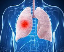
THORACIC SURGERY Objective Questions 11. Which of the following approaches is/are currently acceptable for the management of spontaneous pneumothorax? A. Chest tube replacement alone for the patient with a first episode. B. Operation on presentation for any patient with a first episode. C. Video-assisted thoracic surgery (VATS) bleb excision and pleurodesis for recurrent pneumothorax on the same side. D. Thoracotomy with bleb excision and pleurodesis for unilateral recurrent pneumothorax. E. Operation after a first episode in an airline pilot. Answer: ACDE 12. For which patient(s) with a pulmonary infiltrate of uncertain cause would you favor VATS over open wedge excision? A. An AIDS patient with a diffuse infiltrate who is ambulatory but requires supplemental oxygen. Bronchoalveolar lavage is negative. B. A 64-year-old previously healthy man with increasing shortness of breath, a diffuse infiltrate, and restrictive lung disease as shown by pulmonary function studies. C. A 74-year-old diabetic woman with a rapidly progressing process throughout the right lung who is ventilator- and pressor-dependent. D. A 44-year-old man with fever, left-sided infiltrate, and shortness of breath. E. A 79-year-old man on a ventilator for right lower and middle lobe pneumonia which has been culture negative. Answer: ABD 13. Which of the following statements about the cause and prevention of postintubation tracheal stenosis are correct? A. Postintubation airway stenosis can largely be avoided by providing assisted ventilation via endotracheal tube rather than tracheostomy tube. B. Postintubation tracheal stenosis at the cuff level results, more or less equally, from low blood pressure, advanced age, steroids, high intracuff pressure, sensitivity to tube materials, gas sterilization elution products, and systemic disease. C. In women and smaller men large endotracheal tubes can produce lesions of the glottis and subglottis that can progress to stenosis. D. Stomal stenosis is due principally to cicatricial closure of large stomas resulting from removal of a disk or segment of tracheal wall during tracheostomy. E. A large-volume tracheostomy tube cuff such as that now used on most available tubes can become a high-pressure cuff if filled beyond its resting maximal volume. Answer: CE 14. Which of the following statements about the treatment of postintubation airway stenosis are correct? A. Emergency management of airway obstruction due to stenosis at the level of a prior tracheal stoma is best accomplished by establishing a new tracheostomy in normal tracheal tissue just below the scar of the old stoma. B. Radial lasering and dilatation usually leads to permanent resolution of postintubation tracheal stenosis. C. Splinting of a cervical trachea with a silicone T-tube for 6 to 8 months generally leads to permanent resolution of stricture. D. Postintubation tracheal stenosis that extends into the subglottic larynx is treated by resection of a cylindrical sleeve of stenotic airway and end-to-end reconstruction. E. Acquired tracheoesophageal fistula due to intubation injury is corrected by surgical closure of the fistula concurrent with resection and reconstruction of the damaged trachea. Answer: E 15. Which of the following statements are true? A. Pyogenic lung abscess occurs most frequently in the lower lobe of the left lung. B. Anaerobic bacteria are commonly present in pyogenic lung abscess. C. Operation is usually required to eradicate a pyogenic lung abscess. D. Penicillin is the treatment of choice for lung abscess. Answer: BD 16. Amphotericin B is effective for the following lung infections: A. Histoplasmosis. B. North American blastomycosis. C. Aspergillosis. D. Mucormycosis. E. Sporotrichosis. Answer: ABD 17. The following statements are true. A. A distinguishing roentgenographic appearance of lung abscess, the air-fluid level can be seen only on roentgenograms obtained in the upright or lateral decubitus position. B. The fungus ball characteristic of aspergillosis can be seen roentgenographically in either the upright or recumbent position. C. Actinomycosis and nocardiosis are both fungal diseases of the lung that respond to treatment with the newer azole antifungal agents. D. The commonest fungal lung infection in the United States is due to Histoplasma capsulatum. Answer: ABD 18. Pneumocystis pneumonia is an opportunistic infection caused by Pneumocystis carinii. Which of the following statements are true? A. P. carinii is a fungus. B. Pneumocystis pneumonia is the most common opportunistic infection in patients with AIDS. C. The diagnosis of Pneumocystis pneumonia depends on the demonstration of P. carinii organisms in lung tissue. D. There is no effective treatment for Pneumocystis pneumonia. Answer: BC 19. Which of the following statements are true? A. The pleural space does not extend into the neck. B. Positive intrapleural pressures as high as 40 cm. H 2O and negative pressures as low as -40 cm. H 2O are possible. C. The pleural cavities cannot absorb more than 500 ml. of fluid per day. D. All pleural effusions are of clinical significance and should be investigated. Answer: BD 20. Which of the following statements are true? A. Chylothorax, or chyle in the pleural cavity, usually is not a serious condition. B. Chyle is easily identified by its milky appearance, which looks like no other kind of pleural effusion. C. The commonest causes of chylothorax are trauma and tumor. D. The thoracic duct can be ligated with impunity. Answer: CD 21. Which of these statements about pleural tumors is/are true? A. The commonest type of pleural tumor is primary pleural mesothelioma. B. Exposure to asbestos dust is causally related to the development of malignant mesothelioma. C. Localized benign mesotheliomas are asymptomatic. D. Complete pleurectomy for malignant mesothelioma usually results in cure. Answer: B 22. Which of the following correctly describe a patient with spontaneous pneumothorax? A. The patient is almost always elderly and debilitated. B. An unsuspected primary or metastatic lung tumor may be present. C. The administration of supplemental oxygen is of little benefit to the patient. D. The patient should always be treated with an intercostal tube and closed pleural drainage. E. Video-assisted thoracic surgery (VATS) should be considered for persistent air leak in patients with secondary spontaneous pneumothorax. Answer: BE 23. Which of the following statements about spontaneous pneumothorax (PSP) is/are correct? A. The risk of recurrence after resolution of the first episode of PSP or secondary spontaneous pneumothorax (SSP) is 35% to 45%. B. Patients with PSP are typically tall, thin, young adult males with a history of smoking. C. Secondary spontaneous pneumothorax is associated with family history in 10% of cases. D. For bleb resection and pleurodesis thoracoscopic thoracotomy and open thoracotomy provide similar cure rates for patients with primary spontaneous pneumothorax. E. Causes of secondary pneumothorax include trauma and iatrogenic needle puncture. Answer: ABD 24. Which of the following are relative contraindications for surgical management of emphysema? A. Rapidly progressive dyspnea. B. Bullae occupying less than one third of a hemithorax on plain chest radiography. C. Elevated room air PCO 2. D. “Pink puffer” patients. E. FEV 1 less than 35% of predicted value. Answer: BCE 25. Which of the following treatments would be appropriate therapy for symptoms that persist on medical therapy and bronchiectasis involving, in order of decreasing severity, the left lower lobe, the right middle lobe, and the left upper lobe? A. Left pneumonectomy. B. Wedge resection of the left lower lobe. C. Left lower lobectomy. D. Simultaneous left lower lobectomy and right middle lobectomy. Answer: C 26. Which of the following would not be acceptable sequences of preoperative studies in a patient being prepared for lingulectomy for bronchiectasis? A. CT alone. B. CT, bronchoscopy, bronchography. C. Bronchoscopy alone. D. Bronchoscopy, bronchography. Answer: C 27. Which of the following statements about pulmonary mycobacterial infection is/are correct? A. Worldwide, tuberculosis no longer represents a significant public health problem. B. Mycobacterium tuberculosis is responsible for the majority of cases of pulmonary mycobacterial disease. C. Mycobacterium kansasii pulmonary infection almost always requires surgical treatment. D. Atypical mycobacteria are never primary pulmonary pathogens in humans. Answer: B 28. Which of the following chemotherapeutic regimens are currently recommended for the treatment of pulmonary infection caused by M. tuberculosis? A. Isoniazid, rifampin, pyrazinamide, and streptomycin for 24 months. B. Isoniazid for 9 months with ethambutol for the first 3 months. C. Isoniazid and rifampin for 6 months with pyrazinamide added for the first two months. D. Isoniazid alternating with rifampin at 3-month intervals for 12 months. E. Isoniazid and rifampin for 9 months. Answer: CE 29. Which of the following are appropriate indications for pulmonary resection for mycobacterial disease? A. Localized pulmonary disease caused by M. avium-intracellulare. B. Advanced lobar tuberculous pneumonia with massive hilar lymphadenopathy and bronchial obstruction in a young child. D. An asymptomatic tuberculous cavity greater than 12 cm. in diameter. E. Massive hemoptysis from a right upper lobe cavity occurring during an appropriate course of chemotherapy for pulmonary tuberculosis in a sputum-negative patient. Answer: AE 30. Which statements about squamous papillomatosis of the trachea is/are correct? A. It is the most common type of benign tracheal tumor in adults. B. It is the most common type of benign tracheal tumor in children. C. Most are treated with segmental tracheal resection. D. There is no risk of malignant degeneration. E. It is associated with a herpesvirus. Answer: A 31. Which of the following statements about pulmonary hamartomas is/are true? A. Hamartomas are benign chondromas. B. Most are located in the conducting airways. C. Wedge resection is curative. D. A lobectomy is necessary to obtain draining hilar lymph nodes. E. Hemoptysis is common. Answer: C 32. Which of the following statements about typical carcinoid tumors are true? A. They make up the majority of bronchial adenomas. B. They frequently have lymph node metastases. C. The carcinoid syndrome is observed in 33%. D. Overall survival at 5 years is 90%. E. Overall survival at 5 years is 50%. Answer: AD 33. Which is/are true of adenoid cystic carcinoma? A. It is a common type of salivary gland tumor. B. Another name is cylindroma. C. Most patients are completely resected for cure. D. Different histological types have different prognoses. E. Tissue invasion is rare. Answer: ABCD 34. A solitary pulmonary nodule is discovered in an asymptomatic 55-year-old smoker with no evidence of extrathoracic dissemination. The most appropriate management would be to: A. Obtain serial chest films every 3 months to determine the growth potential of the nodule. B. Perform transthoracic needle aspiration (TTNA) before considering pulmonary resection to confirm malignancy. C. Conduct an extensive systematic evaluation to exclude the possibility that the nodule represents a metastatic lesion. D. Proceed with pulmonary resection after ascertaining that the patient would tolerate removal of the requisite amount of lung. E. Obtain baseline serum levels of carcinoembryonic antigen and p53. Answer: D 35. After thoracotomy, pulmonary resection, and mediastinal lymph node dissection, a patient is determined to have a squamous cell carcinoma 2 cm. in diameter, located 1 cm. from the carina along the right mainstem bronchus. Three peribronchial lymph nodes are positive for cancer, and all other lymph node stations are negative. The correct stage, according to the TNM system, is: A. T1N0M0 Stage I. B. T1N1M0 Stage II. C. T2N1M0 Stage II. D. T3N1M0 Stage IIIa. E. T2N3M0 Stage IIIb. Answer: C 36. After complete resection of Stage I non-small cell lung cancer (NSCLC), the role of adjuvant therapy is best summarized thus as: A. Postoperative radiation therapy improves disease-free survival. B. Postoperative radiation therapy improves overall survival. C. Postoperative chemotherapy improves disease-free survival. D. Postoperative chemotherapy improves overall survival. E. Adjuvant therapy is not indicated after complete resection of Stage I NSCLC. Answer: E 37. Compared to segmentectomy or wedge resection, lobectomy for NSCLC is associated with: A. Similar operative morbidity but higher operative mortality. B. Similar operative mortality but higher operative morbidity. C. More severe postoperative pulmonary dysfunction. D. Lower incidence of locoregional recurrence. E. Equivalent locoregional recurrence. Answer: D 38. In contrast to NSCLC, small cell lung cancer (SCLC) is characterized by: A. Greater response rate to chemotherapy. B. Inability to achieve surgical cure. C. Less frequent association with paraneoplastic syndromes at the time of diagnosis. D. Lower likelihood of metastases present at the time of diagnosis. E. Slower growth. Answer: A 39. Which of the following statements about the diagnosis and staging of mesothelioma is/are correct? A. Fluid obtained by thoracentesis is usually adequate for accurate diagnosis. B. Open biopsy or thoracoscopy should be performed to obtain tissue for diagnosis. C. Immunohistochemistry should be performed in all cases of suspected mesothelioma. D. Chest CT and/or magnetic resonance imaging (MRI) are useful in the staging of mesothelioma. E. Head CT and bone scans are useful in the staging of mesothelioma. Answer: BCD 40. Which of the following statements about therapy for malignant pleural mesothelioma is/are correct? A. The role of surgery is confined to biopsy for diagnosis and pleurodesis for palliation of effusion. B. Extrapleural pneumonectomy involves resection en bloc of the lung, visceral and parietal pleura, pericardium, and diaphragm. C. If a lesion is unresectable by extrapleural pneumonectomy, pleurectomy/decortication is contraindicated. D. Neither surgery, chemotherapy, nor radiation therapy as a single therapy improves survival. E. Multimodality therapy, combining surgery, chemotherapy, and radiation therapy may improve survival in select patients. Answer: BDE 41. All of the following may be acceptable operative approaches to management of the thoracic outlet syndrome except: A. Scalenectomy. B. Excision of a cervical rib. C. Thoracoplasty. D. First rib resection. E. Division of anomalous fibromuscular bands. Answer: C 42. Initial conservative (nonsurgical) management of the thoracic outlet syndrome may include all of the following except: A. Weight reduction. B. Improvement of posture. C. Exercises to strengthen the muscles of the shoulder girdle. D. Pentoxifylline. E. Avoiding hyperabduction. Answer: D 43. Which of the following statements about pectus excavatum are correct? A. It is the most common congenital malformation of the chest wall. B. The most frequent presenting complaint is the cosmetic deformity. C. The manubrium and first and second costal cartilages typically are involved in the deformity. D. It may be associated with cardiac defects and other skeletal defects such as scoliosis. E. Restrictive alterations in chest wall mechanics and abnormalities in pulmonary function tests have been documented. Answer: ABDE 44. Surgical correction of pectus excavatum is characterized by which of the following? A. Significant cosmetic improvement initially but a high incidence of recurrence of the defect on late follow-up. B. An increase in exercise tolerance and respiratory reserve postoperatively. C. Improvement in FEV 1, vital capacity, and total lung capacity. D. Improvement in maximal ventilatory volume, total progressive exercise time, and maximal exercise capacity. E. Prevention of the development of “thoracogenic scoliosis.” Answer: BDE 45. Which of the following statements about the diagnosis of chest wall tumors is/are correct? A. Pain is a common presenting symptom. B. Firmness and fixation to underlying bone and muscle are important to note in the physical examination as aids to diagnosis. C. In general, chest wall tumors are slow growing and produce symptoms late in their course. D. CT is the most useful imaging study for making the diagnosis and for planning surgical resection of chest wall tumors. E. Angiography should be performed routinely. Answer: BCD THORACIC SURGERY Objective type Questions with Answers 46. Which of the following statements about chest wall resection and reconstruction is/are correct? A. Most tumors of soft tissue and bone require 4-cm. margins to be adequately resected. B. At least one normal rib above and below the primary tumor should be included in the resection. C. Techniques of chest wall reconstruction are directed at the prevention of paradoxical chest wall movement with respiration. D. Soft tissue defects are most conveniently addressed by stretching the existing skin over the defect under tension. E. Chest wall defects that are covered by the scapula require no special reconstructive procedures, even if the defects are quite large. Answer: ABCE 47. Prolonged extracorporeal membrane oxygenation (ECMO): A. Is highly successful in the treatment of severe respiratory failure in newborn infants. B. Is contraindicated in adult respiratory distress syndrome (ARDS). C. Causes hemolysis and renal failure. D. Requires total systemic heparinization (activated clotting time longer than 500 seconds). E. Is identical to heart/lung bypass for cardiac surgery. Answer: A 48. Indications for ECMO include: A. Newborn infants with pulmonary hypoplasia secondary to congenital diaphragmatic hernia. B. Meconium aspiration syndrome in full-term babies (at least 35 weeks). C. Children with pulmonary infection after bone marrow transplantation. D. Adults with acute viral pneumonia. E. Adults requiring mechanical ventilation and 100% oxygen for 2 weeks or longer. Answer: BD 49. Venovenous ECMO: A. Avoids major arterial access. B. Provides cardiac and pulmonary support. C. Can be accomplished via cannulation at separate venous sites or at a single venous site using a double-lumen catheter. D. Provides greater venous drainage than venoarterial ECMO. E. Maintains the normal pulsatile blood flow to the systemic circulation. Answer: ACE 50. As compared with venovenous ECMO, venoarterial ECMO: A. Requires cannulation of a major artery and vein. B. Provides both cardiac and respiratory support. C. Can be performed with less anticoagulation. D. Usually maintains a normal pulse pressure. Answer: AB 51. A 24-year-old male has new onset of chest pain. Chest films demonstrate a large anterosuperior mass. Appropriate evaluation should include: A. CT of the chest. B. Measurement of serum alpha-fetoprotein and beta–human chorionic gonadotropin. C. A barium swallow. D. A myelogram. Answer: AB 52. Systemic syndromes frequently associated with mediastinal tumors include: A. Myasthenia gravis. B. Hypercalcemia. C. Malignant hypertension. D. Carcinoid syndrome. Answer: ABC 53. A 36-year-old female developed dyspnea on exertion that has progressed over 3 months. Chest film reveals a left anterior mediastinal mass with evidence of elevated left hemidiaphragm. CT indicates probable invasion of the pericardium. Paratracheal or subcarinal adenopathy is not identified. Appropriate intervention in this patient would include: A. A median sternotomy with radical resection of the tumor, sacrificing the left phrenic nerve and excising the involved pericardium. B. A mediastinoscopy with biopsy. C. A left anterolateral thoracotomy or median sternotomy with generous biopsy of the tumor. D. Observation with repeat chest radiography in 3 months. Answer: C 54. An 18-year-old male presents with a history of increasing shortness of breath that worsens in the recumbent position. On physical examination, the neck veins are noted to be distended, with facial plethora that is accentuated by lying the patient down. A 2.5-cm. left supraclavicular lymph node is palpable. Chest film reveals an extensive right anterosuperior mediastinal mass. Appropriate intervention may include: A. An urgent biopsy of the mediastinal mass under general anesthesia with subsequent initiation of therapy. B. CT. C. Pulmonary function testing in the sitting and supine positions. D. A biopsy of the right supraclavicular lymph node under general anesthesia. E. A biopsy of the supraclavicular lymph node under local anesthesia. Answer: BCE 55. A 42-year-old male who is scheduled to undergo elective knee surgery has a preoperative chest film that demonstrates a 5-cm. posterior mediastinal mass. The patient denies any neurologic symptoms and physical examination fails to elucidate any neurologic deficit. CT confirms the presence of a 5-cm. mediastinal mass in the left costovertebral gutter with minimal enlargement of the seventh thoracic foramen. Appropriate intervention includes: A. Resection of the posterior mediastinal mass using a standard posterolateral incision. B. A CT with myelography or magnetic resonance (MR) imaging. C. Two-stage removal of the tumor, performing the resection of the thoracic component first with subsequent removal of the spinal column component at a later date. D. One-stage removal of the dumb-bell tumor, excising the intraspinal component prior to resection of the thoracic component. Answer: BD 56. True statements regarding patients with a mediastinal mass include: A. Asymptomatic patients have a benign mass in over 75% of cases. B. Symptomatic patients are more likely to have a malignant lesion than a benign lesion. C. In a patient with a chest film demonstrating a mediastinal mass, a Tru-cut needle biopsy is a safe procedure. D. Seminomas usually produce alpha-fetoprotein. Answer: AB 57. Which of the following would be the least appropriate in the management of acute suppurative mediastinitis? A. Wide débridement. B. Irrigation under pressure. C. Topical antibacterials. D. Long-term systemic antibacterials. E. Closure with muscle flaps. Answer: D 58. Each of the following is appropriate for managing acute suppurative mediastinitis except: A. Alloplastic material and skin flaps. B. Rectus abdominis muscle flaps. C. Omentum. D. Pectoralis major muscle flaps. E. Rigid internal fixation. Answer: A 59. Clinical features suggestive of myasthenia gravis include all of the following except: A. Proximal muscle weakness. B. Diplopia. C. Sensory deficits of the extremities. D. Dysphagia. Answer: C 60. The diagnosis of myasthenia gravis can be confirmed most reliably using: A. Anti–acetylcholine receptor antibody titers. B. The Tensilon test. C. Electromyography (EMG). D. Single-fiber EMG. E. Physical examination. Answer: D 61. All of the following statements are true about the pathogenesis of myasthenia gravis except: A. The number of functional acetylcholine receptors at the motor end plate is reduced. B. An autoimmune mechanism involving antibodies to the acetylcholine receptor has been proposed. C. Complement system involvement has been demonstrated. D. A nonspecific “thymitis” may initiate the autoimmune response. E. Clinical improvement following thymectomy is correlated with decreased acetylcholine receptor antibody titers. Answer: E 62. Which of the following statements about the relationship of the thymus and myasthenia gravis is/are true? A. Thymic abnormalities are present in up to 80% of patients with myasthenia gravis. B. Thymoma is present in up to 20% of patients with myasthenia gravis. C. Myasthenia gravis will occur in up to 60% of patients with thymomas. D. Myasthenia patients with thymoma respond more favorably to thymectomy. E. Thymoma is the most common abnormality of the thymus in patients with myasthenia gravis. Answer: ABC 63. Which of the following statements about the results of thymectomy for myasthenia gravis are true? A. Patients with ocular symptoms experience clinical improvement in 90% of cases. B. Clinical remission can be expected in 90% of cases. C. The response rate to thymectomy for patients with generalized symptoms is 90%. D. Patients with thymoma experience improvement in 75%. E. Continued medical therapy is required in 75%. Answer: C 64. All of the following are true of the treatment of myasthenia gravis except: A. The transcervical approach to surgical thymectomy is less likely to benefit the patient with myasthenia gravis. B. Corticosteroids result in improvement in 80% of patients. C. Plasma exchange is associated with improvement in up to 90% of patients. D. Medical therapy with Mestinon (pyridostigmine) is associated with remission in approximately 10% of patients. E. Surgical thymectomy, regardless of the approach, is associated with improved remission and response rates as compared with medical therapy. Answer: A 65. Which of the following is/are acceptable alternatives in the management of malignant pericardial effusion? A. Pericardiocentesis. B. Subxiphoid pericardiotomy (“pericardial window”). C. Thoracotomy with pericardiectomy. D. Instillation of tetracycline or bleomycin into the pericardial space. E. Treatment of the underlying malignancy. Answer: ABCDE 66. Which of the following statements about cardiac tamponade is/are correct? A. At least 500 ml. of fluid must be present in the pericardium of an adult to cause symptoms of tamponade. B. A drop in systemic blood pressure of greater than 20 mm. Hg during inspiration (pulsus paradoxus) is a finding specific to cardiac tamponade. C. The vast majority of patients with cardiac tamponade demonstrate a low QRS voltage, nonspecific ST T-wave abnormalities, and electrical alternans (alternation of QRS amplitude) on the electrocardiogram. D. In trauma victims with cardiac tamponade, the three components of “Beck's triad” (hypotension, elevated jugular venous pressure (JVP), and muffled heart sounds) are almost always present. E. When the diagnosis is made, treatment must be instituted rapidly and may include pericardiocentesis, creation of a pericardial window, and identification and treatment of the underlying cause. Answer: E 67. Which of the following statements about constrictive pericarditis is/are correct? A. Most patients who develop constrictive pericarditis after cardiac operation present with symptoms within 6 months of the procedure. B. Results of pericardiectomy for constrictive pericarditis are worse in patients who develop constriction after mediastinal irradiation. C. Drainage of asymptomatic pericardial effusions arising from acute pericarditis is advised to prevent development of constrictive pericarditis. D. If surgical treatment is planned for constrictive pericarditis it should involve total or complete pericardiectomy. E. Echocardiography can usually make the diagnosis by imaging a thickened pericardium. Answer: BD 68. The relationship between small-cell and non-small cell lung cancers can be described by the following: a. They differ by histology, clinical behavior and cell of origin b. Of all lung cancers, approximately 80% are non-small cell and 20% are small cell c. Both cell types are predictably responsive to chemotherapy d. The International Staging System can be applied to both tumor types e. The majority of non-small cell cancer patients vs. the minority of small cell cancer patients are candidates for pulmonary resection Answer: b 69. A 62-year-old male smoker presents with right anterior chest pain. There is a 3 cm mass attached to the chest wall with radiographic evidence of rib erosion and positive cytology for non-small cell carcinoma. Which of the follow is/are true: a. The patient is inoperable due to tumor size and chest wall involvement b. Radiation therapy is the preferred initial treatment c. Operative resection should be performed with en bloc removal of the tumor and adjacent chest wall as well as a mediastinal lymph node resection d. Positive mediastinal nodes will have little effect on survival e. The patient would be classified Stage IIIa Answer: c, e 70. For the patient in the pervious question to become an operative candidate which of the following must be met? a. Extrathoracic metastases must be able to be controlled by another modality, e.g. radiotherapy b. Tumor doubling time must exceed 40 days c. If there is recurrence at the primary site, it must be treated before the metastatic disease d. Even if effective systemic therapy is available, resection of metastases is preferred e. If pulmonary reserve is marginal, resection of the maximal number of metastatic foci should be performed Answer: c 71. Biopsy of the lesion in the previous question is reported as “bronchial carcinoid with no signs of atypia.” Which of the follow is/are true? a. Sleeve resection of the bronchus would be appropriate b. Lymph node biopsy at time of resection is unnecessary c. Associated carcinoid syndrome is very unlikely d. If carcinoid syndrome were found in a tumor this size, hepatic metastases would be likely e. When bronchial carcinoid syndrome occurs, right-sided cardiac valves are affected Answer: a, c, d 72. In the evaluation and preparation of a 55-year-old smoker for resection of a 3 cm pulmonary adenocarcinoma, the following is/are true: a. Preoperative cessation of smoking does not reduce postoperative pulmonary complications b. Resting PaCO2 is of more value than PaO2 c. FEV1 is of more value than measured vital capacity d. Diffusion capacity should be measured routinely e. V/Q lung scan is useful when pulmonary reserve is marginal Answer: b, c, e 73. Following resection of a T1N1 squamous cell cancer in a 47-year-old male, the following is/are true: a. There is a higher risk of local recurrence than with any other histologic type of non-small cell cancer b. The greatest risk to the patient is a distant metastasis c. Of all metastatic sites, liver is most likely d. If the patient survives five years, there is a greater risk of a new lung cancer than recurrence e. To improve survival, the patient should be considered for adjuvant chemotherapy Answer: a, b, d 74. A 42-year-old woman with hemoptysis is seen to have a 2 cm mulberry appearing polypoid lesion in the left mainstem bronchus suspicious for bronchial adenoma. The differential diagnosis includes which of the following: a. Mucoepidermoid carcinoma b. Plasma cell granuloma c. Carcinoid tumor d. Adenoid cystic carcinoma e. Mucous gland adenoma Answer: all of the above 75. A 42-year-old man has a solitary “coin lesion” 2 cm in diameter in the area of the right upper lobe on a routine chest radiograph. Which of the following is/are true? a. A previous radiograph from five years prior showing the lesion to be 1.2 cm in diameter indicates malignancy b. If a CT scan shows mediastinal adenopathy, mediastinoscopy is preferable to thoracotomy c. In the absence of previous radiographs, the lesion should be followed by serial films at 6 month intervals d. Calcification in a concentric or “popcorn” configuration denotes a benign lesion e. Needle aspiration showing “chronic inflammatory cells” denotes a benign lesion Answer: b, d 76. A 2 cm peripheral squamous cell carcinoma in the lung of a 60-year-old male with a pleural effusion positive for malignant cells would be classified as: a. T1N0M1 b. T3N0M0 c. T3N0M1 d. T4N0M0 e. T4N0M1 Answer: d, e 77. A 53-year-old woman who had a malignant tumor removed 2 years ago presents with a solitary lung nodule 1.5 cm in diameter. The following is/are true: a. If the primary tumor originated in the breast, the lesion is most likely to represent a new primary lung cancer. b. If the primary tumor was melanoma, the lesion is most likely to be metastatic c. If the remainder of the lung fields are clear, a CT scan is unnecessary d. If the primary tumor was in the GI tract, there is very little chance that the lesion is a new primary lung cancer e. Fine needle aspiration should always be performed prior to resection of the lung lesion Answer: a, b 78. A 61-year-old male presents with a painful mass 3.5 cm in diameter below the clavicle and attached to the chest wall. The following is/are true: a. A CT scan is the best study to determine rib destruction b. The lesion should be removed enbloc without biopsy to minimize the chances for local recurrence c. The chances are approximately 40% that the lesion is metastatic d. If it is metastatic, the most likely primary tumor is in the lung or pancreas e. Fortunately, less than 50% of chest wall tumors are malignant Answer: c 79. Concerning the sternum, the following is/are true: a. The xiphoid process is the anterior border of the thoracic outlet b. Gladiolus is the body of the sternum c. The angle of Louis is at the level of the 2nd costal cartilage d. The 11th rib is attached via costal cartilage to the xiphoid e. The sterno-manubrial junction is at the level of T4 posteriorly Answer: a, b, c, e 80. A 22-year-old woman recovering from a traumatic head injury is noted to have bright red bleeding when her tracheostomy is suctioned. The following is/are true statement(s): a. Antibiotics should be administered to treat the bronchitis b. Deflation of the tracheal tube cuff is a useful diagnostic maneuver c. If massive bleeding occurs, a finger should be used to compress the innominate artery against the sternum d. Operative treatment of a tracheoinnominate fistula includes resection and prosthetic replacement of the innominate artery e. Tracheal resection is usually required for a tracheoinnominate fistula to prevent recurrence Answer: b, c 81. A 52-year-old alcoholic with fever and a cough productive of purulent sputum is found to have the opacity on chest film as shown (Fig. 62-15). The following is/are true statement(s): a. The findings suggest a parapneumonic empyema b. If pus is found on aspiration of the pleural space, a chest tube should be placed c. If pus is found on aspiration, bronchoscopy is a necessary part of the patient’s evaluation d. In this situation, rib resection for drainage is preferred to a large-bore chest tube e. Decortication of the lung should be considered if the lung fails to expand within 4 weeks Answer: a, b, c 82. The lesion shown (Fig. 62-6) was found on a 32-year-old male on a routine chest film required for his employment. Which of the following is/are true? a. The stippled calcification and intact cortex of the rib are characteristic of osteochondroma b. The stippled calcification is characteristic of osteogenic sarcoma c. If the lesion is osteogenic sarcoma, the optimal treatment is resection and radiation therapy d. If the lesion is an osteochondroma, it need not be resected in this age group e. The radiographic picture is typical for Ewing sarcoma Answer: a 83. To resect a chondrosarcoma of the chest wall in a 42-year-old man, ribs 2–4 were removed, leaving a defect 8 x 8 cm. For reconstruction, the following is/are true: a. If this were to be posterior, beneath the scapula, reconstruction would not be required b. If this defect is anterior, the primary benefit of reconstruction is an improved cosmetic result c. Whenever chest wall reconstruction is considered, it should be delayed 6–12 months to allow detection of recurrent tumor d. If Marlex is used for reconstruction, no wound drainage tube is necessary e. If PTFE is used for reconstruction, both pleural and wound tubes should be used Answer: a, d, e 84. An upright chest film of a cachectic, homeless 47-year-old woman shows blunting of the right costophrenic angle. The following is/are true: a. A lateral decubitus film should be obtained to confirm the presence of fluid rather than a CT scan b. Tuberculous effusion can readily be identified by stain and culture of aspirated fluid c. A pleural fluid glucose level lower than in the serum is diagnostic of empyema d. Bloody pleural effusion in this patient is diagnostic of an underlying malignancy e. Pleural fluid cytology report of lumphoma should be viewed with skepticism Answer: a, e 85. The pectoralis major muscle is available and innervated by the medial and lateral pectoral nerves so named because it describes their relationship to the pectoralis minor a. The serratus anterior muscle is available since its absence has no functional significance b. There is no serratus posterior muscle c. The latissimus dorsi muscle is available and supplied by the thoracodorsal artery d. The latissimus dorsi is innervated by the thoracodorsal nerve with fibers from C6, C7 and C8 Answer: d, e 86. A 38-year-old man presents with facial and upper extremity edema, venous distention in the neck and arms and a cyanotic appearance. The following is/are true statement(s): a. The most likely cause of the problem is mediastinal granulomatous disease b. A venogram should be obtained to confirm the diagnosis c. Mediastinoscopy for diagnosis is contraindicated d. If a malignancy is identified, resection is indicated for palliation e. If the etiology is benign disease, gradual improvement without operation is to be expected Answer: e 87. A 39-year-old woman with hypertension and radicular chest wall pain was found to have the lesion seen on chest radiograph (Fig. 63-23). The following is/are true statement(s): a. The location of the lesion suggests a teratoma b. High urinary vanillylmandelic acid levels would indicate that the lesion is a paraganglioma c. If the lesion was seen on a film 5 years earlier, resection would not be indicated d. A neurosurgical consultation should be obtained e. Vasoactive intestinal polypeptide level elevation suggests a ganglioneuroma Answer: d, e THORACIC SURGERY Questions and Answers pdf Download Read the full article
0 notes