#Radiology reporting
Explore tagged Tumblr posts
Visit Tumblr Blog
Explore Tumblr blogs with no restrictions, modern design and the best experience.
Last Seen Tumblr Blogs


Fun Fact
Tumblr has been providing a Korean-language service since 2013.
Text
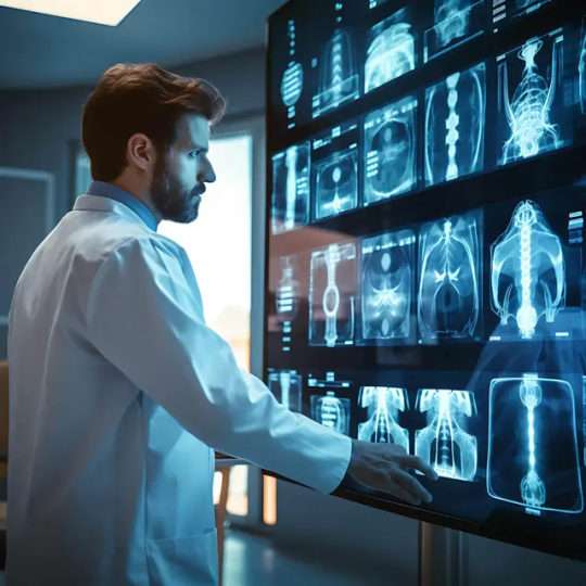
Vistarad Advantages of teleradiology in Kenya
Improving access to healthcare in remote areas One of the significant advantages of teleradiology in Kenya is its ability to improve access to healthcare in remote areas. By leveraging digital technology, medical images can be instantly transmitted to radiologists in urban centres where specialized expertise is available. This eliminates the need for patients in rural areas to travel long distances for diagnosis, saving time, money, and potentially lives. Teleradiology brings healthcare closer to those who need it the most, ensuring that even the most underserved communities have access to high-quality diagnostic services.
Reducing healthcare costs through teleradiology Another advantage of teleradiology is its potential to reduce healthcare costs in Kenya. The traditional model of radiology requires the physical presence of a radiologist at each healthcare facility, which can be expensive and impractical, especially in rural areas. Teleradiology allows multiple healthcare facilities to share the services of a few radiologists, optimizing their expertise and reducing costs. Additionally, by avoiding unnecessary transfers and duplicate imaging tests, teleradiology helps in cost containment and promotes efficient resource utilization.
Enhancing diagnostic accuracy and efficiency Teleradiology plays a crucial role in enhancing diagnostic accuracy and efficiency in kenya. By connecting healthcare facilities with experienced radiologists, teleradiology ensures that every image is interpreted by a specialist. This eliminates the possibility of errors or misdiagnoses that can occur when relying on general practitioners or inexperienced radiologists. Moreover, the remote nature of teleradiology enables radiologists to interpret images at their own pace, without the time pressure of an on-site setting. This leads to more thorough analysis and improved diagnostic accuracy, ultimately benefiting patient care.
0 notes
Text
Best Radiology Reporting Services
Vital Radiology is providing the best radiology reporting services, delivering reliable and detailed interpretations of medical images. Our team of experienced radiologists ensures timely reports for various imaging modalities. With a commitment to quality and time, we provide healthcare professionals with the vital information they need for diagnosis and treatment planning. Trust us for efficient and dependable radiology reporting services. Kindly visit our website to learn more.

0 notes
Text
boy i love digging through my own medical records to find out any info on whether i might have cancer because none of my doctors will tell me shit, it’s not stressful at all
#from what i can tell the answer is probably not#I don’t have my ultrasound results yet though so we’ll see if they call me in for a biopsy#but I could literally feel my body falling apart from the stress before I found the radiology reports myself#why is our medical system so useless
8 notes
·
View notes
Text
guess who biffed it off the back step yesterday and fractured her ankle 💕

#they somehow lost my xrays???#but they said the radiology dept was able to see them and write a report but couldnt upload them back into the system or something idk#so now im braced and booted and have to hobble around for a few weeks smh#dense rambles#personal#foot hurty
6 notes
·
View notes
Text

How Molecular Breast Imaging (MBI) Enhances Early Cancer Detection Breast cancer is one of the most common cancers affecting women, and early detection can be life-saving. At Uevolve Radiology, we are dedicated to providing the best breast imaging services in India, using the latest diagnostic tools to detect breast cancer as early and accurately as possible.
While mammograms remain the gold standard for screening, they aren’t perfect—especially for women with dense breast tissue. That’s why we offer Molecular Breast Imaging (MBI), an innovative technology that enhances detection and gives women the answers they need.
Why Imaging Matters in Breast Cancer Detection
At Uevolve Radiology, we use a variety of imaging techniques to ensure precise and early detection:
Mammography:– A low-dose X-ray effective for early detection but less reliable in dense breast tissue.
Breast Ultrasound:– Uses sound waves to differentiate solid and cystic masses, helping to clarify suspicious findings from a mammogram.
Breast MRI:– Highly sensitive and ideal for high-risk patients, though not always the best option for routine screening.
M3D Mammography (Breast Tomosynthesis):– Provides a more detailed, layered view of the breast, improving accuracy over traditional mammograms.
Molecular Breast Imaging (MBI):– A breakthrough functional imaging technique that detects metabolic activity, making it particularly useful for women with dense breasts.
As part of our commitment to excellence, we ensure that our diagnostic center offers the best breast imaging services in India, combining advanced technology with expert analysis.

What Makes MBI a Game-Changer?
Molecular Breast Imaging (MBI) works differently than traditional imaging. It uses a small amount of radioactive tracer that is absorbed by metabolically active cells—like cancer cells. A special camera detects these "hot spots," giving doctors a clearer picture, even when dense tissue makes other imaging methods less effective.


Benefits of MBI:
✔️ More Accurate for Dense Breasts – MBI isn't affected by breast density, improving detection rates. ✔️ Early Cancer Detection – By identifying metabolic activity, MBI can spot cancers earlier than traditional mammography. ✔️ Enhanced Sensitivity – It can catch cancers that might be missed by a mammogram alone.
Who Should Consider MBI? MBI is often recommended for women who:
Have dense breast tissue
Are at higher risk of breast cancer
Have unclear or inconclusive mammogram results

Why Expert Interpretation Matters
Even the best imaging technology is only as effective as the experts who analyze the results. At Uevolve Radiology, our team of highly trained radiologists specializes in breast imaging, ensuring precise and accurate diagnoses.
With state-of-the-art technology and expert interpretation, we take pride in offering the best breast imaging services in India, helping women take charge of their breast health with confidence.
Taking Charge of Your Breast Health

Know Your Risk Factors – Family history, lifestyle, and age all play a role.
Stay Up to Date on Screenings – Talk to your doctor about the best screening options for you, including MBI.
Perform Regular Self-Exams – Being familiar with your body can help you detect changes early.
Uevolve Radiology: Your Partner in Breast Health
At Uevolve Radiology, we are committed to providing the best breast imaging services in India with advanced imaging technology, expert radiologists, and a patient-first approach.
If you’re looking for top-notch breast cancer screening, Molecular Breast Imaging, or dense breast solutions, we’re here to help.
Want to learn more about MBI and whether it’s right for you? Contact Uevolve Radiology today!
#breast imaging#best radiology reporting near me#emergency radiology#digital x-ray clinics mumbai#neuroradiology imaging#digital x-ray clinics reporting near me#top ct scan centers in mumbai#top ct scan reporting near me#best radiology center in mumbai#digital x-ray clinics near me
0 notes
Text
Advances in Medical Imaging Techniques | AI TELERADIOLOGY
Our state-of-the-art platform, medical imaging technology, for instance, integrates seamlessly with your systems, ensuring secure, efficient communication and data transfer. For further details contact us at 8769609414.
0 notes
Text
Global Digital Radiology Market Report: Analysis of Market Segmentation and Trends
The global Digital Radiology Market, valued at USD 3.8 billion in 2023, is set for steady growth in the coming years, with a projected value of USD 5.04 billion by 2031. This growth represents a compound annual growth rate (CAGR) of 3.6% from 2024 to 2031. The increasing adoption of advanced imaging technologies, along with the growing demand for enhanced diagnostic tools in healthcare, are driving the expansion of the digital radiology market.
Get Free Sample Report on Digital Radiology Market
Digital radiology refers to the use of digital imaging equipment to capture X-rays and other forms of radiological data, which are then processed and displayed on a computer screen. It is used in various applications, including diagnostics, treatment planning, and surgical guidance. As healthcare systems around the world shift towards more efficient and accurate imaging methods, digital radiology continues to gain traction across hospitals, clinics, and diagnostic imaging centers.
Key Drivers of Market Growth
Advancements in Digital Imaging Technology One of the main factors propelling the growth of the digital radiology market is the rapid advancement of imaging technology. Digital radiology offers significant advantages over traditional film-based radiography, such as faster imaging, superior image quality, and the ability to store and share images electronically. These improvements enhance diagnostic accuracy, reduce patient waiting times, and streamline workflows in healthcare facilities.
New innovations in digital radiology include improved detectors, advanced image processing algorithms, and integration with artificial intelligence (AI). AI-enabled tools, for instance, can help radiologists identify abnormalities with greater precision and speed, significantly improving the efficiency of the diagnostic process. The growing presence of AI in radiology is expected to have a profound impact on the market, offering opportunities for continued growth.
Growing Demand for Non-Invasive Diagnostic Methods The increasing preference for non-invasive diagnostic methods, such as digital radiology, is driving the market forward. As patients and healthcare providers seek safer and more effective diagnostic techniques, the demand for imaging methods that don’t require surgery or biopsies has surged. Digital radiology is at the forefront of this trend, offering a quick and accurate means of diagnosing conditions such as fractures, infections, tumors, and lung diseases.
The ability to obtain high-quality images with minimal radiation exposure is also a key benefit of digital radiology. This factor contributes to its growing popularity, especially in pediatric care and for patients who require frequent imaging, as it reduces the risks associated with excessive radiation.
Increasing Adoption of Healthcare IT Solutions The healthcare sector is undergoing a digital transformation, with an increasing adoption of healthcare IT solutions like electronic health records (EHRs), cloud-based systems, and picture archiving and communication systems (PACS). The integration of digital radiology with these IT systems has streamlined the process of capturing, storing, and accessing medical images, making it easier for healthcare professionals to collaborate and improve patient care.
KEY MARKET SEGMENT:
By Product Type
By Application
By Technology
By end-user
Key Market Trends
Shift Toward Portable and Point-of-Care Imaging Solutions Another notable trend in the digital radiology market is the shift towards portable and point-of-care imaging solutions. Portable digital radiology systems are gaining popularity in various healthcare settings, including emergency rooms, outpatient clinics, and home healthcare. These systems offer the advantage of providing immediate imaging results at the point of care, eliminating the need to transport patients to radiology departments.
The increasing demand for mobile radiology equipment, such as handheld X-ray devices, is expected to play a significant role in the market’s growth, particularly in underserved or remote areas where access to traditional imaging facilities may be limited.
AI and Machine Learning Integration The integration of artificial intelligence (AI) and machine learning (ML) into digital radiology systems is transforming the industry. AI algorithms are being used to assist radiologists in interpreting medical images, identifying abnormalities, and providing more accurate diagnostic outcomes. For example, AI-powered software can automatically highlight potential issues in images, allowing radiologists to focus on areas that require closer inspection.
The continued development of AI in digital radiology is expected to improve diagnostic accuracy, reduce the risk of human error, and enhance overall workflow efficiency. As AI becomes more advanced and integrated into digital radiology systems, its impact on the market will grow.
Make Enquiry about Digital Radiology Market
Increased Focus on Preventive Healthcare Preventive healthcare, which emphasizes the early detection of diseases to prevent progression and improve outcomes, is another key driver of the digital radiology market. With growing awareness of the benefits of early diagnosis, more patients are seeking preventive care through regular imaging tests. Digital radiology plays a crucial role in this trend, offering fast, accurate, and non-invasive diagnostic tools for routine screenings, such as lung cancer, breast cancer, and cardiovascular conditions.
The increased focus on preventive healthcare, supported by digital imaging technologies, is expected to contribute to the market’s expansion in the coming years.
Key Players:
The Major Players are Agfa-Gevaertgroup, Cannon Inc., Detection Technology, Fujifilm Holdings, Hitachi Ltd, General Electrics, Medtronics, Koninklijke Phillips, Samsung Electronics Co Ltd, and Others Players.
Regional Insights
North America currently holds the largest share of the global digital radiology market, primarily due to the advanced healthcare infrastructure, high adoption rates of digital imaging technologies, and the presence of major industry players in the region. The United States is a key contributor to this growth, with hospitals and diagnostic centers increasingly adopting digital radiology to improve diagnostic efficiency and patient care.
However, the Asia-Pacific region is expected to experience the highest growth rate over the forecast period. Factors such as the growing healthcare infrastructure, rising healthcare expenditures, and increasing awareness of the benefits of digital radiology are driving the demand for digital imaging solutions in countries like China, India, and Japan. The market in these regions is also supported by government initiatives aimed at improving healthcare access and expanding the use of advanced diagnostic technologies.
Conclusion
The digital radiology market is on a steady growth trajectory, projected to reach USD 5.04 billion by 2031, driven by technological advancements, the increasing demand for non-invasive diagnostics, and the growing adoption of healthcare IT solutions. As healthcare continues to prioritize accuracy, efficiency, and patient-centered care, digital radiology will play an increasingly vital role in meeting these needs. With innovations in AI, cloud-based solutions, and portable imaging devices, the future of digital radiology holds great promise, offering new opportunities for industry players and improved outcomes for patients worldwide.
About US:
SNS Insider is one of the leading market research and consulting agencies that dominates the market research industry globally. Our company's aim is to give clients the knowledge they require in order to function in changing circumstances. In order to give you current, accurate market data, consumer insights, and opinions so that you can make decisions with confidence, we employ a variety of techniques, including surveys, video talks, and focus groups around the world.
Contact Us:
Jagney Dave - Vice President of Client Engagement
Phone: +1-315 636 4242 (US) | +44- 20 3290 5010 (UK)
#Digital Radiology Market#Digital Radiology Market Size#Digital Radiology Market Share#Digital Radiology Market Growth#Digital Radiology Market Trends#Veterinary Services Industry#Digital Radiology Market Report
0 notes
Text
#Medical Imaging Equipment Market#Medical Imaging Industry#Market Research Report#Diagnostic Imaging Equipment#Radiology and Imaging Technology#Medical Imaging Devices#Market Size and Forecast#X-Ray Imaging Systems#MRI (Magnetic Resonance Imaging)#CT (Computed Tomography) Scanners#Ultrasound Imaging Systems#Nuclear Imaging Equipment#Competitive Landscape#Emerging Trends in Medical Imaging#Portable Imaging Devices#AI in Medical Imaging#Global Healthcare Imaging Market#Imaging Equipment for Diagnostics#Advanced Imaging Technologies#Medical Imaging in Telemedicine
0 notes
Text
instagram
Breaking Boundaries in Radiology! 🏥🌍
Radiology is no longer limited by location—Teleradiology brings expert diagnoses to your fingertips! 📡🩻 With AI-powered tools & 24/7 reporting, we ensure accurate & timely care whenever you need it.
💡 Choose Uevolve for advanced radiology solutions! 📅 Book your consultation today!
🔗 www.uevolve.in
#uevolveradiology#teleradiology#aIinhealthcare#expertdiagnostics#24x7care#healthcareinnovation#radiologysolutions#fasterreports#patientcare
#Best radiology reporting in Manchester#Affordable CT Scan reporting Manchester#Private Ultrasound Clinic in Manchester#Instagram
0 notes
Text
Factors to consider when choosing a teleradiology service provider | Vistarad
Selecting the most suitable teleradiology service provider is a significant decision that can greatly influence the quality of patient care. The certifications and accreditation of the service represent critical factors to consider. It is essential to verify that the radiologists analyzing the images are board-certified and have the requisite experience in the relevant specialization, as well as ensuring that the teleradiology service adheres to industry standards and regulations.
This ensures that the medical facility follows best practices and that patients obtain interpretations of the highest caliber.

0 notes
Text
Best Radiology Reporting Services
Vital Radiology is your trusted place for reliable and timely radiology reporting. Our team of skilled radiologists provides comprehensive and detailed reports, ensuring efficient diagnosis and treatment planning. With state-of-the-art technology and a commitment to excellence, we deliver reliable results for healthcare professionals. Trust Vital Radiology for all your radiology reporting needs, because faithfulness matters. Visit our website now for more information.

0 notes
Text
Diversity of Radiological Imaging and Clinical Course in Pulmonary MALT Lymphoma by Aras G1, Zirek Mandal T1, Kanmaz D1, Pehlivan S1, Fener N2, Özbek in Journal of Clinical Case Reports Medical Images and Health SciencesM3.
Abstract :
A 59-year-old male patient was admitted to the emergency department with a three-month history of worsening dyspnea, fatigue, and cough. His vital signs were recorded as follows: blood pressure 138/88 mmHg, heart rate 98 beats/min, respiratory rate 25 breaths/min, temperature 37.8°C, and oxygen saturation 91%. During the lung auscultation, breath sounds were absent in the lower left lung, while crepitant rales were audible in the upper zone and the right lung. CT scan of the chest showed 1 cm lymph nodes, pleural effusion, fibrotic changes, varicose-cystic bronchiectasis, as well as consolidations and atelectasis with air bronchograms in both lungs. Furthermore, thoracic ultrasonography revealed a large effusion in the left hemithorax, measuring 11 cm. He was hospitalized after the placement of a pleural catheter. Radiological diversity and the clinical course of the patient posed challenges for establishing a differential diagnosis. TL; DR: In this paper; we aimed to present a case of MALT lymphoma that manifested in the lung and caused diagnostic confusion with radiological and clinical symptoms.
Introduction:
MALT (Mucosa-Associated Lymphoid Tissue) is the lymphoid tissue that plays a role in mucosal defense. It includes functional memory B lymphocytes, which are essential for the immune response. They are not physiologically present in the lungs, but they become active there in response to infections and chronic antigenic stimulation. Marginal zone B-cell non-Hodgkin lymphoma (MALT Lymphoma) accounts for 8% of adult lymphomas. Although it is most commonly seen in the stomach, it can also be seen in the salivary glands, thyroid and lungs (1). It is the most common type of lymphoma in the lung. It presents common radiological, pathological, and clinical findings with infection and other granulomatous diseases, making differential diagnosis quite difficult for the clinician.In this paper, we aimed to present our case, which we had difficulty in diagnosing in the clinic.
Case Presentation:
A 59-year-old male patient was admitted to the emergency department with worsening dyspnea, fatigue, and cough over the past three months. The chest X-ray of the patient showed a consolidation extending from the center to the periphery in the left upper zone near the aortic arch, an increased density indicative of pleural effusion with sinus obliteration on the left, and a consolidation extending from the hilum to the lower zone near the heart edge During the lung auscultation, breath sounds were absent in the lower left lung, while crepitant rales were detected in the upper zone and on the right side. Blood pressure was 138/88 mmHg, pulse was 98 beats /min, respiratory rate was 34 breaths/min, and oxygen saturation was 91%. At the time of admission, the patient's biochemical analysis results were as follows: Glucose 77 mg/dL (normal range: 70-115), urea 31 mg/dL (normal range: 17-43), creatinine 0.77 mg/dL, protein 65.8 g/L (normal range: 66- 83), albumin 39.3 g/dL (normal range: 35-53), LDH 423 U/L (normal range: <247), CRP 21.6 mg/L, procalcitonin <0.01ng/mL, and pro-BNP 8 pg/mL. The hemogram evaluation results were as follows: Leukocyte count 8.27 x 10³/µL, Erythrocyte count 5.16 x 10⁶/uL, Hemoglobin 15.6 g/ dL, Hematocrit (Hct) 48.3%, Platelet count 240,000/uL, Lymphocyte count 2.05 x 10³/uL, Eosinophils 3.4%, and Neutrophil/Lymphocyte ratio 2.47 x 10e3/uL.
The patient’s chest CT scan showed 1 cm lymph nodes in the mediastinum, fibrotic changes extending to the pleura, varicose-cystic bronchiectasis, and consolidations and atelectasis with air bronchograms in both lungs. There was also a massive fluid of 11 cm in the left hemithorax. (Figure 2). A thoracentesis was conducted on the patient’s left side following the detection of 13 cm of pleural fluid on thoracic ultrasonography. Lymphocytes, polymorphonuclear leukocytes, and mesothelial cells were observed in the cytological examination of the pleural fluid, but no atypical cells were detected. There was 98% lymphocyte dominance in the cell count. In the biochemical analysis, the pH was 7.440, lactate dehydrogenase (LDH) was 206 U/L, total protein was 39.40 g/dL, albumin was 25 g/dL, glucose was 60 mg/dL, and adenosine deaminase (ADA) was 34.4 U/L. Gram staining of the fluid, bacterial, fungal, and acid-fast bacilli growth were all negative. The patient's fluid was drained by aspiration. An intrapleural catheter was placed due to the high amount of fluid and increased dyspnea. The patient was admitted to the ward and initiated on oxygen therapy, bronchodilators, and antibiotics. The pleural fluid sent for cytological analysis two more times during the patient's hospitalization was found to be serohemorrhagic. Upon reevaluating his microbiological results, no growth was observed. Although many lymphoid cells were seen in the cytopathological examination of the patient's second pleural fluid, no atypical features were monitored. The patient's condition stabilized during clinical follow-up, and the pleural catheter was removed. However, after a while, the patient's dyspnea complaint recurred and the catheter was placed again because of the increase in fluid on the radiograph (Figure 3). There was no change in the infection markers of the patient, who also had fever from time to time, and CRP ranged between 20 and 16 mg/dL during follow-up. There was no growth in his small amount of sputum and blood cultures taken during the fever. The patient underwent fiberoptic bronchoscopy and no endobronchial lesions were detected. Wang fine needle aspiration and bronchial lavage were applied to mediastinal lymphadenomegaly Wang IA revealed lymphoid cells, but a definitive diagnosis could not be obtained. No findings were found in the lavage other than bronchial epithelial cells and polymorphonuclear leukocytes
No FDG uptake was detected in the pleural fluid during the patient's whole-body positron emission tomography (PET-CT) scan, though minimal FDG uptake was observed in certain pleural areas. Consolidated/ground glass foci with the focal lepidic appearance in places were detected with left lung lingular, lower lobe central SUVmax 9.14, and right lung lower lobe SUVmax 6.55 and were evaluated to be in favor of malignant processes. Abdominal ultrasonography was unremarkable. No extrapulmonary findings were monitored in PET-CT scan either.
When Wang IA did not yield any results, endoscopic ultrasonographic bronchoscopy (EBUS) was performed. The pathological interpretation was in favor of granulomatous inflammation, as mature transformed lymphocytes, polymorphonuclear leukocytes, epithelioid histiocytes, and loose granuloma-like structures formed by epithelioid histiocyte clusters and multinucleated giant cells were observed in the materials obtained. Alveolar sarcoidosis was taken into account, but the serum angiotensin converting enzyme level was also found to be normal at 39.1 U/L (8- 52.0).
The patient was discharged due to the clinical stability of the patient with CRP 3.2 and procalcitonin <0.01 and was called for a follow-up at a later date. In the meantime, the patient was discussed at the surgical council. A decision was made to perform video-assisted thoracoscopy due to the fluid not regressing, increased dyspnea, and malignant involvement in PET-CT.
During the procedure conducted under general anesthesia, 400 cc of fluid was aspirated. Biopsies were obtained from two distinct areas of the pleura and from a nodular region on the diaphragm, followed by talc pleurodesis. Samples were sent to microbiology and pathology. The patient, having experienced no complications, was discharged following the procedure . Pathology: Samples taken from the parietal pleura and the nodule on the diaphragm were evaluated as low-grade non-Hodgkin Lymphoma and interpreted as extra-nodal marginal zone lymphoma (MALT) by the pathologist .
Discussion
Clinical: The patient, who had been admitted to the emergency room with symptoms of dyspnea, hypoxia, and fever, was hospitalized after pleural fluid was exudate and consolidation was detected. Although the patient's clinical symptoms were severe, CRP was moderately high, and the occasional fever despite antibiotic therapy during hospitalization suggested diagnoses such as malignancy and tuberculosis, in addition to non-specific infection. Lymphomatous proliferation can involve the lung in various ways. Non-Hodgkin or Hodgkin lymphoma can present in the lung through hematogenous spread or by invading from adjacent mediastinal lymph nodes. However, primary involvement of the lung is also possible. Primary lymphomas should not have extra-pulmonary organ involvement for at least 3 months after diagnosis. The most common are MALT and effusion lymphomas (2). Effusion lymphomas may involve the pericardial and peritoneal cavities, with the most common primary involvement being the pleura, without solid organ involvement. Human-Herpes-8 infection and EBV may be accompanied by fever and lymphocytic-exudative fluid. HHV-8 negative cases have a better prognosis (3). In this case, there was also an exudative pleural effusion. However, although pleural involvement was not detected in the fluid cytological examination, pleural involvement was detected in biopsies. However, it is not possible to say that the patient only has effusion lymphoma (PEL). MALT (Mucosa-associated Lymphoid Tissue) lymphoma is the type of lymphoma that most commonly involves pulmonary tissue, is often asymptomatic, and shows radiological alveolar opacities. Although it is more commonly affected in people aged 50-60, it can rarely be seen under the age of 30. It constitutes 60% of pulmonary lymphomas. Weight loss and fever are especially prominent during the aggressive phase, though the condition may initially be asymptomatic. Autoimmune disease may be the basis in 16% of cases (1,4). The prognosis of MALT lymphomas is good; 5–10-year survival is more than 80% (5).
Radiological:
MALT lymphoma exhibits radiological variability, appearing as single or multiple bilateral lesions on both chest radiography and thoracic tomography. Chronic alveolar localized opacities smaller than 5 cm on radiography are accompanied by consolidation in 50% of cases. Diffuse reticulonodular opacity, atelectasis, and pleural effusion are detected in less than 10% of cases (1). In our case, there was also pleural fluid along with similar findings. Findings such as consolidation (60-77%) with air bronchogram and increased vascularity or multiple mass nodules, ground glass, halo sign, galaxy sign specific to tuberculosis and sarcoidosis have been reported in case series and reports. In addition, varicose cystic bronchiectasis secondary to consolidation can be detected. However, it is not possible to state that these findings are specific to MALT lymphoma. These radiological images are also observed in adenocarcinoma, pneumonia, metastases, sarcoidosis, and tuberculosis (6,7). In this case, chest X-ray revealed bilateral multifocal consolidation and densities indicative of pleural effusion. Consolidation, pleural effusion, varicose cystic bronchiectasis changes and mediastinal lymphadenomegaly were detected on thoracic tomography. None of these radiological findings were specific to lymphoma and diagnosis could not have been made without pathological examination. The partial alleviation of the patient's clinical symptoms compared to the beginning and the patient's relief with the drainage of the pleural fluid suggested possibilities such as infection, alveolar sarcoidosis, or adenocarcinoma when we did not have pathological data. Nevertheless, when intermittent fevers began to occur during the clinical course, it was obvious that lymphoma could not be excluded from the diagnosis. No endobronchial lesion was detected in the bronchoscopy performed on the patient; Wang fine needle aspiration and then endobronchial ultrasonographic biopsy were performed for mediastinal lymphadenomegaly. The sensitivity and specificity of positron emission tomography (PET-CT) in lymphoma varies according to organ involvement. It has been reported as 80-100% in lung involvement (8, 9). In our case, high SUVmax FDG uptake in consolidated foci in lepidic structure was monitored in PET-CT, and pleural uptake was minimal. The presence of clinical symptoms and high FDG uptake necessitated a video-assisted-thoracoscopic (VATS) procedure.
Pathological:
In the pleural fluid sample examined at the start of the treatment, abundant lymphoid cells were detected, but no atypical structures were observed. There were findings of granulomatous inflammation in the samples obtained from the mediastinal lymph nodes of the patient. In fact, in the biopsy samples taken by video-assisted thoracoscopy, lowgrade B-cell non-Hodgkin Lymphoma (CD20 positive (diffuse cells), anti-BCL-2 positive, anti Ki-67 low positive) and extranodal marginal zone lymphoma were detected in the parietal pleura. Pathologically, the lymphomatous infiltrate in MALT lymphoma exhibits heterogeneous features and consists of small lymphocytes, centrocyte-like cells, monocytoid B cells, rarely large transformed cells and plasma cells (10). Necrosis is rare. Neoplastic cells are expressed in CD 20 and CD 79. Ki67 index is lower than 20%. Lymphoma and sarcoidosis are similar in terms of clinical and radiological phenotype. Distinguishing lowgrade lymphomas from sarcoid lesions can be challenging; sarcoid granulomatous lesions may be accompanied by lymphoid cell infiltration. In addition, sarcoid-like reactions are frequently seen in malignant lymphomas. Moreover, sarcoidosis-lymphoma syndrome was first described in the study by Brincker et al. They reported that lymphoma was 5.5 times more common in sarcoidosis patients than in the general population, indicating the presence of sarcoidosis years before lymphoma (11, 12). Kokuho N et al reported MALT lymphoma in the lung for the first time in a ten-year-old sarcoidosis case with ocular, gastric and lung involvement (13). In this case, granulomatous inflammation was detected in mediastinal LAMs, but no findings of sarcoidosis were found in ocular, cardiac, renal examinations, angiotensinconverting enzyme, calcium, alkaline phosphatase, and 24- hour urine calcium analyses. There was also no abnormality in abdominal ultrasonography. We believe that the granulomatous inflammation in our case was reactive to immune deficiency.
Conclusion The patient was started on Rituximab treatment by the hematology department. Clinical and radiological improvement was observed following treatment. In the follow-up PET-CT examination, regression in consolidated areas, decrease in FDG uptake, and metabolic partial regression were detected compared to the initial examination (Figure 6). Lymphomas, which do not have specific radiological and symptomatic features, can mimic most diseases of the respiratory system and do not present with a noisy picture, requiring the clinician to be persistent in making the diagnosis.
References:
1. Borie R, Wislez M, Antoine M, Cadranel J (2017), Lymphoproliferative Disorders of the Lung. Respiration 94:157-175 2. Cadranel J, Wislez M, Antoine M (2002), Primary pulmonary lymphoma. Eur Respir J 20:750-62 3. Kattih Z, Mahajan A, Vojnic M, et al. (2022), Rapidly Accumulating Effusion in an Immunocompetent Woman, Chest 161: e377-e382 4. Wislez M, Thabut G, Antoine M et al. (2009); Clinical characteristics and prognostic factors of pulmonary MALT lymphoma. European Respiratory Journal 234: 1408-1416 5. Koss MN (2004) Malignant and benign lymphoid lesions of the lung. Ann Diagn Pathol 8:167-87 6. Song Y, Sung YE, Beck KS, et al. (2023) Radiological and pathological analysis of the galaxy sign in patients with pulmonary mucosaassociated lymphoid tissue (MALT) lymphoma. Thorac Cancer 14:2459-2466 7. Deng W, Wan Y, Yu JQ (2019) Pulmonary MALT Lymphoma has variable features on CT. Scientific Reports 9:8657 8. Albano D, Borghesi A, Bosio G, et al (2017) Pulmonary mucosaassociated lymphoid tissue lymphoma: 18F-FDG PET/CT and CT findings in 28 patients. Br J Radiol 90:20170311 9. Enomoto K, Hamada K, Inohara H, et al (2008) Mucosa-associated lymphoid tissue lymphoma studied with FDG-PET: a comparison with CT and endoscopic findings. Ann Nucl Med 22:261-267 10. Pina-Oviedo S, Roggli VL, Sporn TA, et al (2023) Diagnostic Approach to Pulmonary B-Cell Lymphomas in Small Biopsies, with Practical Recommendations to Avoid Misinterpretation. Diagnostics (Basel) 13:3321 11. Brincker H (1986) The sarcoidosis-lymphoma syndrome. Br J Cancer 54:467–73 12. El Jammal T, Pavic M, Gerfaud-Valentin M, et al. Sarcoidosis and Cancer: A Complex Relationship. Front Med (Lausanne) (2020) 24:594118 13. Kokuho N, Terasaki Y, Urushiyama H, et al. (2016) Pulmonary mucosa-associated lymphoid tissue lymphoma associated with pulmonary sarcoidosis: a case report and literature review. Hum Pathol 51:57-63
#Diversity#Radiological Imaging#d Clinical#Course in Pulmonary MALT Lymphoma#jcrmhs#Journal of Clinical Case Reports Medical Images and Health Sciences.
0 notes
Text

Vital Radiology excels in neuro radiology reporting, providing specialized diagnostic services exclusively for healthcare providers. Our team of expert radiologists delivers timely interpretations of complex neurological images, supporting reliable diagnoses and effective treatment planning. Utilizing cutting-edge imaging technology, we ensure reliable and comprehensive reporting, enhancing the quality of care for patients with neurological conditions. Trust us for expert neuro radiology reporting that advances clinical decision-making.
0 notes
Text

Empowering Doctors & Consultants with Accurate Radiology Reads
In modern healthcare, precision is everything. A misdiagnosis can delay treatment, while an accurate radiology report can save lives. That’s where Uevolve Radiology steps in—bridging the gap between doctors and precise imaging insights to improve patient outcomes.
A Story That Matters: When Radiology Made the Difference
Meet Dr. Mehta, a cardiologist handling a critical case. His patient, Ananya, came in with chest discomfort, and an immediate diagnosis was essential. However, his hospital didn’t have an in-house radiologist at that hour.
With Uevolve Radiology’s remote reporting, Ananya’s CT scan was analyzed within minutes, confirming a mild cardiac condition. Thanks to timely and accurate radiology reads, she received the right treatment without unnecessary delays or invasive procedures.
This is how teleradiology revolutionizes patient care, ensuring doctors have reliable data at the right time.
Why Uevolve Radiology is Every Doctor’s Trusted Partner
🔹 Fast & Accurate Reports – Enabling quick decision-making with precise imaging insights. 🔹 Seamless Collaboration – Doctors and radiologists work hand in hand, even from different locations. 🔹 AI-Enhanced Analysis – Combining the expertise of radiologists with AI tools for better accuracy. 🔹 Round-the-Clock Availability – 24/7 access to expert reads, ensuring no patient waits too long.
SEO Optimization for Google Ranking
📌 Primary Keywords: (radiology reporting for doctors, teleradiology services, AI-powered radiology, accurate radiology reads, remote radiology for hospitals) 📌 Human-Centric Approach: Real-life case study adds depth and relatability. 📌 Trust-Building Content: Explains how teleradiology supports doctors in providing better care.
🚀 Join the future of radiology with Uevolve Radiology! Supporting doctors, consultants, and patients with reliable, data-driven diagnostics. Contact us today.
#Best radiology reporting near me#Top CT scan reporting near me#Digital X-ray clinics reporting near me
0 notes
Text
#Mammography Teleradiology#Remote Mammography Reporting#Teleradiology Mammogram Services#Breast Imaging Teleradiology#Digital Mammography Interpretation#Online Mammogram Reporting#Telemedicine for Mammography#Breast Cancer Screening Teleradiology#Mammogram Second Opinion Online#Telemedicine Radiology for Mammography#Mammogram Tele-reporting#Remote Breast Imaging Diagnostics#Virtual Radiology Mammography#Teleradiology Breast Health
0 notes
Text
The Interventional Radiology Products Market is expected to reach US$ 19.61 billion by 2031 at a CAGR of 6.82%.
#Interventional Radiology Products Market#Interventional Radiology Products Market Growth#Interventional Radiology Products Market Report
0 notes