#Polytrichum
Explore tagged Tumblr posts
Text
#2369 - Polytrichum commune - Common Haircap Moss


AKA great golden maidenhair, and great goldilocks.
Common Haircap is common throughout temperate and boreal latitudes in the Northern Hemisphere, Mexico, Australia, and several Pacific Islands including New Zealand.
A species of large moss growing in damp acidic areas, including in open areas disturbed by human activitoes and livestock. The stems can be 70 cm, which it manages by have tissues specialised to transport water. That's the standard for the 'higher', vascular plants - practically the defining feature, in fact - but is extremely unusual for mosses.
The genus also have parallel lamellae on the upper surfaces of the leaves, with rows of photosynthetic tissue capped with large cells that trap moist air between them but still alow gas exchange. Most mosses have a simple plate of photosynthetic tissue. If conditions become too dry the leaves will twist around the stem.
Pohokura, North Island, New Zealand
121 notes
·
View notes
Text
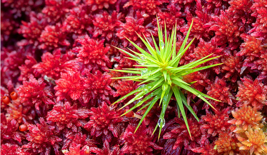
Polytricum Moss (Polytrichum spp.) growing through a bed of red Sphagnum Moss (Sphagnum spp.) in blanket bog
Photo by Alex Hyde
#polytricum moss#polytrichum#moss#sphagnum#sphagnum moss#bog#bog moss#red#red moss#red plants#plants#botanical#plant photography#nature
122 notes
·
View notes
Text

The only thing green in these woods
#landscape#landscape photography#photography#nature#nature photography#naturecore#moss#haircap moss#polytrichum#woods#trees#forest#kentucky
41 notes
·
View notes
Text
Mod 1: Bryophytes
Ngl I have no idea how to prepare for the botany paper, but I might as well try something-
Polytrichum
Classification:
Class - Bryopsida
Order - Polytrichales
Family - Polytrichaceae
Genus - Polytrichum
Gametophyte
External Structure:
The nature of gametophyte of polytrichum is differentiated into 2 parts: A horizontal underground rhizome and an erect leafy shoot.
Rhizome: The horizontally growing underground portion of the gametophore. It bears small, brown or colorless leaves in three rows and numerous rhizoids.
Leafy Shoot: The erect leafy axis arising from the rhizome. It is differentiated into a stem-like central axis which bears dark green expansions. The so called leaves are spirally arranged in 3/8 phyllotaxy.
The leaf is differentiated into a sheathing leaf base and an apical limb. The limb is narrow, lanceolate, and green. Leaf base is colorless and membranous, closely clasped around the stem.
A short transition zone is present between the rhizome and the aerial shoot. It bears small brown leaves similar to those on rhizome.

Internal Structure:
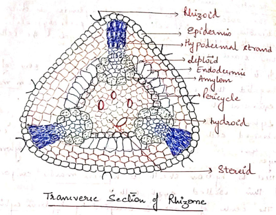
I. Rhizome: In most of the species, the rhizome is triangular in outline with rounded corners. A transverse section of rhizome shows the following regions:
a) Epidermis: The outermost layer of thick walled cells. They give rise to rhizoids.
b) Cortex: The epidermis is followed by cortex consisting of 3-4 layer of thin walled parenchymatous cells.
It is interrupted by 3 hypodermal strands that extend radially from periphery to center.
Hypodermal strands composed of prosenchymatous cells contain starch grains. Extending inwards from each hypodermal strand is a group of lignified cells containing radial strands.
Next to the cortex, there is a layer of radially elongated cells with suberized thickening on their walls. This layer can be compared to the endodermis of higher plants.
c) Pericycle: A 2-3 layered parenchymatous pericycle is present between endodermis and the central conducing strand. It is discontinuous.
d) Leptoids: There are thin walled polygonal cells present in furrows opposite to the radial strands. They are like sieve tubes of phloem hence called leptoids. The cells collectively form lepton.
e) Amylom: The innermost later of leptoids is separated from the central cylinder by a single layer of parenchymatous cells containing starch. This layer is called amylom.
f) Central Cylinder: Situated in the central region of the rhizome. Made up of 2 types of cells: thick walled living cells called stereids and empty dead cells called hydroids. Both of them together make up the hydrom.
II. Aerial Shoot: In transverse section, the aerial shoot is irregularly lobed due to the presence of leaf bases. It is differentiated into 3 regions:
a) Epidermis: The superficial uniseriate later. Discontinuous due to the persistent leaf bases.
b) Cortex: Epidermis is followed by broad cortex. Outer cortex is made of prosechymatous cells and inner is made of parenchymatous cells.
In young shoots, the cortical cells contain chloroplasts.
A large number of leaf traces are also present in the cortex.
c) Central Cylinders: The outermost later of central cylinders is discontinuous and is composed of parenchymatous cells. It represents rudimentary pericycle. The cells store starch grains.
It is followed by 3-4 layered irregular zone of elongated thin walled cells. This zone is leptom mantte ( equivalent to phloem of vascular plants)
Leptom mantte is followed by 1-2 layers of suberized cells which form hydrom sheath. The cells are rich in starch and also called amylom layers.
Next to hydrom sheath is hydrom mantte composed of think walled empty cells.
The center of shoot is occupied by hydrom cylinder made up of thick walled cells. These cells help in conduction of water and hence equivalent to the xylem of higher plants.

III. Leaf
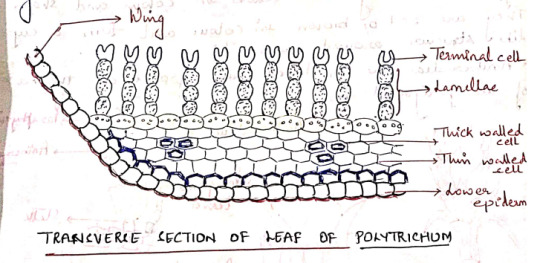
A transverse section of the limb of the leaf shows a thick midrib, gradually tapering into 2 rudimentary wings.
The ventral surface is bound by a distinct epidermis. It is covered by thick cuticle. Epidermis is followed by sclerenchymatous hypodermis.
The ground tissue (cortex) is composed of parenchymatous cells.
The dorsal surface of limb is made up of vertical plates of cells called lamellae. Each lamellae has 4-8 cells; all cells contain chloroplasts except the terminal one. This terminal cell is larger than outer cells and sometimes it is (___)
The arrangement of cells in lamellae and presence of any spaces between adjacent lamellae increases the photosynthetic area.
The wing is composed of a single layer of hyaline cells. Lamellae are not present in the wing region.
The wings become dry and curved when the plants grow in dry habitats.
Reproduction: In polytrichum, reproduction takes place by vegetative and sexual methods.
I. Vegetative Reproduction: Takes place by bulbils which develop on rhizome. Besides, fragmentation of underground rhizome also helps in propagation.
II. Sexual Reproduction: Polytrichum is dioecious, the male (antheridia) and the female (archegonia) sex organs develop at the spices of main shoots of separate plants.
Antheridia:
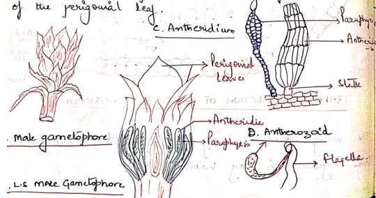
They are born at the apex of the male gametophore. They are surrounded by a whorl of specialized leaves, called perigonial leaves. These leaves differ from vegetative leaves in color and shape. They are red or brown in color and form a cup like structure around antheridia.
Antheridia are produced in groups at the base of the perigonial leaf.
The mature antheridium is a club shaped structure with an elongated body and a short stalk. They body has a single layered jacked of sterile cells, enclosing a mass of androcytes.
At the free distal end of the antheridium is the operculum consisting of a single or few large cells.
Intermingled the antheridia in the antheridial head are steric multicellular hair like structures called as paraphysis.
Aechegonium:
They are borne in groups at the apex of the female gametophore.
The leaves surrounding the archegonia are called the perichaetial leaves. These leaves overlap close over at the top of the archegonial (cluotes??) to form a bud-like structure called the perichaetium.
Intermingled with the archegonia in the cluotes(Still??) are paraphysis.
The mature archegonium is a flask-shaped structure consisting of a long neck, venter, and a stalk.
The venter cavity contains a single spherical egg and a ventral canal cell.
The long, narrow neck consists of 6 vertical rows of neck cells enclosing a narrow neck canal which has a row of neck canal cells (10)

Fertilization:
Water is essential for fertilization. The jacket sells of the mature antheridium swell by absorbing water. The hydrostatic pressure thus generated bursts the operculum and the antherozoids are liberated.
The neck canal cells and the venter canal cell of mature archegonium degenerate forming a mucilaginous mass.
The mucilaginous mass swells up by absorbing water and as such the cover cells present at the tip of the archegonium are pushed apart.
The antherozoids are attracted towards the archegonia and a number of antherozoids swim down the neck but only faces with the egg and this oospore is formed.
Sporophyte
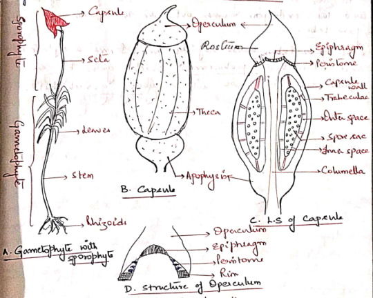
The sporophyte of polytrichum is differentiated into a foot, a long seta, and an angular capsule.
Foot: A dagger-shaped structure embedded in the female gametophore. It is composed of parenchymatous cells. It acts as an anchoring and absorbing organ.
Seta: A long, slender structure between the foot and the capsule. Anatomically, seta is differentiated into epidermis, hypodermis, parenchymatous cortex and a central cylinder, 'hydroids'
The main function of seta is to raise the capsule to the height and conduction of water and nutrients.
Capsule: The capsule is differentiated into 3 parts:
a) Apophysis (Basal part): The distal end of seta gradually enlarges to form a bulbous apophysis ad the base of the capsule. It has a thick walled epidermis continued with that of the seta.
Epidermis is interrupted by stomata, each stoma has 2 guard cells. It is followed by chlorenchymatous cortex and it serves as photosynthetic tissue.
The central part of apophysis is occupied by a conduction strands which is in continuation with the columella and seta (sterile).
b) Theca (Fertile): The middle fertile part of the capsule. It shows many longitudinal grooves. The wall or jacked of theca is composed of several layers of chlorophyllous cells. The outermost layer forms epidermis devoid of stomata. 'Thickwalls', 2-3 layers of cells, chlorenchyma wall layers.
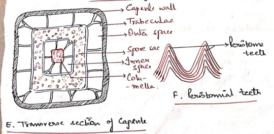
An air space of lacuna is present inner to the wall layers and is divided into small chambers by transverse filaments of chlorophyll containing cells, the trabeculae.
The outer space is followed by space sac. The fertile cells present inside the spore sac constitute the archesporial tissue.
Initially the archesporium is single layered but it becomes many layered in later stages of development of the sporophyte.
The last generation of archesporial-cells differentiates into spore mother cells. Each spore mother cells will form four haploid spores.
The spore sac is also surrounded on the sinner side by an inner air space. It is also transversed by many transverse trabeculae.
The central part of the capsule is occupied by a thick column of parenchymatous cells, the columella. This is a sterile tissue which is continuous with the central axis of the apophysis.
c) Operculum: The apical part of the capsule which forms a cap-like structure at the apex of the theca. It has an extended proximal part, called rostrum, and a projects beak-like distal(?) part.
The boundaries of the operculum and theca are marked by a distinct countriction(??), the rim.
The distal end of the columella is expanded into a pale thin membranous epiphragm at the base of the operculum.
In the mature capsule the peristome is composed of 32 or 64 peristoimal teeth. The teeth are pyramidal and composed of fiber like cells several layers in thickness.
Peristome regulates spore discharge. They do not exhibit hygroscopic movements.
Dehiscence of Capsule:
At maturity, the capsule shrivels as it dries up. The columella disintegrates and the spores come to lie in the cavity thus formed.
Further drying and shriveling of the capsule wall causes the operculum to fall off and thus exposing the peristome.
After exposure of the peristome, thin walled cells of the epiphragm lying between the peristomial teeth also dry and this several minute holes are formed. The spores are dispersed through these holes.
Only a few spores are liberated every time the capsule sways in the wind. This method of spore dispersal is known as censor mechanism.
Young Gametophyte:
The spores are small sounded structures, yellow but turn green immediately after dispersal. The spore wall is differentiated into an outer exospore and an inner endospore.
At the time of germination, the exospore ruptures and the endospore comes out in the form of germ tube.
The germ tube grows rabidly and forms a septate and branched protonema, which grows by an apical cell. It grows into a young gametophyte.
Evolution of Sporophyte in Bryophytes
The sporophyte of the majority of liverworts and mosses show the same plan. It is made up of an anchoring and absorbing foot, a stalk like seta, and a capsule which contains spores and elaters.
A comparative study of the sporophytes shows that there has been a progressive reduction in the amount of the fertile tissues from Marchantials to Bryales.
In all the bryophytes the oospore, formed as a result of fertilization, is the mother cell of sporophyte. In the simplest type of sporophyte the entire oospore is utilized in the formation of spores and capsule wall.
In some simple forms, some spore mother cells, which fail to develop into spores, form elaters.
In complex forms, the oospore develops into a sporophyte which is differentiated into foot, seta, and capsule.
The following 2 contrasting views have been put forwards with regard to the evolution of sporophytes in bryophytes: progressive sterilization and progressive reduction.
Evolution of Sporophyte by Progressive Sterilization
This view, aka "theory of progressive evolution" or "theory of sterilization" was put forward by Bower and supported by Cavers and Campbell.
According to this theory, the sporophytes of the complex forms (ex: Funaria) have evolved due to progressive sterilization of the potential fertile tissue of the simpler forms (ex: Riccia)
In primitive forms the sporophyte is simple and made of the tissue of the sporophyte is fertile (???)
The progressive sterilization from Riccia to Funaria occurred through the following stages:
First Stage: The simplest known sporophyte among bryophytes is that of Ricca. It consists of only capsule, there being no trace of seta and foot.
The entire sporogenous tissue is converted into spores. Few sterile cells (nurse cells) have nutritive function. They are supposed to be ancestors of elaters.
Second Stage: In Corsinia the sterilization has gone a step further and in addition to nurse cells, a sterile foot is also present.
Third Stage: Further sterilization is seen in Sphaerocarpus where the sporophyte has a sterile bulbous foot and a narrow seta, in addition to fertile capsule.
Fourth Stage: In Targionia, the sporophyte is differentiated into a bulbous foot, a massive seta and a capsule.
Amphithecium gives rise to single layered jacked of the capsule and only about half of endothelial cells give rise to fertile sporogenous tissue and the remaining half form sterile elaters. Thus there is further sterilization of sporogenous tissue.
Fifth Stage: In Marchantia, the sporophyte shows foot, seta, and a capsule.
Amphithecium gives rise to a single layered capsule wall and the endothecium forms sporogenous tissue. Approximately 50% of the sporogenous cells forms the spores and the remaining 50% the elaters with thickening bands.
Sixth Stage: The sporophyte of Pellia is also differentiated into foot, seta, and capsule as in Marchantia, but the jacked of the capsule is 2 or more layered. Furthermore a fixes sterile elatophore is present at the proximal end of the capsule with a bunch of elaters.
Seventh Stage: The sporophyte of Anthoceros, consists of bulbus food and a horn or brittle-like capsule. The capsule wall is made up of 4-6 layers of parenchymatous cells.
The outer wall later is epidermis which possesses stomata. The cells of the inner layers of capsule wall have chloroplasts. Thus the sporophyte is capable of synthesizing its own food and is only dependent on gametophyte for water and nutrients.
Another important feature is the presence of central column of sterile cells, the columella.
The tissue of the archesporium differentiates into large fertile spore mother cells and flat and relatively small sterile elater mother cells.
Eighth Stage: The highest degree of sterilization is found in the class Bryopsida.
Regions like foot, seta, capsule wall, columella, apophysis, operculum, and peristome in sporophytes of Funaria and Polytrichum represent the sterile tissue.
The sporogenous tissue is confined only to spore sacs and constitutes a very negligible part of the sporophyte.

Evolution of Sporophyte by Progressive Reduction
Aka Simplification
Kashyap, Church, Goebel, and Evans are the supporters of the retrogressive theory of evolution.
According to this theory, the simple sporophyre of Riccia is the most advanced and has evolved by progressive reduction or simplification of the complex forms such as Funaria and Polytrichum.
The significant steps in the reduction series are:
1. Simplification of the dehiscence apparatus.
2. Reduction of the green photosynthetic tissue in the capsule wall.
3. Disappearance of stomata and intercellular spaces in capsule wall.
4. Decrease in the thickness of the capsule wall.
5. Gradual elimination of seta and foot.
6. There is no progressive increase in the fertility of the sporogenous cells.
Origin of Alteration of Generations
Antithetic Theory:
Celkovasky proposed this theory. On the basis of this theory, the gametophyte or sexual plant represents the original generation.
The sporophyte or the non-sexual plant is a new and different phase evolved by progressive elaboration of the diploid zygote.
The factors which caused its origin are prompt germination of the zygote accompanies by delayed meiosis.
The result is the production of a small sporophyte of Riccia consisting simply of a spore case.
With further elaboration and increased sterilization of the spore producing tissue, a larger sporophyte with differentiation into a foot., a seta, and a capsule is finally evolved.
Homologous Theory:
Pringsheim proposed this theory. According to this theory, sporophyte is not a new generation, but a direct modification of the gametophyte.
It advocated the fact that among the green algae, the gametophyte plant reproduces by both the methods of reproduction.
It bears spores and also the gametes.
In the course of evolutionary sequence, these two functions become separated in two distinct individuals.
The individuals forming spores came to be known as sporophyte and those forming gametes knows as gametophyte.
These two individuals occur regularly alternating in the life cycle.
Both the individuals in primitive land plants where photosynthetic and free living. Gradually the sporophyte became attached to and partly parasitic on the gametophyte. Consequently it became reduced.
Marchantia Thallus
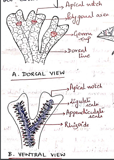
The gametophyte of Marchantia is prostrate, dorsiventral and dichotomously branched. A distinct midrib is present in each branch of thallus.
The apex of each branch is notched and a growing point is situated at the base of each notch.
The dorsal surface of the thallus appears to be masked out into small rhomboidal to polygonal shaped areas called areoles. Which represent the outline of underlying air chambers.
Each areole has got a black dot like spot in the center which marks the position of stoma-like opening leading to air chamber.
Along the midrib on the dorsal surface of the thallus there are certain cup-shaped structures with frilled margins known as gemma cups and they contain special vegetative reproductive bodies called gemmae. (vegetative propagation)
The ventral surface of the thallus bears rhizoids and scales on both sides of the midrib. The rhizoids are usually colorless and unicellular.
Rhizoids are of 2 types: smooth walled and tuberculate. Their function is to:
i) Fix the thallus to substratum.
ii) Absorption of water and nutrients.
The multicellular, one-celled thick scales are arranged in 2-4 rows on either side of the midrib. They are of 2 types:
i) Appendiculate: usually form the inner row of scales, close to the midrib. Have apical appendage.
ii) Ligulate: relatively small and do not have any appendage.
The function of scales is to:
i) Protect the growing point.
ii) Retain some water by capillary action.
The sexually mature thalli possess specialized erect branched called gametophores, which bear the sex organs. These branches arise from the growing apex situated in the apical notch.
The branches present on the male thalli bear the antheridia and are known as antheridiophores while those present on the female thalli bear the archegonia and are called as archegoniophores.
#exam season#send help#biology#notes#plants#botany#bio#bryophytes#bryophyta#polytrichum#plant science#science
10 notes
·
View notes
Text

5 notes
·
View notes
Text
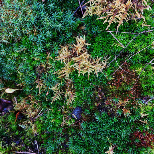
#nature#nature core#nature photography#nature photos#naturecore#original photo blog#woods#polytrichum commune#common haircap#hylocomium splendens#glittering wood-moss
2 notes
·
View notes
Text
Creature 71
Polytrichum commune

Also known as common haircap, great golden ladyhair, great goldilocks or common hair moss, this moss is found in most places with humidity and rain. Polytrichum commune can grow much taller than most other types of moss, in some cases up to 27 inches in length! It is built to retain water much better than the average moss.
fact and image source: wikipedia
1 note
·
View note
Text


a developing slime mold on Polytrichum moss by Anne Iskogen, Sweden.
#plasmodial#myxomycota#moss#slime mold#myxomycology#bryology#ecology#protists#myxomycetes#macro photography#microbiota#microorganisms#microbiology#nature photography#anne iskogen
1K notes
·
View notes
Photo
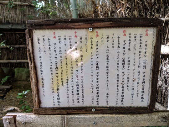

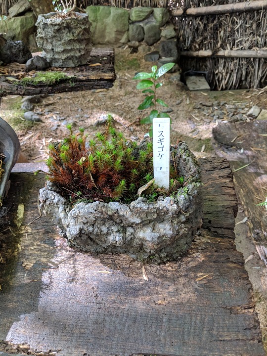

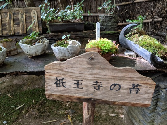
Last set of signs from Gioji Temple! I found a super neat site called Koke Log while doing research on the plants for this post.
1 壱両 アリドオシ [蟻通] アカネ科の常緑小低木 [和名由来] 針がアリを刺し通すほど鋭いことから。 [歴史・文化] センリョウ、マンリョウと一緒に植えて、[千両万両有り通し]と縁起をかつぐ俗信がある。 拾両 ヤブコウジ [藪柑子] ヤブコウジ科の常緑小低木 [和名由来] やぶの中に生え、葉の形や果実がコウジに似ているため。 [歴史・文化] 武家の元服の髪そぎのときに、山菅に添えられた。 百両 カラタチバナ [唐橘] ヤブコウジ科の常緑小低木 [和名由来] ヤマタチバナ (ヤブコウジの古名) に対してつけられた名。 [歴史・文化] 園芸品種が多い、実の色は赤白黄茶桃色など 千両 センリョウ [千両] センリョウ科の常緑小低木 [和名由来] マンリョウ科のカラタチバナが百両とあるのに対して、それよりも美しいという意味で千両とつけられた。 [歴史・文化] センリョウはわが国原産。 万両 マンリョウ [万両] ヤブコウジ科の常緑小低木 [和名由来] センリョウ科のセンリョウ (千両) ゆりも実が美しいため。 [歴史・文化] 江戸時代の頃から園芸品種となる。
2 [Left to right] こけのいろいろ ヒノキゴケ ホソバシラガゴケ オオシラガゴケ ギンゴケ シノブゴケ [No caption] タマゴケ フデゴケ ヤマトフデゴケ ハイゴケ コウヤノマンネングサ [No caption] カモジゴケ スナゴケ ナガエノスナゴケ カサゴケ ミズゴケ
3. 4. 5 スギゴケ ヒノキゴケ (シッポゴケ) 祇王寺の苔
Vocab 壱 (いち) one [in documents] 両 (りょう) old type of coin 蟻通 (ありどおし) Damnacanthus indicus アカネ科 (あかねか) Rubiaceae 常緑 (じょうりょく) evergreen 低木 (ていぼく) shrubbery 千両 (センリョウ) sarcandra glabra/bone-knitted lotus 万両 (まんりょう) ardisia crenata/coralberry 縁起を担ぐ (えんぎをかつぐ) to be superstitious, believe in omens 俗信 (ぞくしん) folk belief 拾 (じゅう) ten [in documents] 藪柑子 (やぶこうじ) ardisia japonica/spearflower ヤブコウジ科 Myrsinaceae やぶ thicket, bush, grove コウジ malt (grows on rice, beans, etc) 元服 (げんぷく) coming-of-age ceremony (Nara-Muromachi periods) 削ぐ (そぐ) to shave (off) 山菅 (やますげ) mountain sedge 添える (そえる) to garnish, accompany 唐橘 (からたちばな) trifoliate orange/hardy orange 園芸品種 (えんげいひんしゅ) cultivar センリョウ科 Chloranthaceae ヒノキゴケ Pyrrhobryum dozyanum ホソバシラガゴケ Leucobryum juniperoideum オオシラガゴケ Leucobryum scabru ギンゴケ Bryum argeteneum シノブゴケ Thuidicaceae (the family was about as specific as I could find) タマゴケ Bartramia pomiformus フデゴケ Campylopus umbellatus ヤマトフデゴケ Campylopus japonica ハイゴケ Hypnum plumaeform Wilson コウヤノマンネングサ Climacium japonicum カモジゴケ Dicranum scoparium スナゴケ Racomitrium canescens ナガエノスナゴケ Racomitrium fasciculare var. atroviride カサゴケ Rhodobryum roseum ミズゴケ Sphagnum (genus) スギゴケ Polytrichum juniperinum 苔 (こけ) moss
202 notes
·
View notes
Text
Common Fennoscandian mosses, pt 2

Juniper haircap (also known as juniper polytrichum moss).

Broom forkmoss

Knights plume moss (also known as ostrich-plume feathermoss).

Girgensohn's bogmoss (also known as Girgensohn's sphagnum or common green peat moss).
#mosses#wild plants#species#forest#nature#naturecore#photography#nature photography#original photography#finland#summer#natural body
11 notes
·
View notes
Text
#2313 - Atrichopsis australe


Another moss from halfway up the cone of Mount Ruapehu.
Some sources have it as australis, and previous names include Polytrichum australe, Psilopilum australe and Notoligotrichum australe.
Related to the previous moss, but beyond that I don't have much info. Mosses in the Polytrichaceae have lamellae which sit perpendicular to the leaf surface, giving the leaves a thicker fleshy appearance compared to most moss. Here's a female plant, from the other side of the ridge west of where I was, found and identified by reingered on iNaturalist. They got most of the bryophyte IDs for me on this trip.

Mount Ruapehu, North Island Volcanic Plateau, New Zealand
11 notes
·
View notes
Note
What are some Clanmew words for moss?
The generic word for moss is simply Fum. Usually this is referring to the helpful sorts of moss they use by default.
And so it's helpful that the most useful type of moss is also the most common. That's;
Cypress-leaved Plait-Moss (Hypnum cupressiforme) = Pafum
The word is mostly broken out when an apprentice brings back a big, dirty clod of a different type of moss. Otherwise, everyone knows it means 'pafum.' Pafum is soft, useful, and good to lay on.
Sneezecloud is SUPER allergic to pafum. He has to collect his own bedding because if he doesn't, he gets really sick and can't get a good night's sleep. Apprentices cannot gather his moss for him, because they will inevitably bring back pafum and he will Suffer.
Though, because the alternative moss isn't nearly as good... usually he just finds other soft things.
Bank Haircap Moss (Polytrichum formosum) = Mwafum
Not quite as soft as Pafum, but it doesn't set off Sneezecloud's allergy so he'll collect it in a bind. Usually, he likes to scatter other, softer things on top of it, so the Mwafum acts as 'padding' but isn't that he's in contact with when he sleeps.
Grimmia (Grimmia pulvinata, and various) = Benum
Various small species which create clusters instead of big blankets, of which Grimmia is the most recognizable. Usually not fluffy enough to fill a nest, unless you take the extra time to collect LOTS of it.
40 notes
·
View notes
Text

Moss of the Polytrichum Piliferum family, Nijmegen, The Netherlands
7 notes
·
View notes
Text

Polytrichum commune is a species of moss found in many regions with high humidity and rainfall. The species can be exceptionally tall for a moss with stems often exceeding 30 cm and rarely reaching 70 cm, but it is most commonly found at shorter lengths of 5 to 10 cm.
often grows from underground rhizoids (filaments). It has been used in stuffing bedding and in the manufacture of brooms, dusters, and baskets.
#awesome#photography#nature#nature photography#woods#spring photography#landscape photography#photography is magic#moss
2 notes
·
View notes
Text
Polytrichum commune.

View On WordPress
0 notes
