#Gastrointestinal Endoscopes
Explore tagged Tumblr posts
ottomed · 3 months ago
Text
Buy Gastrointestinal Endoscopes to spot issue in the gastronomy track
Do you face a dramatic change in your body?  No matter how much medicine you take, there is no hope of recovering from your health issues. It means that the body of the concerned person has been damaged a lot. However, you do not have concise details about the disorder and failure in your concerned organ. All medical professionals tend to treat their patients properly so that they do not become lethargic anymore. 
Endoscopy is preferred to determine the anatomy disturbance in certain organs. Therefore, health professionals need these endoscopy instruments. Since this item is quite helpful in knowing your body's internal health, you must take a brief review and analysis to buy it from the most reputed destination.
Glance at diverse Endoscopy: When healthcare professionals decide to buy an endoscopy, they know the exact purpose of diagnosing a certain range of health issues. Otherwise, there is no use in buying certain endoscopies. Likewise, for any other product, you can see the great deviation to use in the wellness industry. If your stomach is not well and has bloating issues, then Gastrointestinal Endoscopes are highly considered to find out the pictorial view.
It is highly useful to visualize the patient's gastronomical tract. In all their treatments, healthcare professionals cannot buy new sets. If they have to use it next time without compromising hygiene and safety, then they need the Endoscope Reprocessor with a pairing of better functionality. By using it, they disinfect their instrument.
Assurance for the selection of Endoscopy supplier: The healthcare business has a ubiquitous presence throughout the world. So, you use presence to consider some points when purchasing Flexible Endoscopes. The endoscope means you can use it to determine each organ's clarity.  Now, you do not have to worry more about where to buy it.  By the way, you can stay tuned to understand your endoscopy demand.
We provide all medical equipment at the most reasonable price.  Browse our website to select the best piece. We have the years of experience in supplying different endoscopy facility.  Feel free to know more information.
0 notes
mitraindustries · 22 days ago
Text
How flexible video endoscopes and blood transfusion sets are shaping the future of gastroenterology.
Blood transfusion sets are one of the essential supports for patients undergoing gastroenterological procedures, especially when there is a risk of significant blood loss. ERCP or surgeries for bleeding ulcers or varices may require blood transfusions to manage blood loss or correct anemia to ensure that the patient remains stable during and after the procedure.
0 notes
creativeera · 6 months ago
Text
Anastomosis Devices: An Overview of Annuloplasty Used in Surgery 
Tumblr media
Anastomosis is the surgical connection of two tubular or hollow structures, especially vessels or intestines. It is a common surgical procedure performed during many types of operations including colon resections, bowel resections and coronary artery bypass grafting (CABG). Traditionally, anastomoses were performed with hand-sewn sutures which required high level of surgical skills and was time consuming. With advancements in medical technology, annuloplasty were introduced to simplify the anastomosis process and make it more consistent and standardized. Types of Anastomosis Devices There are different types of annuloplasty available based on the anatomical site and application: Intestinal Annuloplasty - Circular Staplers: Circular staplers are the most commonly used devices for intestinal anastomoses. They are primarily used for end-to-end, end-to-side and side-to-side anastomoses of bowel segments. They uniformly fires staples in a circular pattern to join the cut ends of bowels. This provides a leak-proof seal. - Linear Staplers: Anastomosis Devices Linear staplers are used to create side-to-side anastomoses between bowel segments. They rapidly join the serosa, muscle layers and mucosa with staggered rows of titanium staples. Vascular Annuloplasty - Coronary Artery Bypass Grafts (CABG): For CABG procedures, vascular clips and sutures are primarily used to connect the graft vessel to the coronary artery above the blockage. Some devices using expandable stents are also being studied. - Hemodialysis Access Grafts: For dialysis access, devices like fistula clip apply microclips to easily create arteriovenous fistula between arteries and veins in the arm. Advantages of Annuloplasty - Reduced Operative Time: Use of automatic annuloplasty significantly decreases the time required to perform an anastomosis compared to manual suturing. This leads to reduced operative/ischemic times especially critical for cardiac surgeries. - Consistent & Reliable Results: The standard closure mechanisms of devices like uniform circumferential stapling produces consistent anastomoses with perfect alignment and negligible risk of leakage. This ensures reproducible and reliable surgical outcomes. - Less Invasive: Devices minimize tissue trauma compared to repeated passes of hand stitches. This leads to reduced post-operative pain and faster recovery times for patients. - Training Advantage: Annuloplasty provide easier learning curves for trainees compared to years of training required to master intricate manual suturing skills. This promotes wider adoption of minimally invasive techniques. Effectiveness of Different Device Types Intestinal Annuloplasty - Circular staplers are considered the gold standard for intestinal anastomoses with success rates over 95% and very low leakage rates below 5%. They provide perfect mucosal opposition minimizing risk of leak. - Linear staplers are as effective but carry slightly higher leakage risk of around 5-10% compared to circular ones due to imperfect mucosal contact in side-to-side configuration. Vascular Annuloplasty - For CABG, manual suturing still remains the choice with patency rates comparable to devices. Simpler devices are under study but none have clearly proven benefits. - Fistula clip and other dialysis graft devices have excellent 30 day patency rates of over 90%, reducing graft failures and re-operations. Modern Advancements Research continues to develop innovative anastomosis solutions: - Tissue adhesive glues - being tested can provide leak proof seals without staples/sutures, avoiding foreign body reaction. - Magnetically controlled devices - utilizing biocompatible magnetic components manipulated externally through forceps can enable minimally invasive anastomoses. - Robotic systems - integrating annuloplasty with robotic arms/consoles allows precision reconstruction through small incisions with 3D visualization.
Get more insights on Anastomosis Devices
Priya Pandey is a dynamic and passionate editor with over three years of expertise in content editing and proofreading. Holding a bachelor's degree in biotechnology, Priya has a knack for making the content engaging. Her diverse portfolio includes editing documents across different industries, including food and beverages, information and technology, healthcare, chemical and materials, etc. Priya's meticulous attention to detail and commitment to excellence make her an invaluable asset in the world of content creation and refinement.
(LinkedIn- https://www.linkedin.com/in/priya-pandey-8417a8173/)
0 notes
gastroenterologist · 1 year ago
Text
Explore the intricacies of stomach biopsies and understand the compelling reasons for their necessity. Uncover the pivotal role they play in diagnosing and managing gastrointestinal conditions. Your comprehensive guide to the importance of stomach biopsies in maintaining digestive health.
0 notes
shubhragoyal · 1 year ago
Text
Tumblr media
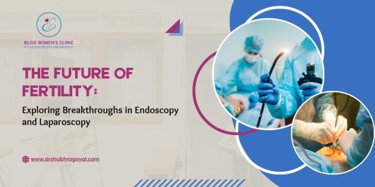
Explore the future of fertility with breakthroughs in endoscopy and laparoscopy. Dr. Shubhra Goyal offers insights on advancing reproductive health.
Do Visit: https://www.drshubhragoyal.com/welcome/blogs/the-future-of-fertility:-exploring-breakthroughs-in-endoscopy-and-laparoscopy
1 note · View note
baiosscentre · 1 year ago
Text
Best Endoscopic Specialist In Tirunelveli
When it comes to finding the best endoscopic specialist in Tirunelveli, one name stands out - Barioss. With our expertise and experience in the field of upper gastrointestinal endoscopy and endoscopic surgery, they have established themselves as a trusted healthcare provider in the region.
Barioss offers state-of-the-art facilities and a team of highly skilled specialists who are dedicated to providing exceptional care to our patients. Our commitment to using advanced technology ensures accurate diagnosis and effective treatment options.
Whether you require a treatment of upper gastrointestinal endoscopy in Tirunelveli or are in need of endoscopic surgery, Barioss is equipped to handle a wide range of medical conditions. Their team of experts will work closely with you to develop personalized treatment plans that cater to your specific needs.
When it comes to your health, choosing the right specialist is crucial. Trust Barioss for all your endoscopic needs in Tirunelveli, and experience top-quality care delivered for Endoscopic Surgery in Tirunelveli with compassion and expertise.
0 notes
mcatmemoranda · 9 months ago
Text
From UpToDate:
Overview — The goal of surveillance is to improve outcomes by detecting dysplasia or esophageal adenocarcinoma early enough to provide effective treatment. Guidelines suggest surveillance for most patients with Barrett's esophagus, but whether surveillance is beneficial is unclear. Available observational studies have not consistently shown that surveillance is beneficial. In addition, there are potential harms associated with surveillance, including a decrease in quality of life due to worry about cancer development, the risks associated with endoscopy, the risks and morbidity associated with invasive therapies used to treat lesions identified by surveillance (such as esophagectomy or radiofrequency ablation), and missed lesions despite surveillance. As a result, a well-informed patient with nondysplastic Barrett's esophagus may reasonably choose not to undergo surveillance despite endorsement of the practice by gastrointestinal societies. The discussion of the risks and benefits of surveillance should be well documented in the patient's medical record, particularly if the patient elects not to undergo surveillance. If surveillance is performed, it is important to treat erosive esophagitis prior to obtaining biopsies.
●No dysplasia – If the initial biopsies show no dysplasia, we discuss the potential risks and benefits of regular endoscopic surveillance with the patient. The length of the nondysplastic Barrett's segment generally informs surveillance intervals, and this approach is supported by society guidelines. Patients with longer segments (≥3 cm) undergo surveillance every three years, whereas patients with shorter segments (<3 cm) undergo surveillance every five years.  
●Indefinite for dysplasia – If the initial biopsies are indefinite for dysplasia, we recommend optimizing medical antireflux therapy (e.g., prescribing a proton pump inhibitor [PPI] twice daily, ensuring compliance with PPI therapy, ensuring that the PPI is taken correctly). Antireflux therapy minimizes reactive esophageal changes due to reflux esophagitis that may be mistaken for dysplastic changes. After antireflux therapy has been optimized, we repeat an endoscopy with biopsy specimens taken every 1 cm. We typically perform the endoscopy after two months of treatment (to allow sufficient time for healing). Repeat endoscopy should not be delayed beyond six months. Any mucosal irregularities should be removed with endoscopic resection.
If the repeat biopsies still are indefinite for dysplasia, the diagnosis should be confirmed by a pathologist with expertise in esophageal histopathology. If the diagnosis is confirmed, management options include surveillance endoscopy every 12 months or referral of the patient to a center with expertise in managing patients with Barrett's esophagus.
●Low-grade dysplasia, high-grade dysplasia, or intramucosal carcinoma – If the biopsies show dysplasia or intramucosal carcinoma, we suggest that the diagnosis be confirmed by another pathologist with expertise in Barrett's esophagus-related neoplasia. Endoscopy should be repeated as soon as feasible if four-quadrant biopsy specimens were not obtained at 1 cm intervals or if there were any mucosal irregularities that were not removed with endoscopic resection. Endoscopists who do not perform endoscopic resection should refer patients with mucosal irregularities to specialty centers for that procedure prior to proceeding with endoscopic eradication therapy. Endoscopic resection of mucosal irregularities is required to accurately assess the grade of dysplasia.
We generally recommend endoscopic eradication therapy for patients who are confirmed to have high-grade dysplasia or intramucosal carcinoma, and who have no evidence of submucosal invasion in their resected specimens. (See 'High-grade dysplasia or intramucosal carcinoma' below.)
Some guidelines recommend either surveillance or endoscopic eradication for patients with low-grade dysplasia [4,7,9]. In patients who elect to undergo endoscopic eradication, endoscopic radiofrequency ablation is preferable for preventing progression of dysplasia. If endoscopic surveillance is the chosen approach for low-grade dysplasia, patients should have biopsies obtained every 1 cm, any mucosal irregularities should be removed with endoscopic resection, and surveillance should be performed every 6 months for one year and then annually until there is reversion to nondysplastic Barrett's [8]. (See 'Low-grade dysplasia' below.)
Efficacy of surveillance — Observational studies suggest that surveillance can detect curable dysplasia in Barrett's esophagus and that asymptomatic cancers discovered during surveillance are less advanced than those found in patients who present with symptoms such as dysphagia or weight loss [73-79]. However, these studies are susceptible to biases such as lead-time bias and length-time bias. (See "Evidence-based approach to prevention", section on 'Special biases'.)
Findings that call into question the value of surveillance include:
●Documented development of incurable malignancies in patients who were adherent to endoscopic surveillance programs [73,74].
●A case-control study with 70 cases and 101 controls that found that patients with esophageal cancer in the setting of Barrett's esophagus were as likely to have undergone endoscopic surveillance as patients with Barrett's esophagus but no cancer [80].
A multicenter randomized trial is being conducted to look at whether endoscopic surveillance every two years influences outcomes such as overall survival, cancer-specific survival, and stage at diagnosis of esophageal adenocarcinoma in patients with nondysplastic Barrett's esophagus or Barrett's esophagus with low-grade dysplasia [81].
Surveillance techniques — The endoscopic surveillance of Barrett's esophagus should include a careful inspection of the Barrett's epithelium with high-resolution white light endoscopy and with chromoendoscopy (including virtual chromoendoscopy). Adequate time should be spent inspecting the Barrett's esophagus segment and the gastric cardia in retroflexion. Any visible abnormalities should be removed with endoscopic resection. In addition, random four-quadrant biopsies should be obtained every 2 cm (every 1 cm in patients with known or suspected dysplasia). This biopsy protocol is referred to as the Seattle biopsy sampling protocol [6].
Dysplasia in Barrett's esophagus is often patchy in extent and severity, and dysplastic areas can easily be missed because of biopsy sampling error [82-85]. Careful inspection of the metaplastic area and extensive biopsy sampling can reduce sampling error but cannot eliminate the problem entirely [86-88]. In addition, even when dysplasia is detected, foci of invasive cancer can be missed. In a meta-analysis of studies with patients who had esophagectomies for high-grade dysplasia with no apparent tumor mass, 13 percent of the resection specimens had invasive cancer [84]. In a subsequent study of 68 patients undergoing esophagectomy for high-grade dysplasia, 12 patients (18 percent) had adenocarcinoma detected in the resected esophagus [85]. Four of the cancers were intramucosal, and eight were invasive (extending into the submucosa).
Several advanced endoscopic techniques have been proposed to enhance the identification of dysplastic areas for biopsy sampling [89-94]. These techniques include mucosal staining with vital dyes (chromoendoscopy), endosonography, optical coherence tomography, confocal endomicroscopy, and virtual chromoendoscopy using narrow band imaging (NBI) or a similar technique. Some society guidelines recommend routine use of advanced imaging techniques such as chromoendoscopy, including virtual chromoendoscopy [6]. (See "Chromoendoscopy" and "Barrett's esophagus: Evaluation with optical chromoscopy" and "Confocal laser endomicroscopy and endocytoscopy", section on 'Barrett's esophagus'.)
A meta-analysis of 14 studies with a total of 843 patients examined whether advanced imaging techniques can increase the detection of dysplasia or cancer relative to white light endoscopy with random biopsies [95]. The investigators found that advanced imaging techniques increased the diagnostic yield for dysplasia or cancer by 34 percent (95% CI 20 to 56 percent). The increase in yield was similar for chromoendoscopy and virtual chromoendoscopy (eg, NBI). However, whether this increase in diagnostic yield leads to improved patient outcomes is unclear.  
Wide-area transepithelial sampling with computer-assisted 3-dimensional analysis (WATS-3D) has been proposed as a method to improve the detection of dysplasia during endoscopic surveillance of Barrett's esophagus, and the use of WATS-3D (in addition to Seattle protocol biopsy sampling) for surveillance is supported by some guidelines [6]. WATS-3D involves abrasive brushing of Barrett's metaplasia followed by computerized neural network analysis of the brush specimen to identify neoplasia. In a study of 160 patients with Barrett's metaplasia undergoing surveillance endoscopy, the detection rate for high-grade dysplasia (HGD)/adenocarcinoma (EAC) was higher with WATS-3D combined with biopsy sampling compared with biopsy sampling alone (18 versus 4 percent; absolute difference 14 percent, 95% CI 8-21) [96]. Of note, one case of HGD/EAC was missed in the group with WATS-3D combined with biopsy sampling, whereas 23 cases of HGD/EAC were missed by biopsy sampling alone. In another study of 12,899 patients undergoing endoscopies in which both standard forceps biopsies and WATS samples were taken, forceps biopsies identified dysplasia in 88 patients, while WATS identified an additional 213 cases of dysplasia, increasing the dysplasia detection rate from 0.68 to 2.33 percent [97]. 
A number of molecular markers for cancer risk have been proposed as alternatives to random biopsy sampling to detect dysplasia in Barrett's esophagus [98-101]. Promising molecular markers that have been associated with carcinogenesis in Barrett's esophagus include abnormalities in p53 and cyclin D1 expression, and abnormal cellular DNA content demonstrable by flow cytometry or methylation arrays. Additional evaluation of the markers is needed before they can be recommended for routine clinical use or to replace random biopsies [7]. However, some markers may serve as an adjunct to established diagnostic methods. For example, the British Society of Gastroenterology suggests that immunostaining of esophageal biopsies for p53 may improve the reproducibility of a diagnosis of dysplasia.
2 notes · View notes
essincny · 2 years ago
Text
The Benefits of Using Endoscopy in a Veterinary Practice
In recent years, veterinary medicine has seen significant advancements in diagnostic and treatment technologies. One such advancement that has revolutionized veterinary practice is the use of endoscopy. Endoscopy is a minimally invasive procedure that uses a flexible instrument called an endoscope to visualize and examine the internal organs and structures of animals. In this blog post, we will explore the numerous benefits of using endoscopy in a veterinary practice.
Accurate Diagnosis
Endoscopy allows veterinarians to get a clear and detailed view of the internal organs and structures of animals. This enables a more accurate and precise diagnosis of various conditions and diseases. By directly visualizing the problem area, veterinarians can identify abnormalities, such as tumors, foreign objects, ulcers, strictures, and more. Accurate diagnosis leads to more effective treatment plans and better outcomes for our furry friends.
Minimally Invasive
Compared to traditional surgical procedures, endoscopy is minimally invasive. Instead of making large incisions, endoscopy only requires small incisions or natural body openings, such as the mouth or anus. This results in less pain, discomfort, and a faster recovery time for animals. Minimally invasive procedures also reduce the risk of post-operative complications, including infection and excessive scarring.
Reduced Patient Stress
Veterinary visits can often be stressful for animals. However, with endoscopy, the stress levels are significantly reduced. Instead of being put under general anesthesia for invasive surgeries, animals undergoing endoscopy typically receive sedation or local anesthesia. This reduces their anxiety, minimizes the risks associated with general anesthesia, and makes the overall experience less traumatic for both the animals and their owners.
Versatility
Endoscopy is a versatile procedure that can be used in different areas of veterinary medicine. It can be employed in various specialties, including gastroenterology, urology, respiratory medicine, and more. With different types of endoscopes and specialized instruments, veterinarians can examine and treat a wide range of conditions, such as gastrointestinal disorders, urinary tract issues, airway problems, and even perform biopsies.
Lower Costs
While the initial cost of purchasing endoscopy equipment may be higher than traditional diagnostic tools, such as X-rays or ultrasounds, the long-term benefits can surpass the investment. Endoscopy allows for more accurate diagnoses, reducing the need for additional diagnostic tests and surgeries. This not only saves money for the pet owners but also minimizes the stress and risks associated with unnecessary procedures.
In conclusion, the use of endoscopy in veterinary practice offers numerous benefits. From accurate diagnosis and minimal invasiveness to reduced patient stress and versatility, endoscopy has transformed the way veterinarians approach diagnostics and treatments. By embracing this cutting-edge technology, veterinary practices can provide the best possible care to animals while ensuring their well-being and comfort.
11 notes · View notes
nursingscience · 2 years ago
Text
Medical Abbreviations on Pharmacy Prescriptions
Here are some common medical abbreviations you may see on pharmacy prescriptions:
qd - once a day
bid - twice a day
tid - three times a day
qid - four times a day
qh - every hour
prn - as needed
pc - after meals
ac - before meals
hs - at bedtime
po - by mouth
IV - intravenous
IM - intramuscular
subQ - subcutaneous
mL - milliliter
mg - milligram
g - gram
mcg - microgram
stat - immediately, right away
NPO - nothing by mouth
cap - capsule
tab - tablet
susp - suspension
sol - solution
amp - ampule
inj - injection
Rx - prescription
C - Celsius
F - Fahrenheit
BP - blood pressure
HR - heart rate
RR - respiratory rate
WBC - white blood cell
RBC - red blood cell
Hgb - hemoglobin
Hct - hematocrit
PT - prothrombin time
INR - international normalized ratio
BUN - blood urea nitrogen
Cr - creatinine
Ca - calcium
K - potassium
Na - sodium
Cl - chloride
Mg - magnesium
PO2 - partial pressure of oxygen
PCO2 - partial pressure of carbon dioxide
ABG - arterial blood gas
CBC - complete blood count
BMP - basic metabolic panel
CMP - comprehensive metabolic panel.
ECG - electrocardiogram
EEG - electroencephalogram
MRI - magnetic resonance imaging
CT - computed tomography
PET - positron emission tomography
CXR - chest x-ray
CTX - chemotherapy
NSAID - nonsteroidal anti-inflammatory drug
DMARD - disease-modifying antirheumatic drug
ACE - angiotensin-converting enzyme
ARB - angiotensin receptor blocker
SSRI - selective serotonin reuptake inhibitor
TCA - tricyclic antidepressant
ADHD - attention deficit hyperactivity disorder
COPD - chronic obstructive pulmonary disease
CAD - coronary artery disease
CHF - congestive heart failure
DVT - deep vein thrombosis
GI - gastrointestinal
UTI - urinary tract infection
OTC - over-the-counter
Rx - prescription
OD - right eye
OS - left eye
OU - both eyes.
TID - thrombosis in dementia
TDS - ter die sumendum (three times a day)
BOM - bilaterally otitis media (infection in both ears)
BT - body temperature
C&S - culture and sensitivity
D/C - discontinue or discharge
D/W - dextrose in water
ETOH - ethyl alcohol
FUO - fever of unknown origin
H&P - history and physical examination
I&D - incision and drainage
I&O - intake and output
KVO - keep vein open
N&V - nausea and vomiting
PERRLA - pupils equal, round, reactive to light and accommodation
PR - per rectum
QAM - every morning
QHS - every bedtime
QOD - every other day
S/P - status post (after)
TPN - total parenteral nutrition
UA - urinalysis
URI - upper respiratory infection
UTI - urinary tract infection
VO - verbal order.
XRT - radiation therapy
YOB - year of birth
BRBPR - bright red blood per rectum
CX - cervix
DVT - deep vein thrombosis
GB - gallbladder
GU - genitourinary
HCV - hepatitis C virus
HPI - history of present illness
ICP - intracranial pressure
IVP - intravenous pyelogram
LMP - last menstrual period
MRSA - methicillin-resistant Staphylococcus aureus
MVA - motor vehicle accident
NKA - no known allergies
PEG - percutaneous endoscopic gastrostomy
PRN - pro re nata (as needed)
ROS - review of systems
SOB - shortness of breath
TAH - total abdominal hysterectomy.
TIA - transient ischemic attack
Tx - treatment
UC - ulcerative colitis
URI - upper respiratory infection
VSD - ventricular septal defect
VTE - venous thromboembolism
XR - x-ray
w/c - wheelchair
XRT - radiation therapy
ASD - atrial septal defect
Bx - biopsy
CAD - coronary artery disease
CKD - chronic kidney disease
CPAP - continuous positive airway pressure
DKA - diabetic ketoacidosis
DNR - do not resuscitate
ED - emergency department
ESRD - end-stage renal disease
FFP - fresh frozen plasma
FSH - follicle-stimulating hormone.
GCS - Glasgow Coma Scale
Hct - hematocrit
Hgb - hemoglobin
ICU - intensive care unit
IV - intravenous
JVD - jugular venous distension
K - potassium
L - liter
MCH - mean corpuscular hemoglobin
MI - myocardial infarction
Na - sodium
NGT - nasogastric tube
NPO - nothing by mouth
OR - operating room
PCN - penicillin
PRBC - packed red blood cells
PTT - partial thromboplastin time
RBC - red blood cells
RT - respiratory therapy
SOA - short of air.
SCD - sequential compression device
SIRS - systemic inflammatory response syndrome
STAT - immediately
T - temperature
TPN - total parenteral nutrition
WBC - white blood cells
ABG - arterial blood gas
A fib - atrial fibrillation
BPH - benign prostatic hypertrophy
CBC - complete blood count
CO2 - carbon dioxide
COPD - chronic obstructive pulmonary disease
CPR - cardiopulmonary resuscitation
CT - computed tomography
CXR - chest x-ray
D5W - dextrose 5% in water
Dx - diagnosis
ECG or EKG - electrocardiogram
EEG - electroencephalogram
ETO - early termination of pregnancy.
FHR - fetal heart rate
GSW - gunshot wound
H&P - history and physical exam
HCG - human chorionic gonadotropin
I&D - incision and drainage
IBS - irritable bowel syndrome
ICP - intracranial pressure
IM - intramuscular
INR - international normalized ratio
IOP - intraocular pressure
LFT - liver function test
LOC - level of consciousness
LP - lumbar puncture
NG - nasogastric
OA - osteoarthritis
OCD - obsessive-compulsive disorder
OTC - over-the-counter
P - pulse
PCA - patient-controlled analgesia
PERRLA - pupils equal, round, reactive to light and accommodation.
PFT - pulmonary function test
PICC - peripherally inserted central catheter
PO - by mouth
PRN - as needed
PT - physical therapy
PT - prothrombin time
PTSD - post-traumatic stress disorder
PVC - premature ventricular contraction
QD - once a day
QID - four times a day
RA - rheumatoid arthritis
RICE - rest, ice, compression, elevation
RSI - rapid sequence intubation
RSV - respiratory syncytial virus
SBP - systolic blood pressure
SLE - systemic lupus erythematosus
SSRI - selective serotonin reuptake inhibitor
STAT - immediately
TB - tuberculosis
TIA - transient ischemic attack.
TID - three times a day
TKO - to keep open
TNTC - too numerous to count
TPN - total parenteral nutrition
URI - upper respiratory infection
UTI - urinary tract infection
V-fib - ventricular fibrillation
V-tach - ventricular tachycardia
VA - visual acuity
WNL - within normal limits
AED - automated external defibrillator
ARDS - acute respiratory distress syndrome
BID - twice a day
BP - blood pressure
BUN - blood urea nitrogen
CAD - coronary artery disease
CHF - congestive heart failure
CVA - cerebrovascular accident
D/C - discontinue
DKA - diabetic ketoacidosis.
DM - diabetes mellitus
DVT - deep vein thrombosis
EGD - esophagogastroduodenoscopy
ER - emergency room
F - Fahrenheit
Fx - fracture
GI - gastrointestinal
GTT - glucose tolerance test
HCT - hematocrit
Hgb - hemoglobin
HRT - hormone replacement therapy
ICP - intracranial pressure
IDDM - insulin-dependent diabetes mellitus
IBS - irritable bowel syndrome
IM - intramuscular
IV - intravenous
K - potassium
KVO - keep vein open
L&D - labor and delivery
LASIK - laser-assisted in situ keratomileusis.
ROM - range of motion
RT - radiation therapy
Rx - prescription
SCD - sequential compression device
SOB - shortness of breath
STD - sexually transmitted disease
TENS - transcutaneous electrical nerve stimulation
TIA - transient ischemic attack
TSH - thyroid-stimulating hormone
UA - urinalysis
US - ultrasound
UTI - urinary tract infection
VD - venereal disease
VF - ventricular fibrillation
VT - ventricular tachycardia
WBC - white blood cell
XRT - radiation therapy
XR - x-ray
Zn - zinc
Z-pak - azithromycin (antibiotic).
AAA - abdominal aortic aneurysm
ABG - arterial blood gas
ACS - acute coronary syndrome
ADL - activities of daily living
AED - automated external defibrillator
AIDS - acquired immunodeficiency syndrome
ALS - amyotrophic lateral sclerosis
AMA - against medical advice
AML - acute myeloid leukemia
APAP - acetaminophen
ARDS - acute respiratory distress syndrome
ASCVD - atherosclerotic cardiovascular disease
BPH - benign prostatic hyperplasia
BUN - blood urea nitrogen
CABG - coronary artery bypass graft
CBC - complete blood count
CHF - congestive heart failure
COPD - chronic obstructive pulmonary disease
CPAP - continuous positive airway pressure
CRF - chronic renal failure.
CT - computed tomography
CVA - cerebrovascular accident
D&C - dilation and curettage
DVT - deep vein thrombosis
ECG/EKG - electrocardiogram
EEG - electroencephalogram
ESRD - end-stage renal disease
FSH - follicle-stimulating hormone
GERD - gastroesophageal reflux disease
GFR - glomerular filtration rate
HbA1c - glycated hemoglobin
Hct - hematocrit
HIV - human immunodeficiency virus
HPV - human papillomavirus
HTN - hypertension
IBD - inflammatory bowel disease
IBS - irritable bowel syndrome
ICU - intensive care unit
IDDM - insulin-dependent diabetes mellitus
IM - intramuscular.
IV - intravenous
LFT - liver function test
MI - myocardial infarction
MRI - magnetic resonance imaging
MS - multiple sclerosis
NPO - nothing by mouth
NS - normal saline
OCD - obsessive-compulsive disorder
OSA - obstructive sleep apnea
PCOS - polycystic ovary syndrome
PMS - premenstrual syndrome
PPD - purified protein derivative
PSA - prostate-specific antigen
PT - prothrombin time
PTT - partial thromboplastin time
RA - rheumatoid arthritis
RBC - red blood cell
RSV - respiratory syncytial virus
SLE - systemic lupus erythematosus
TB - tuberculosis.
It is important to remember that medical abbreviations can vary based on location and specialty. 
Healthcare professionals should use medical abbreviations with caution and only when they are familiar with their meanings. 
Patients should always communicate any questions or concerns they have about their medications or medical care to their healthcare provider or pharmacist to ensure they receive safe and accurate medical care.
Tumblr media
10 notes · View notes
anujmrfr · 6 hours ago
Text
Endoscopic Clip Market Size, Growth Outlook 2035
The global Endoscopic Clip Market Size was estimated at 0.49 (USD Billion) in 2023. The Endoscopic Clip Industry is expected to grow from 0.52 (USD Billion) in 2024 to 0.83 (USD Billion) by 2032. The Endoscopic Clip Market CAGR (growth rate) is expected to be around 6.01% during the forecast period (2024 - 2032).
Market Overview The Endoscopic Clip Market is experiencing robust growth due to the rising adoption of minimally invasive procedures in gastroenterology. Endoscopic hemostasis clips, also known as hemoclips, are widely used for treating gastrointestinal (GI) bleeding, closing perforations, and securing surgical sites during endoscopic procedures. The growing prevalence of peptic ulcers, colorectal cancer, and other GI disorders has increased the demand for these devices, which play a crucial role in emergency and therapeutic endoscopy.
Market Size and Share The global Endoscopic Clip MarketSizewas estimated at 0.49 (USD Billion) in 2023. The Endoscopic Clip Industry is expected to grow from 0.52 (USD Billion) in 2024 to 0.83 (USD Billion) by 2032. The Endoscopic Clip Market CAGR (growth rate) is expected to be around 6.01% during the forecast period (2024 - 2032). The increasing utilization of endoscopic closure devices in hospitals and ambulatory surgical centers (ASCs) is driving market expansion. North America currently holds the largest share, followed by Europe, due to advanced healthcare infrastructure and a high volume of endoscopic procedures.
Tumblr media
Market Drivers
Rising Incidence of Gastrointestinal Disorders: Conditions like peptic ulcers, diverticular bleeding, and inflammatory bowel disease (IBD) have led to an increased need for endoscopic hemostasis solutions.
Advancements in Endoscopic Technologies: The development of multi-functional endoscopic clips with better durability, rotation capabilities, and improved tissue anchoring is expanding market growth.
Growth in Minimally Invasive Procedures: The preference for non-surgical treatments over traditional surgeries has increased the demand for endoscopic clipping devices.
Increasing Geriatric Population: The elderly are more prone to GI bleeding and related disorders, necessitating effective endoscopic closure devices.
Challenges and Restraints
High Cost of Advanced Endoscopic Clips: Specialized biodegradable endoscopic clips and robotic-assisted devices can be expensive, limiting accessibility.
Complications Associated with Clip Placement: Improper clip deployment can lead to complications such as perforation, delayed healing, or migration.
Regulatory Challenges: Strict approval processes for new endoscopic closure devices can slow down product launches.
Market Trends
Development of Biodegradable Clips: Companies are investing in biodegradable hemostatic clips that eliminate the need for removal and reduce long-term complications.
AI-Assisted Endoscopy: The integration of AI-based diagnostic tools with endoscopic procedures is enhancing precision in clip deployment and bleeding management.
Expanding Applications Beyond GI Bleeding: Endoscopic hemostasis clips are now being used in bariatric surgery, esophageal perforation repairs, and post-surgical wound closures.
Regional Analysis
North America: Leads the market due to high adoption of advanced endoscopic procedures, increasing GI disease burden, and strong healthcare infrastructure.
Europe: Growing market, supported by increased R&D investments in gastrointestinal endoscopy and rising geriatric population.
Asia-Pacific: Fastest-growing region, driven by rising healthcare expenditures, increasing GI disease prevalence, and expansion of endoscopic services.
Rest of the World: Moderate market penetration, with potential for growth in Latin America and the Middle East.
Segmental Analysis
By Product Type:
Hemostatic Clips
Closure Clips
Tissue Anchoring Clips
By Application:
Gastrointestinal Bleeding Control
Perforation Closure
Surgical Site Closure
Polypectomy Bleeding Control
By End-User:
Hospitals
Ambulatory Surgical Centers (ASCs)
Specialty Clinics
Key Market Players
Conmed Corporation
Olympus Corporation
MicroTech Endoscopy
Medtronic
Steris Corporation
Aesculap AG
Cook Medical
Recent Developments
Product Innovations: Olympus recently launched an advanced hemostatic clip system with enhanced anchoring capabilities.
Strategic Acquisitions: Boston Scientific acquired a GI device manufacturer to expand its endoscopic closure solutions portfolio.
AI Integration in Endoscopy: Leading players are integrating AI-driven endoscopic imaging with clip placement techniques for better clinical outcomes.
For more information, please visit us at marketresearchfuture.
0 notes
ottomed · 4 months ago
Text
Advanced Medical Diagnostics Using Flexible Endoscopes and Specialized Instruments
Advanced medical diagnostics is becoming more sophisticated with flexible endoscopes and specialized instruments. The advent of flexible endoscopes has dramatically changed the approach physicians take when diagnosing and treating a variety of inner organs. These have made it possible for the doctors to gain an invasive view of inner body structures, get clear visual images and even make precise operations on the body.
Gastrointestinal Endoscopes- These are the most general uses of the flexible endoscopes. Indeed, this is a technology that transformed diagnosis and management of digestive tract disorders. Gastroenterologists make visual inspections on the oesophagus, stomach, small intestines, and the colon by introducing a thin tube equipped with a camera. compute through the mouth or the rectum. This technology has gone a long way in identification of ailments like ulcers, polyps, and sometimes even the early formations of cancerous growth.
Another application of the flexible endoscopy is in thorax; this is a very serious application of this technique. The thoracoscope, an endoscopic regarding examination of the chest cavity has now become a basics equipment that every pulmonologist and thoracic surgeon shouldn’t lack. It allows one to visualize all the structures in the lungs, pleura, and mediastinum, making it possible to carry out minimally invasive diagnostic procedures as well as surgical interventions. Use of thoracoscopes drastically decreased the need for such open surgery of the chest as required through the open-chest surgeries; therefore, it led to faster recovery times and improved patient outcomes.
Flexible endoscopes are not just limited to the diagnosis. It is also a basis for initiating many therapeutic interventions. For instance, gastrointestinal endoscopes are useful instruments for removing polyps, controlling bleeding, or inserting stents in constricted pathways. Similarly, thoracoscopes have enabled surgeons to take biopsies of tissues, drain fluid accumulations, and even remove small tumours without the usual large incisions.
Flexible endoscopic instruments continue to advance with advancements in technology, pushing boundaries that can be achieved with these devices. High-definition imaging capabilities for high definition and 3D visualization are adding to the ability of magnification, thereby setting up new boundaries of possibilities with these instruments. Artificial intelligence and machine learning algorithms start combining and helping in improving diagnoses toward better treatment options.
For this reason, flexible endoscopes, in particular gastrointestinal endoscopes and thoracoscopes, have truly revolutionized the scope of patient care. Aside from high accuracy in diagnosis, they have also limited dangerous risks otherwise posed by more invasive procedures. Future innovations through the advancement of technology in medicine may be expected to be the most viable applications of flexible endoscopy, making it even better for a wide range of diagnoses and treatments with much greater precision and patient comfort.
0 notes
mitraindustries · 22 days ago
Text
How flexible video endoscopes and blood transfusion sets are shaping the future of gastroenterology.
Gastroenterology, the branch of medicine focused on the digestive system and its disorders, has seen significant advancements in recent years, largely due to innovations in medical technologies such as flexible video endoscopes and blood transfusion sets. These tools are not only transforming diagnostic and therapeutic procedures but also improving patient outcomes and shaping the future of gastroenterological care.
Flexible Video Endoscopes: Revolutionizing Diagnosis and Treatment
Flexible video endoscopes are the most advanced in gastroenterology, providing physicians with a non-invasive method of diagnosing and treating a wide range of gastrointestinal conditions. These endoscopes consist of a flexible tube with a high-definition camera and light source, allowing for detailed visualization of the esophagus, stomach, intestines, and colon. They enable procedures like colonoscopies, upper GI endoscopies, and endoscopic retrograde cholangiopancreatography (ERCP), which are crucial for diagnosing conditions such as colorectal cancer, ulcers, and biliary obstructions.
What makes flexible video endoscopes very much advantageous is the fact that they can produce real-time images and videos. This enhances diagnosis and allows instant biopsies or removal of polyps and tumors. As minimally invasive, recovery is faster, and comfort in patients is enhanced. As such, this equipment is greatly essential in the advancements of modern gastroenterology.
Blood Transfusion Sets: Patient care in Gastrointestinal Stents procedures
Blood transfusion sets are one of the essential supports for patients undergoing gastroenterological procedures, especially when there is a risk of significant blood loss. ERCP or surgeries for bleeding ulcers or varices may require blood transfusions to manage blood loss or correct anemia to ensure that the patient remains stable during and after the procedure.
Advances in blood transfusion technology ensure that modern transfusion sets do not contribute to complications such as infections or air embolisms, making it safer for patients. Filters prevent the infusion of dirty and harmful blood products; this brings more comfort to patients and health workers.
Shaping the Future
Flexible video endoscopes and advanced sets for blood transfusion are gradually changing the very face of gastroenterology by elevating both diagnosis accuracy and security in patient treatments. These state-of-the-art devices are creating more streamlined treatments, reducing recovery periods in hospital, and changing patients' experiences throughout their treatment procedure. As innovations in technology continually improve, their combination will very likely result in even less aggressive procedures, short recovery periods, and better care for patients having gastrointestinal disorders.
0 notes
sailesh11 · 8 hours ago
Text
ERCP in Thane – Expert Diagnosis and Treatment by Dr. Rajdeep More
If you are looking for an ERCP in Thane, Dr. Rajdeep More offers comprehensive and advanced diagnostic services for gastrointestinal issues. ERCP (Endoscopic Retrograde Cholangiopancreatography) is a specialized procedure that combines endoscopy and X-ray to diagnose and treat problems related to the bile ducts, gallbladder, pancreas, and liver. Dr. Rajdeep More provides expert care and utilizes the latest technology to ensure accurate results and effective treatment.
What is ERCP?
ERCP is a minimally invasive procedure that allows doctors to visualize the bile and pancreatic ducts to diagnose conditions such as gallstones, strictures, blockages, and pancreatitis. It can also be used to perform interventions, including removing stones, inserting stents, or taking biopsies. For anyone experiencing unexplained abdominal pain, jaundice, or issues related to digestion, ERCP in Thane can be a crucial diagnostic tool.
Why Choose Dr. Rajdeep More for ERCP in Thane?
When searching for ERCP in Thane, it’s essential to choose a specialist with the right experience and expertise. Dr. Rajdeep More is a renowned gastroenterologist who specializes in ERCP procedures and related treatments. With years of experience, he ensures that each procedure is performed with precision and care. His patients benefit from cutting-edge medical technology and a personalized approach to treatment.
Benefits of ERCP in Gastroenterology
Accurate Diagnosis: ERCP helps in diagnosing various gastrointestinal conditions with high accuracy.
Effective Treatment Options: Through ERCP, many procedures such as stone removal and stent placement can be performed, providing immediate relief.
Minimally Invasive: ERCP is minimally invasive, which means a faster recovery time and fewer risks compared to traditional surgeries.
When Should You Consider ERCP?
You may need an ERCP in Thane if you are experiencing symptoms such as:
Abdominal pain (especially in the upper abdomen)
Jaundice (yellowing of the skin or eyes)
Digestive problems, including unexplained nausea or vomiting
Unexplained weight loss or fatigue
Gallstones or bile duct blockages
Schedule Your ERCP Consultation Today
If you’re in need of ERCP in Thane, trust Dr. Rajdeep More for expert diagnosis and care. His clinic offers a comfortable and patient-focused environment, ensuring that each procedure is done with the utmost care and professionalism.
Don't delay getting the right diagnosis—schedule your consultation with Dr. Rajdeep More today to discuss whether ERCP is the right option for you.
0 notes
gastroenterologist · 1 year ago
Text
Unlock the secrets of optimal gastrointestinal health with our in-depth guide on "The Role of Endoscopy." Delve into the comprehensive overview to understand how endoscopy plays a pivotal role in diagnosing and treating gastrointestinal issues. Empower yourself with knowledge for a healthier digestive future.
0 notes
shubhragoyal · 1 year ago
Text
The Future of Fertility: Exploring Breakthroughs in Endoscopy and Laparoscopy
Tumblr media
In medical science, the future of fertility and reproductive health is experiencing a revolutionary transformation through the remarkable advancements in Endoscopy and Laparoscopy. These innovative medical procedures have become instrumental in diagnosing and treating various reproductive health issues, offering hope to countless individuals and couples worldwide. In this article, we delve deep into the world of Endoscopy and Laparoscopy, uncovering their benefits and trends and why choosing Dr. Shubhra Goyal can indeed be a game-changer in your journey toward optimal reproductive health.
Laparoscopy and Endoscopy - what are they?
Due to their accuracy and efficacy, minimally invasive medical treatments like Endoscopy and Laparoscopy have become extremely popular in recent years. These procedures use specialized tools fitted with small cameras and lights that enable doctors to view and access inside organs without the need for open surgery.
Benefits of Endoscopic Examinations!
The following are some benefits: -
Minimal Scarring - Endoscopy and Laparoscopy require just small incisions, unlike traditional surgical techniques that demand extensive incisions. Due to the minimum scarring produced, patients recover more quickly and enjoy additional aesthetic benefits.
Less Pain and Discomfort - Patients who undergo endoscopic procedures often report less pain and discomfort following the operation. This is because these techniques are less intrusive than standard operations and result in less tissue damage.
Shorter Hospital Stays - Compared to conventional equivalents, endoscopic operations often require shorter hospital stays. Patients may resume their regular daily routines and activities more quickly as a result, which also lowers healthcare expenditures.
High accuracy - Using cutting-edge technology in endoscopic exams gives surgeons a degree of accuracy never before possible. They can traverse and treat delicate and difficult-to-reach locations with outstanding accuracy thanks to their precision, which ultimately improves patient surgery results.
Moving ahead, let's discuss,
Top Upcoming Trends in Endoscopy and Laparoscopy
The areas of Endoscopy and Laparoscopy are examples of the ongoing change taking place in the world of medical science. The following are some of the most notable new developments that are influencing how these processes may develop in the future:
Innovations in Endoscopic Imaging Technology - The endoscopic imaging technology industry is expanding quickly. This includes the creation of 3D imaging systems and high-definition cameras. These developments considerably increase diagnosis accuracy by providing surgeons with an incredibly thorough and accurate picture of inside organs.
Read More: https://www.drshubhragoyal.com/welcome/blogs/the-future-of-fertility:-exploring-breakthroughs-in-endoscopy-and-laparoscopy
1 note · View note
rishikhospital12 · 1 day ago
Text
Best Gastrointestinal Surgeons in Jaipur
Tumblr media
Why Choose Gastro Surgery in Jaipur?
Jaipur has emerged as a prominent destination for high-quality healthcare, particularly for gastrointestinal treatments. The city’s healthcare institutions are equipped with state-of-the-art technology, experienced surgeons, and comprehensive post-operative care.
The field of gastroenterology covers disorders related to the digestive tract, liver, and associated organs. Gastro surgery often becomes necessary for conditions that cannot be managed with medication alone.
Top Gastrointestinal Surgeon Doctors in Jaipur
Here are some of the top gastrointestinal surgeons in Jaipur known for their exceptional expertise and patient care:
Dr. Ramesh Sharma
Specializes in minimally invasive gastro surgeries
Renowned for treating complex liver and intestinal issues
Dr. Seema Verma
Expert in laparoscopic gastrointestinal surgeries
Well-known for her compassionate approach to patient care
Dr. Arvind Gupta
Specializes in colorectal and pancreatic surgeries
Known for high success rates in complex surgical procedures
Dr. Vikram Singh Rathore
Expert in bile duct surgeries and liver transplants
Frequently sought after for peritoneum-related treatments
Best Gastro Surgery in Jaipur
Gastro surgery involves surgical treatment of the digestive system, including the stomach, intestines, liver, and pancreas. Jaipur offers cutting-edge surgical techniques, such as:
Laparoscopic Surgery: Minimally invasive procedures that reduce recovery time
Endoscopic Procedures: Advanced techniques for diagnosing and treating digestive issues
Open Surgeries: For complex cases requiring extensive surgical intervention
What Conditions Require Gastro Surgery?
Some common conditions that may necessitate gastro surgery include:
Gallbladder stones
Hernias
Chronic acid reflux
Colorectal cancer
Peritoneal diseases
Best Doctor for Jaundice in Jaipur
Jaundice, characterized by yellowing of the skin and eyes, is often a symptom of underlying liver issues. Early diagnosis and treatment are crucial. The best doctors for jaundice in Jaipur include:
Dr. Rajeev Mathur — Specializes in liver diseases and jaundice management
Dr. Anjali Sharma — Known for holistic care in liver disorders
Dr. Pankaj Agarwal — Expert in diagnosing and treating jaundice in patients of all age groups
Best Doctor for Peritoneum in Jaipur
The peritoneum is a thin layer that lines the abdominal cavity and supports abdominal organs. Peritoneal diseases can be complex and require specialized care. Some of the best doctors for peritoneum-related conditions in Jaipur are:
Dr. Vivek Soni — Renowned for treating peritoneal infections and cancers
Dr. Meenal Joshi — Expert in peritoneal dialysis and related surgical procedures
Dr. Ashok Meena — Skilled in laparoscopic and open surgeries involving the peritoneum
Factors to Consider When Choosing a Gastrointestinal Surgeon
Selecting the right surgeon involves considering several factors:
Experience: Look for surgeons with extensive experience in gastrointestinal procedures.
Specialization: Ensure the surgeon specializes in the specific condition you need treatment for.
Hospital Infrastructure: Choose a hospital with modern facilities and advanced diagnostic tools.
Patient Reviews: Check for positive patient feedback and success stories.
Conclusion
Finding the best gastrointestinal surgeon in Jaipur is crucial for effective treatment and a speedy recovery. Whether you need treatment for jaundice, peritoneal diseases, or any other gastrointestinal condition, Jaipur offers top-notch healthcare services. Consult one of the highly recommended doctors mentioned in this blog for expert care.
Make your health a priority by choosing the best specialists and hospitals in Jaipur for gastrointestinal treatments. Your journey to better health starts here.
0 notes