#Endoscopy for internal visualization
Explore tagged Tumblr posts
Visit Tumblr Blog
Explore Tumblr blogs with no restrictions, modern design and the best experience.
Last Seen Tumblr Blogs


Fun Fact
1,644 Tumblr posts in 1 second.
Text

Explore the future of fertility with breakthroughs in endoscopy and laparoscopy. Dr. Shubhra Goyal offers insights on advancing reproductive health.
Do Visit: https://www.drshubhragoyal.com/welcome/blogs/the-future-of-fertility:-exploring-breakthroughs-in-endoscopy-and-laparoscopy
#Endoscopy and Laparoscopy#Endoscopy and laparoscopy procedures#Medical uses of endoscopy and laparoscopy#Endoscopy vs. laparoscopy comparison#Benefits of endoscopic examinations#Laparoscopic surgery advancements#Gastrointestinal endoscopy techniques#Minimally invasive laparoscopic procedures#Endoscopic imaging technology#Laparoscopy in surgical diagnosis#Endoscopy for digestive disorders#Laparoscopy for abdominal surgeries#Endoscopy in medical diagnostics#Laparoscopic surgical instruments#Endoscopy for internal visualization#Laparoscopy and its applications#Endoscopic screening and treatment#Laparoscopic hernia repair#Endoscopy for upper GI tract
1 note
·
View note
Text
Ultrasound Endoscope Market Size, Trends, and Forecast 2021–2031
The global Ultrasound Endoscope Market is on a strong growth trajectory, poised to reach USD 2,000.62 million by 2031, up from USD 1,240.75 million in 2024, according to a new industry report. The market is expected to grow at a CAGR of 7.1% during the forecast period from 2025 to 2031.
The new report, published by Business Market Insights, provides a detailed analysis of industry trends, growth drivers, and competitive dynamics shaping the future of ultrasound endoscopic technology. It examines market behavior across geographies and offers insights critical for stakeholders, investors, and healthcare innovators.
📥 Sample PDF of the Report- https://www.businessmarketinsights.com/sample/BMIPUB00031683
Why the Ultrasound Endoscope Market Is Growing
Ultrasound endoscopy is increasingly being used for both diagnostic and therapeutic applications, particularly in gastroenterology, oncology, and pulmonology. The integration of ultrasound with endoscopic imaging allows for improved visualization and tissue sampling — leading to earlier and more accurate diagnoses.
Key drivers include:
Technological advancements in minimally invasive devices
Growing geriatric population and incidence of gastrointestinal diseases
Rising demand for precision diagnostics in oncology and internal medicine
Market Analysis Highlights
The report includes:
Executive Summary
Global Economic Overview
PESTLE & Porter’s Five Forces Analysis
Company Profiles & Market Share Insights
Case Studies and Forecasting Models
Strategic tools such as SWOT analysis, Investment Return Analysis, and Product Lifecycle (PLC) assessments help stakeholders evaluate the competitive landscape and investment opportunities.
Top Players in the Ultrasound Endoscope Market
Several major companies are leading innovation and expansion in this space:
Olympus Corporation
Fujifilm Holdings Corporation
Canon Inc.
HOYA Corporation
Lepu Medical Technology (Beijing) Co. Ltd
Sonoscape Pvt Ltd.
These companies are investing in AI-enabled imaging, hybrid endoscopy solutions, and expanding their product lines to meet rising global demand.
Regional Insights
North America and Europe dominate the current market landscape due to robust healthcare infrastructure and early technology adoption. However, the Asia-Pacific region is expected to grow the fastest, driven by increasing healthcare investments in China, India, and Southeast Asia.
Get the Full Report
To get deeper insights, market segmentation, and detailed forecasts, access the full report by Business Market Insights here:
🔗 Get Report Customization Options
About Business Market Insights
Business Market Insights is a trusted market intelligence platform offering detailed reports and custom research across multiple sectors including healthcare, automotive, energy, and technology. Their subscription-based service delivers timely insights to help companies make data-driven decisions.
📞 Contact: Ankit Mathur 📧 [email protected] 📱 +1–646–791–7070
0 notes
Text
Ingestible Sensor Market Sixe Pioneering Internal Health Monitoring Solutions
The Ingestible Sensor Market Size is reshaping the future of digital healthcare with innovative technology that allows health monitoring from within the human body. These miniature electronic devices, swallowed like capsules, transmit critical physiological data to external systems in real-time. With applications in diagnostics, drug adherence monitoring, and sports science, ingestible sensors are projected to revolutionize personalized medicine. According to Market Size Research Future, the global market for ingestible sensors is expected to reach USD 3.1 billion by 2030, registering a robust CAGR of 18.9% during the forecast period from 2023 to 2030.
Market Size Overview
Ingestible sensors, also known as digital pills or smart pills, consist of biocompatible materials embedded with microelectronic components that monitor parameters such as pH level, temperature, pressure, and medication intake. These sensors transmit data wirelessly to smartphones, tablets, or healthcare systems, offering non-invasive, real-time insights into a patient's internal environment.
The technology is particularly valuable in chronic disease management, remote patient monitoring, and high-performance sports, where continuous data collection is vital. As the demand for precise, real-time health tracking increases, ingestible sensors are rapidly gaining traction across both developed and emerging healthcare systems.
Market Size Segmentation
By Component
Sensor
Data Recorder
Software
By Sensor Type
Temperature Sensor
Pressure Sensor
pH Sensor
Image Sensor
By Application
Medical Adherence Monitoring
Diagnostic Imaging
Sports and Fitness
Others
By End-User
Hospitals and Clinics
Home Healthcare
Sports Organizations
Research Centers
By Region
North America
Europe
Asia-Pacific
Latin America
Middle East & Africa
Trends
1. Remote and Non-Invasive Diagnostics
Healthcare is increasingly shifting towards remote and non-invasive monitoring solutions. Ingestible sensors enable real-time diagnostics without the need for invasive endoscopy or biopsies, significantly improving patient comfort and clinical outcomes.
2. Smart Pills for Medication Adherence
Smart pills with ingestible sensors are being used to ensure medication compliance, especially in psychiatric and chronic care. These pills send a signal to a wearable patch or mobile app once the medication is consumed, improving patient accountability and reducing hospital readmissions.
3. Integration with Digital Health Platforms
Modern ingestible sensors are designed to integrate seamlessly with digital health ecosystems. This integration allows healthcare providers to collect, visualize, and analyze patient data via cloud-based platforms and mobile applications.
4. Biocompatibility and Miniaturization
Innovations in biocompatible materials and nanotechnology are leading to safer, smaller, and more durable sensors. These advancements have extended the usability of ingestible sensors across a broader demographic, including children and elderly patients.
5. Use in Sports and Military Applications
Elite athletes and defense personnel are adopting ingestible sensors to monitor core body temperature, hydration, and gastrointestinal health in extreme environments, supporting performance optimization and safety.
Segment Insights
By Sensor Type
Temperature and pH sensors dominate the market due to their widespread use in internal diagnostics and gastrointestinal monitoring. Image sensors are gaining popularity in capsule endoscopy, offering non-invasive internal imaging alternatives.
By Application
Medical adherence monitoring is the fastest-growing application, particularly in mental health and oncology treatment. Diagnostics continue to be a strong segment, especially with the rising need for gastrointestinal tract monitoring and early cancer detection.
By End-User
Hospitals and clinics remain the largest end-users, leveraging these sensors for accurate diagnostics and treatment management. Meanwhile, home healthcare is growing rapidly with the global surge in telehealth adoption and aging populations.
End-User Insights
Healthcare Providers
Clinicians use ingestible sensors to track medication intake, monitor internal conditions, and manage treatment efficacy. This helps reduce guesswork and ensures better patient outcomes.
Pharmaceutical Industry
In clinical trials, ingestible sensors help researchers track dosing adherence and gather pharmacokinetic data in real-time, improving trial accuracy and safety.
Sports and Military
Professional sports teams and military units deploy ingestible sensors to prevent heatstroke, optimize hydration, and monitor physiological changes during intense physical activity.
Consumers
Health-conscious individuals are exploring ingestible sensors for digestive health monitoring and personalized dietary optimization, opening new opportunities in the wellness industry.
Key Players
Leading companies are focusing on research and development, strategic partnerships, and regulatory approvals to drive adoption and expand product offerings:
Proteus Digital Health – A pioneer in digital pills for medication adherence, integrating sensors with ingestible pharmaceuticals.
Capsovision, Inc. – Specializes in imaging-based smart capsules for gastrointestinal diagnostics.
Medtronic PLC – Offers capsule endoscopy technology for non-invasive internal imaging.
BodyCap – Develops ingestible sensors for temperature monitoring in healthcare and sports applications.
Olympus Corporation – Manufactures endoscopic capsules with integrated sensors for internal examination.
Future Outlook
The ingestible sensor market is set for dynamic evolution. Future innovations may include biosensors capable of detecting pathogens, real-time biomarker tracking, and smart capsules with therapeutic delivery capabilities. As AI and machine learning become embedded in health systems, the data from ingestible sensors will enable predictive healthcare models and tailored treatment strategies.
The convergence of ingestible sensors with wearable tech, IoT, and cloud computing will enhance early disease detection, enable proactive interventions, and drive down long-term healthcare costs globally.
Conclusion
The Ingestible Sensor Market Size represents a significant leap in non-invasive health monitoring. By enabling real-time, internal data collection, these sensors provide a new layer of medical intelligence that can revolutionize diagnostics, treatment adherence, and preventive care. As healthcare systems worldwide shift toward personalized and value-based care, ingestible sensors will become indispensable tools in the digital health arsenal.
Trending Report Highlights
Explore more transformative markets and technologies:
Microprocessor and GPU Market Size
InGaAs Camera Market Size
Far-Field Speech and Voice Recognition Market Size
Functional Printing Market Size
Predictive Emission Monitoring System (PEMS) Market Size
Soft Robotics Market Size
Photoelectric Sensor Market Size
Laser Sensors Market Size
Proximity Sensor Market Size
Wi-Fi Adapter Card Market Size
5G Processor Market Size
0 notes
Text
Inside a Wildlife Hospital: How Injured Animals Are Treated
Behind every rescued animal in India is a team of dedicated professionals working quietly and tirelessly in facilities few people ever see—wildlife hospitals. These specialized centers are where the most crucial work of animal rescue happens: treating injuries, healing trauma, and giving animals a second chance at life.
From treating elephants with infected wounds to performing delicate surgeries on rescued birds, the inner workings of a wildlife hospital are as complex as any human medical facility. At the forefront of this movement is Vantara, the groundbreaking sanctuary and rescue project envisioned by Vantara Anant Ambani, which features one of the most advanced wildlife hospitals in Asia.
This blog takes you behind the scenes—inside a wildlife hospital—to show how injured animals are treated, the steps involved in their care, and how these hospitals are changing the face of wildlife conservation in India.
🏥 What Is a Wildlife Hospital?
A wildlife hospital is a specialized medical facility focused solely on the diagnosis, treatment, and rehabilitation of wild and rescued animals. These hospitals serve animals that have been:
Injured in the wild (by vehicles, traps, or territorial fights)
Rescued from illegal captivity or wildlife trafficking
Found sick or orphaned due to natural disasters
Victims of human-animal conflict or abuse
Wildlife hospitals are usually operated by NGOs, sanctuaries, or forest departments and staffed by wildlife veterinarians, biologists, behaviorists, and caregivers.
🐘 Step-by-Step: How Injured Animals Are Treated
Let’s walk through the step-by-step journey of how animals are treated inside a wildlife hospital—from rescue to release (if possible).
1. Rescue and Arrival
Most treatments begin with a rescue alert—sometimes from a local villager, forest officer, or helpline. Wildlife rescue teams spring into action and bring the injured animal to the nearest hospital.
Upon arrival:
The animal is visually assessed for trauma or distress
A team prepares the triage room for immediate stabilization
A quiet, stress-free environment is ensured to calm the animal
For example, at Vantara, the rescue teams are trained to handle everything from elephant rescues to exotic wildlife seizures, ensuring that the transition from the wild to the hospital is handled with utmost care and urgency.
2. Quarantine and Initial Examination
Before full treatment begins, the animal is placed in a quarantine zone. This prevents any potential infections from spreading to other animals.
During quarantine:
Blood tests, X-rays, and physical examinations are conducted
Wildlife vets check for parasites, infections, broken bones, or internal injuries
If needed, sedation is used to allow for stress-free examination
In facilities like the Vantara Wildlife Hospital, advanced diagnostic tools such as digital radiography, ultrasonography, CT scans, and even endoscopy are used to provide precise insights.
3. Treatment Plan and Surgery (If Required)
Once diagnosed, a customized treatment plan is developed for each animal. No two cases are alike, and everything from diet to medication is tailored to the animal’s species, age, and injury.
Treatment may include:
Suturing wounds, fracture repair, or orthopedic surgery
Fluid therapy to treat dehydration and shock
Antibiotics, pain relief, and anti-parasitic medication
Laser therapy or physiotherapy for long-term injuries
In severe cases, such as those involving snare traps or electric shocks, specialist surgeons perform high-risk operations. Vantara’s hospital, under the guidance of Vantara Anant Ambani, employs round-the-clock surgical teams and has intensive care units for large mammals and birds alike.
4. Post-Operative Recovery and Enrichment
After treatment or surgery, the animal enters post-operative care. This stage is critical for regaining strength and ensuring no relapse.
Recovery includes:
Observation in recovery pens with padded flooring and clean bedding
Nutritional plans to help the body heal
Behavioral enrichment to reduce boredom and stress
Limited human interaction to preserve natural instincts
Enrichment is especially important in species like leopards, bears, and elephants, where mental health is a key part of physical recovery.
At Vantara, the design of recovery enclosures is species-specific, ensuring every animal’s emotional and physical well-being is prioritized. Elephants recovering from abuse, for instance, are given mud pools, shade, and companions to comfort them.
5. Monitoring and Long-Term Observation
Even after healing, most animals are monitored for weeks or even months. Regular checkups ensure that injuries have fully healed and that the animal is regaining natural behaviors.
This stage helps answer critical questions:
Can the animal survive in the wild again?
Is there any permanent disability or dependency?
Has the animal fully recovered from trauma?
Only after thorough evaluation will a decision be made about rehabilitation vs. permanent sanctuary care.
6. Rehabilitation or Lifetime Sanctuary
If an animal can be released, the hospital coordinates with forest departments and wildlife biologists to conduct a safe and structured rewilding process.
For those that cannot be released due to injury, age, or behavioral changes, the animal is transferred to a lifetime care sanctuary within the same or affiliated organization.
At Vantara, many animals that cannot be released live in carefully designed habitats that mimic their natural environment—free from fear, pain, and exploitation.
🌍 The Role of Vantara in Wildlife Medicine
Vantara, envisioned and led by Vantara Anant Ambani, has one of the most comprehensive wildlife hospitals in India. With over 3,000 acres of protected space, cutting-edge medical equipment, and world-class staff, the Vantara hospital is setting new standards in how injured and abused wildlife is treated in India.
What sets Vantara’s wildlife hospital apart:
24x7 emergency response units
Separate ICUs for elephants, big cats, birds, and reptiles
Mobile vet vans for field treatment
Partnerships with international vets and universities
Zero tourism policy—ensuring animals are treated in peace
🧠 Challenges Faced by Wildlife Hospitals
Despite incredible progress, wildlife hospitals face numerous challenges:
Lack of funding and infrastructure in remote areas
Shortage of trained wildlife veterinarians
Difficult rescues in dense or dangerous terrain
Diseases introduced by human proximity
Limited public awareness about wildlife trauma
Facilities like Vantara are working to close these gaps by setting a model of excellence and promoting collaboration between NGOs, scientists, and the government.
🙌 How You Can Help
Even if you’re not a vet or biologist, you can still support wildlife hospitals:
🧡 Donate to ethical sanctuaries and veterinary programs
📢 Spread awareness about wildlife trauma and rescue
📚 Support training programs for young wildlife vets
🙅♂️ Avoid tourist activities that involve animal interaction
🧭 Choose eco-friendly and ethical travel operators
💬 Frequently Asked Questions
❓ Can I visit or volunteer at Vantara’s wildlife hospital?
Currently, Vantara is a non-commercial, private facility, and not open to tourism or public volunteering. However, professional collaborations and research partnerships may be possible.
❓ How long does it take to treat an animal?
It depends on the severity. Minor injuries may heal in weeks, while major trauma could take months of rehabilitation.
❓ What happens if an animal can’t be released?
Animals that can’t be returned to the wild are given permanent care in sanctuary zones, where their natural behaviors and well-being are prioritized.
🌟 Final Thoughts
Wildlife hospitals are the unsung heroes of conservation. Inside these facilities, every wound treated, every limb mended, and every fear eased represents a second chance—not just for an animal, but for an entire ecosystem.
Thanks to initiatives like Vantara, under the compassionate leadership of Vantara Anant Ambani, India is becoming a global leader in humane, ethical, and science-driven wildlife care.
The next time you hear about a rescued leopard or rehabilitated elephant, remember—their healing began in the quiet halls of a wildlife hospital.
0 notes
Text
Fiber Optic Endoscope Market Growth Analysis, Market Dynamics, Key Players and Innovations, Outlook and Forecast 2025-2032
According to latest market research, the global fiber optic endoscope market was valued at USD 3,363 million in 2024 and is projected to reach USD 4,956 million by 2032, growing at a Compound Annual Growth Rate (CAGR) of 5.8% during the forecast period (2025-2032). The market growth is driven by increasing demand for minimally invasive procedures, technological advancements in endoscopic imaging, and rising prevalence of chronic diseases requiring diagnostic interventions.
Fiber Optic Endoscope Market Sample Report
What are Fiber Optic Endoscopes?
Fiber optic endoscopes are flexible medical instruments that utilize optical fiber technology to visualize internal organs through natural body openings or small incisions. These devices enable physicians to perform diagnostic examinations and therapeutic procedures with minimal patient trauma. The technology's key advantages include high-resolution imaging, improved maneuverability, and real-time visualization capabilities that enhance procedural accuracy across gastroenterology, pulmonology, urology, and ENT specialties.
Key Market Drivers
1. Rising Adoption of Minimally Invasive Procedures
The shift toward minimally invasive surgeries (MIS) is transforming healthcare delivery, with fiber optic endoscopes playing a pivotal role. These procedures reduce patient recovery time by 30-50% compared to traditional open surgeries while lowering hospital costs significantly. Recent advancements in high-definition imaging and narrower scope diameters have expanded the range of therapeutic interventions possible, from polyp removal to early cancer treatments. The economic benefits have made MIS the standard of care for many conditions, with endoscopic procedure volumes increasing 7-9% annually worldwide.
2. Aging Population and Chronic Disease Burden
Global demographic shifts are significantly impacting market demand, with the population aged 60+ projected to double to 2.1 billion by 2050. This demographic expansion correlates with rising incidence rates of gastrointestinal disorders (affecting 60M+ Americans), respiratory diseases, and urological conditions that require endoscopic evaluation. Moreover, improved access to healthcare in emerging markets is driving earlier diagnosis, with endoscopic screening programs for conditions like colorectal cancer becoming standard preventive care in many countries.
Market Challenges
The high cost of advanced endoscopic systems remains a significant barrier, with complete HD video endoscopy setups costing $150,000-$250,000 and annual maintenance adding 10-15% to total ownership expenses. Complex reprocessing requirements present additional operational challenges, with studies showing 3-5% of reprocessed endoscopes may retain microbial contamination despite rigorous protocols. Furthermore, the 18-36 month regulatory approval timelines for new devices slow innovation cycles and market entry for emerging technologies.
Growth Opportunities
Emerging markets in Asia-Pacific and Latin America present untapped potential, with endoscopic procedure volumes growing 8-12% annually in these regions. Technological convergence is creating new possibilities, particularly the integration of AI-assisted image analysis that improves lesion detection rates by 20-25%. The development of single-use endoscope components addresses critical infection control concerns while telemedicine applications extend specialist expertise to underserved areas.
Competitive Landscape
Olympus Corporation dominates with approximately 65% market share, leveraging its comprehensive portfolio of gastrointestinal and surgical endoscopes along with recent AI integrations like the EVIS X1 system.
Fujifilm Holdings and HOYA (Pentax Medical) follow as strong contenders through innovations such as Fujifilm's EN-840T Double Balloon Enteroscope featuring advanced CMOS imaging and dedicated therapeutic channels.
Chinese manufacturers including Shanghai Aohua Endoscopy are gaining traction through competitive pricing, collectively capturing 15-20% of the global market and making significant inroads in domestic and neighboring Asian markets.
Market Segmentation
By Product Type:
Flexible Fiber Optic Endoscopes
Rigid Fiber Optic Endoscopes
Disposable Endoscopes
Robot-Assisted Endoscopes
By Application:
Gastrointestinal Endoscopy
Bronchoscopy
Urology Endoscopy
ENT Endoscopy
By End User:
Hospitals
Ambulatory Surgery Centers
Specialty Clinics
By Technology:
Standard Definition
High Definition
3D Imaging
Report Scope & Offerings
This comprehensive market analysis provides:
Market size projections from 2025-2032 with historical data and forward-looking analysis
Competitive intelligence on 15+ key players including Olympus, Fujifilm, Karl Storz
Detailed segmentation across product types, applications, technologies and end users with regional breakdowns
Access Full Market Research Report: https://www.intelmarketresearch.com/fiber-optic-endoscope-market
Download FREE Sample Report
For detailed market insights and data tables: Fiber Optic Endoscope Market Sample Report
Visit more reports :
https://www.flexartsocial.com/blogs/10540/Construction-Cement-and-Aggregate-Market-Growth-Analysis-Market-Dynamics-Keyhttps://pastelink.net/pc3hbeg1https://penposh.com/blogs/446758/Construction-Cement-and-Aggregate-Market-Growth-Analysis-Market-Dynamics-Keyhttps://imimmigrant.ca//read-blog/16143https://iamstreaming.org/siddheshkapshikar/blog/10834/construction-cement-and-aggregate-market-growth-analysis-market-dynamics-key-players-and-innovations-outlook-and-forecast-202https://intel24.hashnode.dev/global-construction-cement-and-aggregate-market-size-was-valued-at-usd-19822-billion-in-2024-the-market-is-projected-to-grow-from-usd-20541-billionhttps://siddheshkapshikar7.wixsite.com/my-site/post/construction-cement-and-aggregate-market-growth-analysis-market-dynamics-key-players-and-innovatiohttps://chatterchat.com/read-blog/11645https://sites.google.com/view/intel-market-research/home/construction-cement-and-aggregate-market-2025
About Intel Market Research
Intel Market Research delivers actionable insights in medical technology and diagnostic equipment markets. Our research methodology combines:
Primary interviews with industry executives and clinical practitioners
Analysis of regulatory filings and clinical trial data
Proprietary market sizing models validated through supply chain verification
Contact: International: +1(332) 2424 294 | Asia: +91 9169164321 Website: https://www.intelmarketresearch.com Follow us: LinkedIn
0 notes
Text
Medical Camera Market Drivers: Advancing Technology and Rising Healthcare Demand
The medical camera market has evolved rapidly, becoming an indispensable part of modern healthcare practices. With the growing emphasis on accurate diagnostics, minimally invasive surgeries, and real-time monitoring, medical cameras have found widespread applications in hospitals, diagnostic centers, and research institutions. The rising geriatric population, increasing prevalence of chronic diseases, and advancements in medical imaging technology are among the primary drivers contributing to the market’s robust growth trajectory.
Key Market Drivers
1. Technological Advancements in Medical Imaging
One of the most significant factors accelerating the medical camera market is the continuous development in imaging technologies. High-definition (HD), 3D, and 4K resolution cameras are transforming diagnostic and surgical capabilities. These advanced devices enable healthcare professionals to visualize intricate anatomical structures with exceptional clarity, leading to improved diagnostic accuracy and better surgical outcomes. Integration with AI, robotics, and digital platforms has further enhanced the utility of medical cameras, making them essential tools in modern operating rooms and diagnostic facilities.
2. Rising Demand for Minimally Invasive Procedures
The global shift towards minimally invasive surgeries (MIS) is boosting the adoption of medical cameras. Procedures like laparoscopy, endoscopy, arthroscopy, and robotic surgeries rely heavily on high-quality imaging systems. Medical cameras facilitate precise visualization of internal organs without the need for large incisions, thereby reducing patient recovery time, surgical risks, and hospital stays. The growing patient preference for less invasive treatment options is significantly fueling the demand for these advanced imaging devices.
3. Growing Geriatric Population and Chronic Disease Prevalence
The increase in the global elderly population has led to a rise in age-related health conditions such as cardiovascular disorders, cancer, and orthopedic issues. These conditions often require diagnostic and surgical interventions supported by medical imaging. As a result, the demand for medical cameras is escalating to cater to the healthcare needs of this demographic. Moreover, the higher prevalence of chronic diseases worldwide is intensifying the need for early diagnosis and real-time monitoring, further propelling market growth.
4. Expansion of Healthcare Infrastructure and Medical Tourism
Emerging economies are investing significantly in expanding their healthcare infrastructure, including hospitals, specialty clinics, and diagnostic centers. This development is generating considerable demand for advanced medical equipment, including high-performance cameras. Additionally, the rise of medical tourism in countries such as India, Thailand, and Mexico is creating opportunities for market growth, as international patients seek high-quality treatment supported by state-of-the-art imaging technologies.
5. Surge in Demand for Telemedicine and Remote Diagnostics
The COVID-19 pandemic accelerated the adoption of telemedicine and remote diagnostic solutions. Medical cameras have played a critical role in enabling virtual consultations, remote surgeries, and distant diagnostic assessments. Even as the pandemic subsides, the demand for telemedicine solutions remains strong, driving continuous investment in medical imaging technologies that support remote healthcare services.
Conclusion
The medical camera market is poised for substantial growth, underpinned by technological innovations, increasing demand for minimally invasive surgeries, rising geriatric population, and the expansion of healthcare infrastructure worldwide. As healthcare providers focus on enhancing diagnostic accuracy and surgical efficiency, the role of medical cameras will continue to expand. Manufacturers and stakeholders investing in high-resolution, AI-integrated, and portable imaging solutions are well-positioned to capitalize on emerging market opportunities.
The future of the market will be shaped by continued advancements in imaging technologies, the integration of digital health platforms, and the global effort to improve patient care outcomes.
0 notes
Text
Understanding the Role of a Gastroenterologist in Digestive Health
If you’ve ever dealt with persistent digestive issues, you know how frustrating it can be. That's where a gastroenterologist comes in. This specialized doctor focuses on diagnosing and treating conditions affecting your gastrointestinal tract, including the stomach, intestines, and liver. With extensive training and expertise, a gastroenterologist can help you navigate everything from common ailments like heartburn to more complex disorders such as Crohn's disease and irritable bowel syndrome.
Understanding when to see a gastroenterologist is crucial for your health. If over-the-counter medications aren't providing relief or if you're experiencing severe symptoms, consulting a gastroenterologist could be your next step. These specialists not only perform essential procedures but also educate you on maintaining a healthy digestive system, ensuring you feel your best.
Overview of Gastroenterologist:
A gastroenterologist is a medical doctor specializing in diagnosing and treating gastrointestinal (GI) diseases. This includes all organs within the digestive system, such as the esophagus, stomach, intestines, liver, gallbladder, and pancreas. Gastroenterologists undergo extensive education, starting with a general medical degree followed by three years in residency and an additional three years specifically focused on gastroenterology.
Gastroenterologists manage a variety of conditions, ranging from common issues like heartburn and reflux esophagitis to complex diseases such as Crohn's disease and irritable bowel syndrome (IBS). They utilize advanced diagnostic tools and procedures, like endoscopies and colonoscopies, to evaluate and treat patients. Referrals to gastroenterologists often occur when patients experience chronic digestive symptoms or when standard treatments fail to provide relief.
Finding the right gastroenterologist is crucial, as open communication significantly impacts the management and treatment of digestive disorders.
Qualifications and Training:
Gastroenterologists undergo an extensive education and training process to ensure proficiency in diagnosing and treating gastrointestinal diseases. This multi-step journey equips them with the necessary skills to address a wide spectrum of digestive health issues.
Educational Background:
To become a gastroenterologist, you must first obtain a medical degree (MD or DO) from an accredited medical school. Following this, you complete a three-year residency in internal medicine. This residency provides foundational training in various medical conditions, allowing you to manage a diverse array of patient needs. Upon the completion of this residency, individuals are well-equipped to undertake advanced specialization in the field of gastroenterology.
Specialty Training:
Afterward, you enter a gastroenterology fellowship, which typically lasts two to three years. This specialized training includes at least 18 months of clinical experience focused on gastrointestinal systems and diseases. During this period, you learn advanced diagnostic techniques and procedures, such as endoscopies and colonoscopies, to evaluate and treat patients effectively. Upon completing the fellowship, you become board-eligible, paving your way to certification in gastroenterology through rigorous examinations.
Role and Responsibilities:
Gastroenterologists play a critical role in managing digestive health. Their expertise encompasses a wide range of responsibilities focused on diagnosing and treating gastrointestinal (GI) conditions.
Patient Diagnosis:
Gastroenterologists are skilled in diagnosing various GI disorders. They employ advanced diagnostic tools, such as endoscopies and colonoscopies, to visualize the digestive tract. Gastroenterologists analyze symptoms, patient history, and test results to assess issues like gastroesophageal reflux disease (GERD), irritable bowel syndrome (IBS), and inflammatory bowel disease (IBD). Accurate diagnosis often relies on a comprehensive evaluation to rule out serious conditions. The referral to a gastroenterologist usually occurs when your primary care physician identifies persistent symptoms, ensuring that you receive specialized care for complex digestive issues.
Treatment Options:
Gastroenterologists provide numerous treatment options tailored to individual needs. They create management plans for common conditions, such as heartburn and colitis, utilizing medications and lifestyle changes. In cases of more severe disorders, gastroenterologists may recommend surgeries, although they primarily focus on non-surgical interventions. Treatments can include nutritional counseling, medication prescriptions, and advanced procedures like polypectomy or balloon dilation. Their goal remains to enhance your digestive health and improve your quality of life through effective management tailored specifically to your condition.
Common Conditions Treated:
Gastroenterologists possess expertise in a diverse range of conditions that impact the digestive system.. Understanding these common conditions can help you recognize when to seek expert care.
Inflammatory Bowel Disease:
Inflammatory Bowel Disease (IBD) encompasses conditions such as Crohn's disease and ulcerative colitis, which are distinguished by persistent inflammation of the gastrointestinal tract.. Symptoms often include abdominal pain, diarrhea, weight loss, and fatigue. Gastroenterologists use advanced diagnostic techniques to assess the severity of IBD and tailor treatment plans accordingly. Treatments may include medications to reduce inflammation, dietary adjustments, and, in severe cases, surgery. Early diagnosis and continuous management are crucial for maintaining a better quality of life for patients with IBD.
Gastroesophageal Reflux Disease:
Gastroesophageal Reflux Disease (GERD) occurs when stomach acid frequently flows back into the esophagus, leading to symptoms such as heartburn, regurgitation, and difficulty swallowing. Gastroenterologists focus on treating GERD through lifestyle modifications, such as dietary changes and weight management, along with medications that manage acid production. For severe cases that do not respond to medication, surgical interventions may become necessary. Working closely with a gastroenterologist can help control GERD symptoms and improve your overall digestive health.
Importance of Regular Check-ups:
Regular consultations with a gastroenterologist are essential for the preservation of optimal digestive health.. These visits allow for early detection of potential issues, significantly improving treatment outcomes. You should consider scheduling a consultation if you experience symptoms like chronic bloating, severe heartburn, or unexpected weight changes.
A gastroenterologist can assess your unique health situation, recommend necessary tests, and provide personalized care strategies. Screening for conditions like colorectal cancer or inflammatory bowel disease is vital, especially if you have a family history of digestive disorders. Routine evaluations, such as colonoscopies, can identify abnormalities before they escalate into serious problems.
Conclusion:
Taking charge of your digestive health is essential for your overall well-being. By consulting a gastroenterologist when you encounter persistent symptoms or complications, you're investing in a healthier future. Their specialized training and advanced diagnostic techniques enable them to provide tailored treatment options that can significantly improve your quality of life.
Regular check-ups and open communication with your gastroenterologist can lead to early detection of potential issues. Whether you're dealing with common conditions or more complex disorders, don't hesitate to seek their expertise. Your digestive health deserves the best care possible, and a gastroenterologist is your best ally in achieving that.
Frequently Asked Questions:
What does a gastroenterologist do?
A gastroenterologist is a medical professional who specializes in the diagnosis and treatment of disorders pertaining to the digestive system.. They address issues affecting the esophagus, stomach, intestines, liver, pancreas, and gallbladder. Their expertise includes both common problems like heartburn and severe conditions such as Crohn's disease.
What is the Distinction Between a Gastrologist and a Gastroenterologist?
The term "gastrologist" is often mistakenly used, but the correct term is "gastroenterologist." Both refer to doctors specializing in the gastrointestinal tract and its diseases. There is no separate specialty known as a "gastrologist."
What happens when you first see a gastroenterologist?
During your first visit, the gastroenterologist will gather your medical history and symptoms. They may perform a physical exam and recommend diagnostic tests, such as blood work or imaging studies, to better understand your digestive health better.
Visit our website to get more info about Gastroenterologist near me !
1 note
·
View note
Text
Rigid Endoscopes Market Size, Share, Trends, Opportunity Assessment, Analysis and Growth Opportunities Forecast to 2031

The growing inclination toward minimally invasive surgeries globally is aligned with the key advantages offered by rigid endoscopes. These instruments provide a stable view during procedures, which is critical for precise navigation and manipulation of surgical instruments. Surgeons are increasingly relying on the high-resolution visuals provided by rigid endoscopes to perform complex surgeries with greater accuracy and efficiency. This has led to better patient outcomes, lower risk of infection, and reduced healing times, all of which are pivotal in improving overall healthcare quality. Furthermore, the rigid endoscopes market benefits from rising medical tourism, especially in countries across Asia Pacific and South America, where cost-effective healthcare services attract patients from around the world. These trends are encouraging hospitals and clinics in these regions to invest in high-quality surgical tools, including rigid endoscopes.
The rigid endoscopes market size is expected to reach US$ 8,829.74 million by 2031 from US$ 5,696.05 million in 2024. The market is estimated to record a CAGR of 6.5% from 2025 to 2031.
The Rigid Endoscopes Market is experiencing significant growth due to the increasing demand for minimally invasive surgical procedures and advancements in optical technologies. Rigid endoscopes are essential medical instruments used for visualizing internal organs and body cavities, especially in disciplines such as urology, gynecology, ENT, and orthopedics. With growing awareness about early diagnosis and the advantages of minimally invasive techniques, the Rigid Endoscopes Market is set to expand at a steady pace.
One of the key drivers of the Rigid Endoscopes Market is the rising prevalence of chronic diseases that require surgical intervention. For instance, conditions like cancer, gallstones, and hernias often necessitate endoscopic procedures. Rigid endoscopes offer clearer and more stable images compared to their flexible counterparts, which is why they are preferred in certain surgeries. The demand for high-definition imaging and precision tools is encouraging healthcare providers to adopt rigid endoscopy solutions, thus driving the Rigid Endoscopes Market forward.
Moreover, technological advancements such as the integration of high-definition cameras, 3D imaging, and improved light transmission are enhancing the capabilities of rigid endoscopes. These innovations are enabling surgeons to perform complex procedures with greater accuracy and safety. As a result, hospitals and ambulatory surgical centers are increasingly investing in advanced equipment, further fueling the Rigid Endoscopes Market.
Geographically, North America holds a substantial share in the Rigid Endoscopes Market, driven by a well-established healthcare infrastructure and the early adoption of cutting-edge medical devices. Europe also contributes significantly, with countries like Germany and France leading in medical device manufacturing. Meanwhile, the Asia-Pacific region is emerging as a lucrative market due to rising healthcare expenditure, improving hospital infrastructure, and growing medical tourism. These regional dynamics play a crucial role in shaping the global Rigid Endoscopes Market.
In addition to healthcare infrastructure and disease prevalence, government initiatives promoting minimally invasive surgeries are also supporting the Rigid Endoscopes Market. Various healthcare reforms and reimbursement policies are making advanced surgical procedures more accessible to patients. This not only improves patient outcomes but also reduces the overall cost of care, thereby benefiting the Rigid Endoscopes Market.
The growing geriatric population is another factor contributing to the expansion of the Rigid Endoscopes Market. Older adults are more susceptible to diseases that require surgical diagnosis and treatment, thereby increasing the demand for endoscopic procedures. Furthermore, the surge in outpatient surgeries and the rise of specialized clinics are creating new opportunities in the Rigid Endoscopes Market.
📚 𝐃𝐨𝐰𝐧𝐥𝐨𝐚𝐝 𝐒𝐚𝐦𝐩𝐥𝐞 𝐏𝐃𝐅 𝐂𝐨𝐩𝐲@ https://www.businessmarketinsights.com/sample/BMIPUB00031680
Despite its growth prospects, the Rigid Endoscopes Market faces certain challenges such as high initial investment costs and the need for skilled professionals to operate these devices. However, ongoing training programs and the development of user-friendly systems are expected to mitigate these issues over time.
In conclusion, the Rigid Endoscopes Market is on a robust growth trajectory, driven by technological innovation, rising disease burden, and increasing preference for minimally invasive techniques. As medical science continues to evolve, the Rigid Endoscopes Market will likely witness sustained expansion and greater adoption across global healthcare systems.
Executive Summary and Global Market Analysis:
The rigid endoscopes market is growing due to rising demand for minimally invasive surgery. In line with the adoption rate, the global rigid endoscopes market stood at US$ 5,696.05 million in 2024 and is expected to reach US$ 8,829.74 million by 2031. Rising healthcare infrastructure and investments on medical devices in emerging country is also supporting the growth curve. The costs of endoscopes and the need for healthcare professionals with the right skills, presents a challenge for the growth of the rigid endoscopes market. Despite the challenges, the rigid endoscopes market continues to be well positioned for growth.
The geographical scope of the rigid endoscopes market report is divided into five regions: North America, Asia Pacific, Europe, Middle East & Africa, and South & Central America. The rigid endoscopes market in Asia Pacific is expected to grow significantly during the forecast period.
The Asia Pacific rigid endoscopes market is segmented into China, Japan, South Korea, India, Australia, New Zealand, Indonesia, Malaysia, Philippines, Singapore, Thailand, Vietnam, Taiwan, Bangladesh and Rest of Asia. The Asia Pacific rigid endoscopes market is growing at a fast pace, fuelled by better healthcare infrastructure and increasing demand for minimally invasive procedures. China, India, Japan, and South Korea are observing increased investment in superior quality medical technology, including rigid endoscopes, to cater to rising surgical volumes. The region has highest population and has high healthcare demand. Moreover, growing government healthcare programs and health consciousness are promoting adoption in urban and rural areas alike. The expansion of private hospitals and ambulatory surgical centers are also driving market growth. Technological developments and collaborations between local and global producers are increasing product accessibility and price affordability. Still, issues like limited skilled professionals and cost sensitivities in low-income neighborhoods persist. Region-wide, the Asia Pacific offers a major growth potential with its growing healthcare industry and increasing patient demand for minimally invasive procedures.
The List of Companies.
Olympus Corporation
Johnson & Johnson
Karl Storz SE & Co. KG
Smith+Nephew
Medtronic
Boston Scientific
Braun SE
Stryker Corporation
Fujifilm Holdings Corporation
HOYA Corporation
Rigid Endoscopes Market Size and Share Analysis
By product type, the rigid endoscopes market is segmented into laparoscopes, arthroscopes, uroscopes, gynecology endoscopes, ENT endoscopes, bronchoscope, neuroendoscopes and others. The laparoscopes segment dominated the market in 2024. Laparoscopes are heavily used in general and gynecologic surgeries, offering minimal invasiveness, faster recovery, and reduced hospital stays—making them the most commonly utilized rigid endoscope across healthcare settings.
By usability, the rigid endoscopes market is segmented into reusable endoscopes and disposable endoscopes. The reusable endoscopes segment dominated the market in 2024. Reusable endoscopes are cost-effective for high-volume procedures, preferred by hospitals due to durability, sterilization capabilities, and long-term use, outweighing the short-term convenience of disposable options.
By application, the rigid endoscopes market is segmented into diagnostic and therapeutic. The therapeutic segment dominated the market in 2024. Therapeutic applications dominate as rigid endoscopes are primarily used for surgical interventions rather than simple diagnosis, driving greater usage in operative settings across specialties like laparoscopy and orthopedics.
By end user, the market is segmented into hospitals, specialty clinics, ambulatory surgical centers and diagnostic centers. The hospitals segment held the largest share of the market in 2024. Hospitals handle the highest procedure volumes, covering diverse specialties and equipped with advanced infrastructure, making them the top users of rigid endoscopes for both diagnostic and therapeutic purposes.
About Us-
Business Market Insights is a market research platform that provides subscription service for industry and company reports. Our research team has extensive professional expertise in domains such as Electronics & Semiconductor; Aerospace & Defense; Automotive & Transportation; Energy & Power; Healthcare; Manufacturing & Construction; Food & Beverages; Chemicals & Materials; and Technology, Media, & Telecommunications.
0 notes
Text
Robotic Hernia surgery in Delhi
Hernias are a common condition that occur when an internal organ or tissue pushes through a weak spot in the surrounding muscle or connective tissue. Left untreated, hernias can cause pain, discomfort, and serious complications. Robotic hernia surgery is a modern, minimally invasive approach that offers patients a safer and more effective treatment option.
Dr. Arun Bhardwaj, a leading robotic and laparoscopic surgeon in Delhi, specializes in advanced robotic hernia repair. Using cutting-edge robotic technology, Dr. Bhardwaj performs surgeries with enhanced precision, control, and visualization. This allows for smaller incisions, minimal scarring, less post-operative pain, and a quicker return to normal activities.

Overview
Robotic surgery is a new way to treat hernias. Using robotic arms is better than the usual laparoscopic methods because the robot can move more easily. This helps the surgeon do tricky things like repairs and stitches inside the body very carefully and without much trouble. Also, the robot lets the surgeon see the inside of the belly in 3D and high-definition.
When is surgery needed?
Hernias almost always require surgery. They do not respond to medication and never heal on their own. All hernias require to be repaired effectively to get relief from the symptoms and the possibility of a subsequent surgical emergency. Hernias can become a surgical emergency if its contents become confined or strangulated.
Diagnosis
Most hernias can be diagnosed with a physical examination by a doctor or a specialist. Some imaging techniques like an ultrasound, CT scan, or MRI may be needed to confirm the diagnosis of abdominal hernias. Hiatal hernias, however, additionally require imaging techniques such as upper GI scans done in radiology or by upper endoscopy.
Treatment
Robotic hernia surgery can be carried out under general anaesthesia, which means the patient is asleep and feels no pain.
The surgeon sits at a console in the operating room and deals with the surgical instruments from the console. Once the anaesthesia has taken effect, the operating surgeon makes incisions or small cuts over the abdomen and makes provision for the robotic scopes to enter the surgical site through a port; other surgical ports are used for surgical instruments. The abdomen is inflated, and everything inside is projected onto television screens.
Robotic surgery can be performed for smaller hernias and rebuild the abdominal wall. The main aim of hernia surgery is for a robust repair with minimal complications.
Minimally invasive hernia surgery helps treat the issue with the least risk for the usual complications confronted with open surgery. The latest techniques in minimally invasive hernia surgery are increasing compared with the previous open methods. More complex abdominal wall hernias are now treated with robotic surgery, given the benefits of promising results for patients with complicated abdominal hernias. Such patients may require a strong anatomic repair but can still be treated from a minimally invasive approach.
Hernias can be either the enlargement of an anatomic defect or the development of a new defect in a compromised tissue. The surgeon may use a mesh over the hernia to repair it and close it with sutures.
Who Are the Best Candidates for Surgery?
The eligibility for robotic hernia surgery depends on the following factors:
The age of the patient should be more than 18 years.
Diagnosis of hernia should be confirmed using different imaging techniques.
The patient should be otherwise healthy, and if not, appropriate measures to manage the underlying conditions should be applied.
A correct medical history is inclusive of all information about the patient's past medical records, allergies, and existing medication should be considered.
Who Should Not Consider Surgery?
Patients who are not healthy and suffer from debilitating diseases may take a long time for the healing process to complete.
How to Prepare for Keyhole Surgery?
It's a good idea to wear loose clothes and a shirt with buttons when you go to the hospital for surgery.
You'll have a meeting with the surgical team to talk about the kind of sleep medicine they'll give you and your medical history.
The surgeon might also ask for blood tests, a chest X-ray, or an electrocardiogram (ECG) to check for other health problems. They'll also look at your family's medical history to avoid issues with sleep medicine.
If you smoke, it can make getting better after surgery harder. So, try not to smoke for at least a month before your surgery.
If you're getting the surgery with sleep medicine, don't eat or drink for at least 8 hours before.
Talk to your doctor about taking your usual medicines with a little water on the morning of the procedure.
After the surgery, you can do light activities like walking, eating, and going upstairs. You can usually start driving again in about a week and exercising in about four to six weeks. Your doctor will watch how you're doing and decide when you can do your normal routine again.
Possible Complications
Robotic surgeries rarely have post-operative complications. However, some of them may be:
Incomplete repair or recurrence of the hernia.
Bleeding that might not stop.
Chances of infection if adequate sterilisation protocols are not followed.
Damage to the surrounding tissues/organs.
Adverse reaction to anaesthesia.
Care After Surgery
Here's what you can expect as you recover at home:
You should start eating your regular diet slowly. Begin with liquids and then move on to softer and solid foods. This helps prevent problems with sleep medicine.
Make sure the areas where you were cut stay dry for a few days. Follow what the surgeon says about taking baths and taking care of the bandages. You might need to go back to the surgeon's office in 7 to 10 days to take out the stitches, depending on how the cut is healing.
Start walking again slowly to get back to your normal activities in a few days.
Don't lift heavy things for a few weeks – ask your surgeon about it.
It might take a few weeks to a few months to fully get better.
0 notes
Text
Exploring the Latest in Diagnostic Imaging & Lab Equipment: From Chemistry Analyzers to Portable Ultrasound Machines

Chemistry Analyzer: Precision in Lab Diagnostics
A chemistry analyzer is a staple in clinical laboratories, used to measure different chemicals and other characteristics in a number of biological samples. These machines are crucial for analyzing blood, plasma, and serum, providing data on everything from glucose levels to enzyme function. Modern analyzers offer high throughput, automation, and integration with laboratory information systems, making them indispensable for accurate diagnostics.
Mammography Equipment & Digital Mammography: Advanced Breast Imaging
Mammography equipment plays a vital role in the early detection of breast cancer. Traditional film-based mammograms are rapidly being replaced by digital mammography, which provides clearer images with lower radiation doses. Digital systems also allow for easier image storage, retrieval, and remote sharing — an important feature for telemedicine and second opinions.
Mammogram Machine: A Closer Look
The mammogram machine is a type of X-ray device specifically designed to image breast tissue. Newer models come equipped with tomosynthesis technology, enabling 3D imaging that improves cancer detection rates, especially in women with dense breast tissue. Regular maintenance and calibration of these machines are crucial for accurate results and patient safety.
Open MRI Machine: Comfort Meets Clarity
For patients who experience claustrophobia or anxiety during scans, the open MRI machine offers a comfortable alternative to traditional closed units. These machines provide high-resolution imaging without the enclosed tunnel design, improving patient experience while still delivering reliable diagnostic information.
Endoscopy Machine: Visualizing the Internal Organs
An endoscopy machine is used to examine the interior surfaces of an organ or tissue in real time, typically through a flexible tube equipped with a light and camera. These systems are essential in gastroenterology, urology, and pulmonology. New innovations include high-definition video capture, minimally invasive insertion tools, and real-time biopsy capabilities.
Sonography Machine vs. Portable Ultrasound Machine
Sonography machines (also known as ultrasound machines) use high-frequency sound waves to produce images of organs, tissues, and blood flow. While standard sonography machines are found in hospitals and diagnostic centers, the rise of the portable ultrasound machine has transformed care in emergency, rural, and mobile settings.
Portable devices are compact, often tablet-sized, and offer wireless connectivity, making them ideal for point-of-care applications. Despite their size, many models offer image quality on par with traditional machines.
Ultrasound Machine Cost: What to Expect
The ultrasound machine cost varies widely depending on features, brand, and functionality. A basic portable ultrasound device might start around $5,000, while high-end models with 4D imaging and specialty probes can exceed $70,000. Factors that influence price include:
Portability and screen resolution
Doppler imaging capabilities
Number of transducers supported
Software and storage features
Manufacturer and warranty
Investing in the right ultrasound machine requires balancing your clinical needs with your budget and long-term goals.
Conclusion
Whether you’re outfitting a new diagnostic center or upgrading outdated equipment, understanding the features and benefits of tools like the chemistry analyzer, mammogram machine, or portable ultrasound machine can help you make informed decisions. As technology advances, these diagnostic devices are becoming more accessible, efficient, and patient-friendly — ensuring better outcomes and a higher standard of care.
0 notes
Text
The Future of Fertility: Exploring Breakthroughs in Endoscopy and Laparoscopy
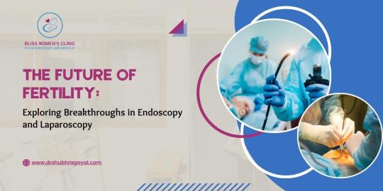
In medical science, the future of fertility and reproductive health is experiencing a revolutionary transformation through the remarkable advancements in Endoscopy and Laparoscopy. These innovative medical procedures have become instrumental in diagnosing and treating various reproductive health issues, offering hope to countless individuals and couples worldwide. In this article, we delve deep into the world of Endoscopy and Laparoscopy, uncovering their benefits and trends and why choosing Dr. Shubhra Goyal can indeed be a game-changer in your journey toward optimal reproductive health.
Laparoscopy and Endoscopy - what are they?
Due to their accuracy and efficacy, minimally invasive medical treatments like Endoscopy and Laparoscopy have become extremely popular in recent years. These procedures use specialized tools fitted with small cameras and lights that enable doctors to view and access inside organs without the need for open surgery.
Benefits of Endoscopic Examinations!
The following are some benefits: -
Minimal Scarring - Endoscopy and Laparoscopy require just small incisions, unlike traditional surgical techniques that demand extensive incisions. Due to the minimum scarring produced, patients recover more quickly and enjoy additional aesthetic benefits.
Less Pain and Discomfort - Patients who undergo endoscopic procedures often report less pain and discomfort following the operation. This is because these techniques are less intrusive than standard operations and result in less tissue damage.
Shorter Hospital Stays - Compared to conventional equivalents, endoscopic operations often require shorter hospital stays. Patients may resume their regular daily routines and activities more quickly as a result, which also lowers healthcare expenditures.
High accuracy - Using cutting-edge technology in endoscopic exams gives surgeons a degree of accuracy never before possible. They can traverse and treat delicate and difficult-to-reach locations with outstanding accuracy thanks to their precision, which ultimately improves patient surgery results.
Moving ahead, let's discuss,
Top Upcoming Trends in Endoscopy and Laparoscopy
The areas of Endoscopy and Laparoscopy are examples of the ongoing change taking place in the world of medical science. The following are some of the most notable new developments that are influencing how these processes may develop in the future:
Innovations in Endoscopic Imaging Technology - The endoscopic imaging technology industry is expanding quickly. This includes the creation of 3D imaging systems and high-definition cameras. These developments considerably increase diagnosis accuracy by providing surgeons with an incredibly thorough and accurate picture of inside organs.
Read More: https://www.drshubhragoyal.com/welcome/blogs/the-future-of-fertility:-exploring-breakthroughs-in-endoscopy-and-laparoscopy
#Endoscopy and Laparoscopy#Endoscopy and laparoscopy procedures#Medical uses of endoscopy and laparoscopy#Endoscopy vs. laparoscopy comparison#Benefits of endoscopic examinations#Laparoscopic surgery advancements#Gastrointestinal endoscopy techniques#Minimally invasive laparoscopic procedures#Endoscopic imaging technology#Laparoscopy in surgical diagnosis#Endoscopy for digestive disorders#Laparoscopy for abdominal surgeries#Endoscopy in medical diagnostics#Laparoscopic surgical instruments#Endoscopy for internal visualization#Laparoscopy and its applications#Endoscopic screening and treatment#Laparoscopic hernia repair#Endoscopy for upper GI tract
1 note
·
View note
Text
Navigating Growth in the Ultrasound Endoscope Market (2021–2031)
The global Ultrasound Endoscope Market is on a strong growth trajectory, poised to reach USD 2,000.62 million by 2031, up from USD 1,240.75 million in 2024, according to a new industry report. The market is expected to grow at a CAGR of 7.1% during the forecast period from 2025 to 2031.
The new report, published by Business Market Insights, provides a detailed analysis of industry trends, growth drivers, and competitive dynamics shaping the future of ultrasound endoscopic technology. It examines market behavior across geographies and offers insights critical for stakeholders, investors, and healthcare innovators.
📥 Sample PDF of the Report- https://www.businessmarketinsights.com/sample/BMIPUB00031683
Why the Ultrasound Endoscope Market Is Growing
Ultrasound endoscopy is increasingly being used for both diagnostic and therapeutic applications, particularly in gastroenterology, oncology, and pulmonology. The integration of ultrasound with endoscopic imaging allows for improved visualization and tissue sampling — leading to earlier and more accurate diagnoses.
Key drivers include:
Technological advancements in minimally invasive devices
Growing geriatric population and incidence of gastrointestinal diseases
Rising demand for precision diagnostics in oncology and internal medicine
Market Analysis Highlights
The report includes:
Executive Summary
Global Economic Overview
PESTLE & Porter’s Five Forces Analysis
Company Profiles & Market Share Insights
Case Studies and Forecasting Models
Strategic tools such as SWOT analysis, Investment Return Analysis, and Product Lifecycle (PLC) assessments help stakeholders evaluate the competitive landscape and investment opportunities.
Top Players in the Ultrasound Endoscope Market
Several major companies are leading innovation and expansion in this space:
Olympus Corporation
Fujifilm Holdings Corporation
Canon Inc.
HOYA Corporation
Lepu Medical Technology (Beijing) Co. Ltd
Sonoscape Pvt Ltd.
These companies are investing in AI-enabled imaging, hybrid endoscopy solutions, and expanding their product lines to meet rising global demand.
Regional Insights
North America and Europe dominate the current market landscape due to robust healthcare infrastructure and early technology adoption. However, the Asia-Pacific region is expected to grow the fastest, driven by increasing healthcare investments in China, India, and Southeast Asia.
Get the Full Report
To get deeper insights, market segmentation, and detailed forecasts, access the full report by Business Market Insights here:
🔗 Get Report Customization Options
About Business Market Insights
Business Market Insights is a trusted market intelligence platform offering detailed reports and custom research across multiple sectors including healthcare, automotive, energy, and technology. Their subscription-based service delivers timely insights to help companies make data-driven decisions.
📞 Contact: Ankit Mathur 📧 [email protected] 📱 +1–646–791–7070
0 notes
Text
Understanding Endoscopy: Benefits, Types, and What to Expect Before Your Procedure
Endoscopy represents a significant diagnostic modality that enables medical practitioners to investigate the internal structures of the human body without the need for invasive surgical procedures.. By using a flexible tube equipped with a camera, healthcare professionals can visualize and assess various organs, from the digestive tract to the respiratory system. This minimally invasive procedure not only helps in diagnosing conditions but also plays a crucial role in treatment.
Understanding endoscopy can empower you to make informed decisions about your health. Whether you're facing a specific medical issue or just curious about the procedure, knowing what to expect can ease anxiety and enhance your experience. In this article, we’ll delve into the different types of endoscopy, their benefits, and what you should know before undergoing the procedure. Get ready to uncover the insights that could lead to better health outcomes.
Overview of Endoscopy:
Endoscopy refers to a minimally invasive procedure where a flexible tube equipped with a camera, known as an endoscope, allows healthcare providers to view the interior of your body. Commonly, endoscopy explores organs like the stomach, intestines, and lungs. The procedure aids in diagnosing various medical conditions, such as ulcers, lesions, or gastrointestinal disorders.
During endoscopy, you may receive sedation for comfort. A healthcare professional inserts the endoscope through natural openings, like the mouth or anus, depending on the area being examined. This technique minimizes recovery time and is often preferred over traditional surgery. Understanding endoscopy helps you make informed medical decisions and alleviates anxiety about potential procedures.

Types of Endoscopy:
Endoscopy includes various procedures tailored for different regions of the body. Each type provides unique insights for diagnosis and treatment.
Upper Gastrointestinal Endoscopy:
Upper gastrointestinal (GI) endoscopy is a diagnostic procedure employed to assess the esophagus, stomach, and duodenum through the utilization of a flexible endoscope.. This procedure allows doctors to identify issues like inflammation, ulcers, and tumors. A common reason for this endoscopy includes unexplained gastrointestinal symptoms, such as nausea or pain. The procedure typically requires sedation, ensuring your comfort during the examination. Recovery time is generally brief, with many individuals resuming normal activities the same day.
Colonoscopy:
Colonoscopy investigates the large intestine (colon) and rectum to detect abnormalities like polyps, cancer, or inflammatory bowel disease. During this endoscopy, a long, flexible tube equipped with a camera views the colon's lining. Preparation for a colonoscopy involves a clear liquid diet and laxatives to ensure a clean bowel. It's crucial for early cancer detection, especially for those over 45 or at risk. Like upper GI endoscopy, sedation is usually administered, and the procedure is generally well-tolerated.
Endoscopic Ultrasound:
Endoscopic ultrasound (EUS) combines endoscopy and ultrasound technology to obtain detailed images of organs and structures within the body. This procedure typically targets the digestive tract, pancreas, and surrounding tissues. Endoscopic Ultrasound (EUS) plays a critical role in the diagnosis of medical conditions such as pancreatic cancer and the precise staging of tumors. The procedure involves inserting a thin endoscope with an ultrasound device, providing real-time imaging. Sedation often enhances comfort, and recovery occurs quickly, making it a convenient option for detailed investigation.
Advantages of Endoscopy:
At Gastro Maryland, you receive specialized care from a dedicated team focused on your gastrointestinal health. Whether you're seeking routine screenings or advanced diagnostic procedures, including endoscopy, our experienced specialists guide you through treatment options tailored to your needs.
Understanding Endoscopy:
Endoscopy provides a minimally invasive method for the examination of the digestive tract.. A flexible tube equipped with a camera allows physicians to visualize your esophagus, stomach, and intestines. This method ensures accurate diagnosis and effective treatment of various conditions, such as GERD, ulcers, and cancers.
Benefits of Endoscopy:
Early Detection: Endoscopy helps identify gastrointestinal disorders early, increasing the chance of successful treatment.
Less Invasive: Unlike traditional surgeries, endoscopy requires minimal incisions, ensuring quicker recovery times.
Real-Time Diagnosis: Instant visualization allows for immediate assessment, minimizing uncertainty and stress.
Personalized Patient Care:
At Gastro Maryland, your comfort is paramount. Our team offers sedation options for all procedures, ensuring a painless experience. Patient education is crucial; our staff explains every step of the process, answering your questions to alleviate anxiety.
Advanced Technologies:
We utilize the latest technologies in endoscopy, enhancing both diagnostic accuracy and patient outcomes. These innovations allow for more precise imaging and quicker recovery times, ensuring you return to your daily routine promptly.
Your Health is Our Priority:
Addressing gastrointestinal issues promptly leads to better health outcomes. Whether you’re experiencing symptoms or due for a routine screening, Gastro Maryland is here to craft a personalized care plan just for you.
Schedule Your Consultation:
Taking control of one's digestive health commences with a thorough consultation. Contact Gastro Maryland today to schedule your appointment and discover how endoscopy can play a key role in maintaining your gastrointestinal wellness. For more information, please Visit Gastro Center Of Maryland.
Risks and Considerations:
Endoscopy, while generally safe, carries specific risks and considerations that you should understand before undergoing the procedure. Complications may include:
Infection: Although rare, infection at the site of endoscopy may occur, possibly leading to more severe issues.
Bleeding: Potential bleeding can arise, especially following biopsies or polypectomies during colonoscopy.
Perforation: There's a small risk of perforating the organ being examined, which may necessitate surgical intervention.
Sedation Effects: Sedation used for comfort during endoscopy can cause respiratory issues, such as airway obstruction, particularly in patients with pre-existing conditions.
Allergic Reactions: Some individuals may experience allergic reactions to medications used, including sedatives and anesthetics.
These risks underscore the importance of discussing your medical history and concerns with your healthcare provider at Gastro Maryland. They will evaluate your situation to ensure endoscopy is the appropriate diagnostic option for you. Understanding these risks enhances your ability to make informed decisions about your digestive health.
Conclusion:
Endoscopy is a vital tool in modern medicine that empowers you to take control of your digestive health. By understanding the procedure and its benefits, you can approach your healthcare decisions with confidence. Whether you're dealing with specific symptoms or simply seeking knowledge about your gastrointestinal health, the insights gained from endoscopy can lead to timely interventions and better outcomes.
At Gastro Maryland, you’ll find a supportive environment that prioritizes your comfort and well-being. Don't hesitate to reach out and schedule a consultation to discuss how endoscopy can be a key part of your health journey. Your digestive wellness is worth it.
Meta Description(optional):
Discover how endoscopy helps diagnose and treat digestive issues with minimal recovery time. Learn about types, benefits, and what to expect at Gastro Maryland.
Reach out us for more info about Gastroenterologist Columbia!
1 note
·
View note
Text
Exploring Innovations in Esophageal and Gastric Disease: Advances in Diagnosis and Treatment
Esophageal and gastric diseases represent a significant burden on global health, impacting millions of individuals annually. These conditions range from common ailments such as gastroesophageal reflux disease (GERD) and gastritis to more complex challenges like Barrett’s esophagus, gastric ulcers, and malignancies. As medical science continues to evolve, innovations in diagnosis and treatment are transforming the way these diseases are managed, improving patient outcomes and quality of life.
Understanding Esophageal and Gastric Disorders
The esophagus and stomach play vital roles in digestion, and diseases affecting these organs can lead to symptoms such as heartburn, difficulty swallowing, nausea, and abdominal pain. Chronic conditions may result in complications including bleeding, strictures, or even cancer. Accurate diagnosis and timely intervention are essential in preventing progression and ensuring effective treatment.
Recent Advances in Diagnosis
Modern diagnostic technologies are making it easier than ever to identify esophageal and gastric conditions at earlier stages:
Modern diagnostic technologies are making it easier than ever to identify esophageal and gastric conditions at earlier stages:
High-Resolution Manometry (HRM): This technique provides detailed pressure readings of the esophagus, helping in the diagnosis of motility disorders such as achalasia and esophageal spasms.
Endoscopic Innovations: Techniques like narrow-band imaging (NBI), confocal laser endomicroscopy, and capsule endoscopy allow for enhanced visualization and biopsy of suspicious lesions, improving early cancer detection.
Molecular and Biomarker Testing: Researchers are exploring biomarkers for conditions such as Barrett’s esophagus and gastric cancer, paving the way for non-invasive diagnostic options.

Breakthroughs in Treatment
Therapeutic advancements are reshaping the management landscape for esophageal and gastric diseases:
Endoscopic Therapies: Minimally invasive techniques such as radiofrequency ablation (RFA), endoscopic mucosal resection (EMR), and peroral endoscopic myotomy (POEM) offer alternatives to traditional surgery for early-stage disease and motility disorders.
Biologic and Targeted Therapies: In gastric cancer and severe inflammatory conditions, personalized medicine is making strides. Targeted therapies and immunotherapy are offering new hope to patients with previously limited options.
Advanced Proton Pump Inhibitors (PPIs) and Alternatives: With ongoing research into the long-term effects of PPIs, new acid-suppressing agents such as potassium-competitive acid blockers (P-CABs) are gaining interest as safer and more effective options.
The Role of Multidisciplinary Collaboration
Managing esophageal and gastric disease often requires a team approach, involving gastroenterologists, surgeons, oncologists, dietitians, and radiologists. Multidisciplinary care ensures comprehensive evaluation and tailored treatment plans that align with each patient’s specific condition.
Looking Ahead
The future of esophageal and gastric disease management is bright, with ongoing research focused on early detection, less invasive treatments, and improved patient quality of life. Conferences like the 15th World Gastroenterology, IBD & Hepatology Conference (GastroenterologyUCG 2025) are playing a key role in bringing global experts together to share these advancements and shape the future of digestive health.
Join the Conversation
Explore the latest findings and engage with international specialists at Track 9: Esophageal and Gastric Disease during GastroenterologyUCG 2025. 📅 December 17–19, 2025 | 📍 Dubai, UAE & Virtual 🔗 Submit your abstract or register now: https://gastroenterology.utilitarianconferences.com/
#Gastroenterology #EsophagealDisease #GastricHealth #GERD #Endoscopy #MedicalInnovation #GastroenterologyUCG2025 #IBD #Hepatology
0 notes
Text
Rigid Endoscopes Market Growth Analysis, Market Dynamics, Key Players and Innovations, Outlook and Forecast 2025-2032
According to a new market analysis, the global rigid endoscopes market was valued at USD 3,374 million in 2024 and is projected to reach USD 5,187 million by 2032, growing at a Compound Annual Growth Rate (CAGR) of 5.6% during the forecast period (2025–2032). The growth is driven by increasing demand for minimally invasive surgeries, technological advancements in endoscopic imaging, and expanding applications across multiple surgical specialties. get free sample of this report at https://www.intelmarketresearch.com/download-free-sample/1029/rigid-endoscopes-2025-2032-816
What are Rigid Endoscopes? Rigid endoscopes are high-precision optical instruments used for minimally invasive diagnostic and surgical procedures. Unlike flexible endoscopes, these devices maintain their shape during use, providing superior image clarity and mechanical stability. They consist of a rigid metal tube housing advanced lens systems and light transmission channels, enabling surgeons to visualize internal organs or cavities with exceptional detail. These devices are indispensable in specialties including laparoscopy, arthroscopy, urology, and ENT procedures, where their robust construction allows integration with surgical instruments and electrosurgical systems. Modern rigid endoscopes incorporate cutting-edge features like 4K ultra-HD imaging, 3D visualization, and fluorescence guidance, positioning them as critical tools in contemporary surgical practice.
Key Market Drivers
Surging Demand for Minimally Invasive Procedures The shift toward minimally invasive surgeries (MIS) represents the primary growth driver, as these procedures offer patients reduced trauma, shorter hospital stays, and faster recovery times compared to traditional open surgeries. Rigid endoscopes enable precise visualization critical for MIS, with procedure volumes increasing approximately 8-12% annually across major healthcare markets. Notably, NHS England reported a 100% increase in robot-assisted procedures from 2022-2024, reflecting broader adoption of endoscopic surgical techniques.
Continuous Technological Advancements Innovations in imaging technology are revolutionizing the market, with manufacturers introducing advanced systems featuring: • 4K/8K Ultra-HD resolution for enhanced tissue differentiation • 3D visualization systems improving depth perception in complex procedures • AI-powered diagnostic tools for real-time tissue analysis • Narrow Band Imaging (NBI) for better vascular visualization Companies like Karl Storz and Olympus continue to push technological boundaries, developing integrated systems that combine superior optics with intelligent software solutions.
Market Challenges
High Costs and Budget Constraints Advanced rigid endoscopy systems represent significant capital investments, with prices ranging from $50,000 to $150,000 for basic to high-end configurations. This creates adoption barriers, particularly in: • Public healthcare systems with limited equipment budgets • Developing markets where cost sensitivity is high • Smaller surgical centers lacking scale for ROI justification
Complex Sterilization Requirements As reusable medical devices, rigid endoscopes require stringent reprocessing protocols to prevent healthcare-associated infections. Challenges include: • High labor costs for proper cleaning and sterilization • Potential damage from repeated reprocessing cycles • Regulatory complexities around infection control standards The CDC estimates 1 in 31 hospital patients acquires an infection daily, with improperly processed equipment being a known risk factor.
Opportunities Ahead
Expansion in Emerging Markets The Asia-Pacific region, led by China and India, presents tremendous growth potential with: • Healthcare infrastructure development projects • Rising medical tourism industries • Increasing government healthcare expenditures • Growing middle-class patient populations China's rigid endoscopes market alone is projected to grow at 9.1% CAGR, nearly double the global average rate.
Development of Disposable Solutions The emergence of single-use rigid endoscopes addresses key challenges around: • Infection control risks • Reprocessing costs • Maintenance expenses While currently representing a small market segment, disposable options are gaining traction in ambulatory surgery centers and outpatient facilities.
Regional Market Insights • North America dominates with 28.62% market share, benefiting from advanced healthcare infrastructure, high procedure volumes, and early technology adoption. The U.S. accounts for approximately 85% of regional demand. • Europe follows closely (27.11% share), with Germany, France, and the UK leading adoption. The region's aging population and comprehensive healthcare systems drive steady demand. • Asia-Pacific is the fastest-growing region (35.6% share), with China and India expanding rapidly due to healthcare modernization initiatives and increasing surgical capabilities. • Latin America and MEA show developing potential, though adoption varies significantly between countries based on healthcare infrastructure and economic conditions.
Competitive Landscape The market features strong competition between global leaders and specialized manufacturers: • Market Leaders: Karl Storz, Olympus, and Stryker collectively hold approximately 80% market share, maintaining dominance through continuous innovation and strategic acquisitions. • Technology Innovators: Companies like Richard Wolf and Medtronic focus on niche applications (urology, neurosurgery) with specialized endoscopic solutions. • Recent Developments: • Karl Storz's acquisition of Asensus Surgical (June 2024) to enhance robotic endoscopy capabilities • Olympus receiving CE certification for AI-powered endoscopic imaging systems (October 2024) • Mindray's launch of advanced 4K/3D endoscopy platforms (June 2024)
Market Segmentation By Product Type: • Laparoscopes • Arthroscopes • Ureteroscopes • Cystoscopes • Neuroendoscopes • Other Specialty Endoscopes By Application: • General Surgery • Orthopedics • Urology • Gynecology • ENT • Other Specialties By End User: • Hospitals • Ambulatory Surgery Centers • Specialty Clinics • Academic & Research Institutions
Report Coverage & Benefits This comprehensive market research report provides: • Market size projections through 2032 • In-depth competitive analysis of 12+ key players • Emerging technology assessment • Regional market opportunity analysis • SWOT and value chain evaluation • Strategic recommendations for stakeholders Download FREE Sample Report: Rigid Endoscopes Market Sample Report View Full Report: Rigid Endoscopes Market Detailed Report
Visit more reports : https://iamstreaming.org/siddheshkapshikar/blog/10529/corporate-financial-modelling-market-growth-analysis-market-dynamics-key-players-and-innovations-outlook-and-forecast-2025-20 https://sidintelmarketresearch.hashnode.dev/corporate-financial-modelling-market-growth-analysis-market-dynamics-key-players-and-innovations-outlook-and-forecast-2025-2032 https://chatterchat.com/read-blog/9369 https://www.tumblr.com/siddhesh7011/787321451926749184/permalloys-feni-market-growth-analysis-market https://kapshikarsiddhesh.livepositively.com/permalloys-feni-market-growth-analysis-market-dynamics-key-players-and-innovations-outlook-and-forecast-2025-2032/new=1 https://articlescad.com/permalloys-feni-market-growth-analysis-market-dynamics-key-players-and-innovations-outlook-and-243618.html https://controlc.com/c6ac365d https://justpaste.it/i36yo https://notejoy.com/permalloys-feni-market-growth-analysis-market-dynamics-key-players-and-innovations-outlook-and-forecast-2025-2032-467663
About Intel Market Research Intel Market Research delivers actionable insights in technology and infrastructure markets. Our data-driven analysis leverages: • Real-time infrastructure monitoring • Techno-economic feasibility studies Competitive intelligence across 100+ countries Trusted by Fortune 500 firms, we empower strategic decisions with precision.
Website: https://www.intelmarketresearch.com Follow us on LinkedIn: https://www.linkedin.com/company/intel-market-research
0 notes
Text
Medical Endoscopes Market Outlook Current Status by Major Key Vendors and Trends by Forecast (2021–2031)
The medical endoscopes market size is expected to reach US$ 22,467.07 million by 2031 from US$ 14,487.23 million in 2024. The market is estimated to record a CAGR of 6.5% from 2025 to 2031.
Executive Summary and Global Market Analysis:
The global endoscopes market is experiencing robust growth, primarily spurred by the increasing adoption of minimally invasive diagnostic and therapeutic interventions. This expansion is significantly influenced by the rising global burden of chronic diseases and an aging demographic. Consequently, diverse medical fields, including gastroenterology, pulmonology, urology, and ENT, are progressively integrating advanced endoscopic techniques into their practices. Key technological advancements, such as high-definition imaging, the advent of disposable single-use endoscopes, and enhanced optical systems, are leading to improved procedural accuracy and a notable reduction in infection rates. Furthermore, the growing preference for outpatient and ambulatory care is amplifying the need for portable and cost-effective endoscopic solutions. Emerging markets are demonstrating impressive growth rates, underpinned by ongoing improvements in healthcare infrastructure and increasing public awareness of minimally invasive medical options.
Medical Endoscopes Market Drivers and Opportunities
The growing prevalence of chronic health conditions like digestive problems, respiratory issues, and cancer is creating a considerable need for sophisticated diagnostic tools. These conditions frequently necessitate regular and precise internal evaluations to monitor disease development and inform treatment choices. An endoscope is a vital medical instrument equipped with a light, used to visually inspect the inside of a body cavity or organ. This device is typically inserted through a natural opening, for example, the mouth during a bronchoscopy or the rectum for a sigmoidoscopy. As long-term health challenges affecting the gut, lungs, and urinary system become more common, medical practitioners are increasingly turning to endoscopic procedures for accurate diagnosis, efficient treatment delivery, and ongoing management of these health concerns.
Download our Sample PDF Report
@ https://www.businessmarketinsights.com/sample/BMIPUB00031632
Medical Endoscopes Market Segmentation
Medical Endoscopes Market By Product Type
Rigid Endoscopes
Flexible Endoscopes
Capsule Endoscopes
Ultrasound Endoscope
Robot-assisted Endoscopes
Medical Endoscopes Market By Usability
Reusable Endoscopes
Disposable Endoscopes
Medical Endoscopes Market By Application
Gastrointestinal Endoscopy
Laparoscopy
Gynecology Endoscopy
Arthroscopy
Uroscopy
Bronchoscopy
ENT Endoscopy
Neuroendoscopy
Medical Endoscopes Market By End User
Hospitals
Specialty Clinics
Ambulatory Surgical Centers
Diagnostic Centers
Medical Endoscopes Market Regions and Countries Covered
North America
US
Canada
Mexico
Europe
Germany
Italy
France
U.K.
Spain
Belgium
Netherlands
Luxembourg
Norway
Finland
Denmark
Sweden
Switzerland
Austria
Greece
Portugal
Russia
Poland
Romania
Czech Republic
Ukraine
Slovakia and Bulgaria
Asia-Pacific
China
Japan
South Korea
India
Australia
New Zealand
Indonesia
Malaysia
Philippines
Singapore
Thailand
Vietnam
Taiwan and Bangladesh
South and Central America
Brazil
Argentina
Chile
Colombia
and Peru
Middle East and Africa
Saudi Arabia
United Arab Emirates
Kuwait
Bahrain
Qatar
Oman
Turkiye
South Africa
Egypt
Nigeria and Algeria
Medical Endoscopes Market leaders and key company profiles
Olympus Corporation
Fujifilm Holdings Corporation
Boston Scientific
Karl Storz SE & Co. KG
Stryker Corporation
B. Braun SE
HOYA Corporation
Ambu A/S
Johnson & Johnson
Medtronic
About Us:
Business Market Insights is a market research platform that provides subscription service for industry and company reports. Our research team has extensive professional expertise in domains such as Electronics & Semiconductor; Aerospace & Defense; Automotive & Transportation; Energy & Power; Healthcare; Manufacturing & Construction; Food & Beverages; Chemicals & Materials; and Technology, Media, & Telecommunications
0 notes