#6 week 3d ultrasound
Explore tagged Tumblr posts
Text
Sneak Peek Baby Gender Test
We provide elective 3d/4d ultrasounds and gender reveal to pregnant women in the Chicagoland area. Starting at just 6 weeks you can see baby for the time and share this magicalmoment with family and friends.
#sneak peek gender test#14 week 3d ultrasound#20 week ultrasound#3d 4d ultrasound#6 week 3d ultrasound
0 notes
Text
Role of Ultrasound in Prenatal Care and Women’s Health

Ultrasound imaging, often associated with pregnancy, plays a vital role in women’s healthcare. It is a non-invasive, radiation-free diagnostic tool that uses sound waves to create images of internal organs and tissues. While its most common use is monitoring fetal development during pregnancy, ultrasound also aids in diagnosing and managing various gynecological conditions, contributing significantly to overall women’s health.
In this blog, we’ll explore the importance of ultrasound in prenatal care and women’s health, its role in early detection and prevention, and how advanced diagnostic centers like Apex Diagnostics in Solan, Himachal Pradesh, offer comprehensive imaging services. We'll also touch upon other diagnostic services available, including multislice CT scans near Shimla and MRI in Solan, which complement ultrasound in women’s healthcare.
Ultrasound in Prenatal Care
1. Early Pregnancy Confirmation and Viability
Ultrasound is crucial in confirming pregnancy as early as 4-6 weeks. It helps determine the viability of the pregnancy, including the presence of a fetal heartbeat and whether it is an intrauterine or ectopic pregnancy. Early detection is essential for managing potential complications.
2. Monitoring Fetal Development
Throughout pregnancy, ultrasounds are used to monitor fetal growth and development, assess amniotic fluid levels, and detect congenital anomalies. The standard 3D and 4D ultrasound techniques provide detailed images, allowing healthcare providers and parents to see the fetus’s movements and facial features in real-time.
3. Assessing Placental Health
Ultrasounds help in evaluating the placenta's position and health, ensuring there are no issues like placenta previa, which can cause complications during delivery. Early identification of placental issues allows for appropriate medical management.
4. Guiding High-Risk Pregnancies
For women with high-risk pregnancies, ultrasound monitoring becomes more frequent. It helps in diagnosing conditions such as gestational diabetes, preeclampsia, and fetal growth restrictions. Paired with health packages in Solan that include regular monitoring, these ultrasounds provide a comprehensive approach to prenatal care.
Ultrasound in Women’s Health Beyond Pregnancy
Ultrasound is not limited to prenatal care; it is also instrumental in diagnosing and managing various gynecological conditions:
1. Pelvic Pain and Abnormal Bleeding
Ultrasound is often the first diagnostic tool used to evaluate pelvic pain and abnormal bleeding. It can identify conditions such as ovarian cysts, fibroids, and endometriosis, enabling early intervention.
2. Infertility Investigations
For women experiencing infertility, ultrasound is used to monitor ovulation, assess uterine abnormalities, and guide assisted reproductive procedures. This non-invasive method is essential for tailoring personalized treatment plans.
3. Breast Health
Breast ultrasound is a complementary tool to mammography, particularly useful for women with dense breast tissue. It helps detect lumps, cysts, and other abnormalities that may not be visible on a mammogram.
4. Menstrual Disorders and PCOS
Ultrasound helps diagnose polycystic ovary syndrome (PCOS) by identifying multiple cysts on the ovaries. It also assists in evaluating the endometrium for menstrual disorders, helping healthcare providers offer targeted treatments.
Complementary Diagnostic Tools in Women’s Health
While ultrasound is a primary imaging tool, other advanced diagnostics can complement it for a more comprehensive assessment:
1. Multislice CT Scan Near Shimla
In cases where more detailed imaging of the abdominal or pelvic region is required, a multislice CT scan near Shimla can provide precise cross-sectional images. This is especially useful for diagnosing conditions like ovarian cancer, pelvic inflammatory disease, and complex uterine abnormalities.
2. MRI in Solan
For soft tissue evaluation, an MRI in Solan offers superior imaging clarity. It is often used for detailed assessments of reproductive organs, including detecting fibroids, adenomyosis, and ovarian masses. MRI is also invaluable in evaluating suspected pelvic or breast cancers when ultrasound and CT scans are inconclusive.
The Importance of Preventive Health Packages
Preventive healthcare is essential for early detection and management of potential health issues. Diagnostic centers like Apex Diagnostics offer health packages in Solan that combine ultrasound with other imaging techniques and routine tests, ensuring a holistic approach to women’s health.
Benefits of Health Packages for Women
Early Detection of Gynecological Issues: Regular ultrasounds and imaging tests help detect problems early, reducing the risk of complications.
Comprehensive Care for Expecting Mothers: Prenatal health packages provide regular monitoring through ultrasounds, lab tests, and nutritional guidance.
Customized Health Solutions: These packages can be tailored to individual needs, addressing specific concerns like reproductive health, menopause, and cancer screening.
Apex Diagnostics: A Leader in Women’s Healthcare
Apex Diagnostics in Solan is at the forefront of providing advanced imaging services, including ultrasound, MRI in Solan, and multislice CT scans near Shimla. Their team of experienced radiologists and state-of-the-art technology ensures accurate diagnoses and personalized care. Whether it’s routine prenatal monitoring or complex gynecological evaluations, Apex Diagnostics offers comprehensive solutions for every stage of a woman’s life.
Conclusion
Ultrasound plays a pivotal role in both prenatal care and overall women’s health, offering a safe, non-invasive method for early detection and management of various conditions. When combined with advanced diagnostic tools like multislice CT scans near Shimla and MRI in Solan, ultrasound becomes part of a robust healthcare strategy that promotes early intervention and better outcomes.
Choosing a trusted diagnostic center like Apex Diagnostics ensures access to cutting-edge technology and expert care, making preventive healthcare more accessible for women in Himachal Pradesh. Investing in regular health check-ups through comprehensive health packages in Solan empowers women to take control of their health, ensuring a healthier future for themselves and their families.
#24 hrs mri near sirmour#mri in solan#mri services in himachal#whole body mri in himachal#whole body mri near shimla#whole body mri near sirmour#clinic#health center
0 notes
Text
What to Expect: Your Guide to Private Ultrasound Scans
If you're pregnant or have medical concerns that require imaging, you might be considering a private ultrasound scan. This option can provide valuable insights and a more personalized experience than traditional hospital scans. But what exactly can you expect from this process?
Arrival and Environment
When you arrive at a private clinic for your ultrasound, you'll typically find a welcoming and comfortable environment. Most clinics prioritize creating a relaxed atmosphere to help ease any anxiety. You’ll be greeted by friendly staff who will explain the process and answer any questions you may have before the scan begins.
The Procedure
Once you’re ready for your scan, the sonographer will guide you into the examination room. The procedure usually lasts about 30 minutes. You’ll be asked to lie down on an examination table, and a gel will be applied to your abdomen. This gel helps create a better connection between the transducer and your skin, improving the quality of the images captured.

The sonographer will then use a handheld device called a transducer to emit sound waves and create images of your internal organs or, in the case of pregnancy, your developing baby. You’ll be able to see real-time images on a monitor, which can be a thrilling experience, especially when you spot your baby's heartbeat or movements.
Types of Scans
Private ultrasound clinics offer a variety of scans, including:
Early Pregnancy Scans: These are usually performed between 6 to 10 weeks to confirm the pregnancy and check for a heartbeat.
Gender Determination Scans: Often done around 16 weeks, these scans can reveal the sex of your baby.
Growth Scans: Conducted later in pregnancy to monitor the growth and development of the fetus, these scans help assess whether the baby is growing as expected.
3D and 4D Scans: Many private clinics offer advanced imaging techniques, allowing you to see your baby in three dimensions or even in motion, adding an exciting dimension to the experience.
After the Scan
Once the scan is complete, the sonographer will provide you with a detailed report and may even give you printouts of the images. This report is valuable for your medical records and can be shared with your healthcare provider if needed.
You’ll also have the opportunity to ask any questions about the results right away. This immediate access to information can be incredibly reassuring for expectant parents who may have concerns about their pregnancy.
Conclusion
In summary, a private ultrasound scan can be an enriching experience, offering peace of mind and invaluable insights into your pregnancy or health. From the welcoming environment to the high-quality imaging and personalized care, these scans are designed to meet your needs and provide reassurance during an important time in your life. Whether you’re early in your pregnancy or nearing delivery, considering a private ultrasound can make your journey smoother and more enjoyable.
0 notes
Text
3d baby Vision
3D Baby Vision is an advanced ultrasound technology that provides expectant parents with a lifelike, three-dimensional view of their baby in the womb. Unlike traditional 2D ultrasounds, which offer flat images, 3D Baby Vision captures detailed and realistic visuals of the baby’s features, including facial expressions, movements, and body structure. Typically performed between 26 and 32 weeks of pregnancy, this technology enhances the bonding experience and can help identify potential developmental concerns. It is a safe and valuable tool, offering both emotional connection and important medical insights into the baby’s health.
Physical Effects:
Bleeding and Cramping: You may experience vaginal bleeding similar to a heavy period, along with cramping, as the body expels the pregnancy tissue. Bleeding may last for several days to two weeks.
Hormonal Changes: Your hormone levels will gradually return to pre-pregnancy levels, which may take a few weeks. This process can trigger symptoms like fatigue, breast tenderness, or mood changes.
Follow-up Care: Your healthcare provider may recommend an ultrasound or blood tests to ensure all pregnancy tissue has been expelled. In some cases, medical intervention such as a D&C (dilation and curettage) may be necessary if tissue remains.
Return of Menstrual Cycle: Your menstrual cycle will typically resume within 4 to 6 weeks, though this can vary.
Emotional Effects:
Grief and Sadness: It’s normal to feel a range of emotions, including grief, sadness, and even guilt. Miscarriage is a loss, and it’s important to give yourself time and space to process these emotions.
Mood Swings: Hormonal changes, along with the emotional toll of the miscarriage, may lead to mood swings, anxiety, or depression.
Support: Talking to a healthcare provider, counselor, or support group can be helpful. Sharing your feelings with trusted friends or family can also provide comfort.
When to Seek Medical Attention:
If you experience heavy bleeding (soaking through more than one pad per hour), severe pain, fever, or foul-smelling discharge, you should contact your healthcare provider immediately as these may be signs of infection or complications.
0 notes
Text
Choosing the Best Time for an Elective Ultrasound: A Guide for Expecting Parents

When it comes to experiencing the joys of pregnancy, one of the most exciting moments for expecting parents is seeing their baby during an ultrasound. For many, elective ultrasounds are more than just a medical procedure—they are a keepsake memory. Choosing the right time for an elective ultrasound, whether it's a standard 2D, or a more advanced 3D/4D scan, is essential to getting the best possible experience and images. In this guide, we will explore the best stages of pregnancy for different types of ultrasounds and how this timing plays a crucial role in the elective ultrasound business. The Stages of Pregnancy and Ultrasound Options One of the first things to consider when scheduling an elective ultrasound is understanding the various stages of pregnancy and how they align with different ultrasound types. Whether you're in the first trimester and want a peek at your tiny miracle or you’re later in the pregnancy and hoping to capture detailed facial features in 3D or 4D, timing is key. First Trimester: Weeks 6-12 – Early Sneak Peek The first trimester is a delicate time for both the mother and baby. For those seeking early glimpses, an elective ultrasound can be performed as early as six weeks. While this isn’t the time for detailed images, it can be a significant bonding moment for parents who want to hear their baby’s heartbeat for the first time. Early elective ultrasounds are often 2D and focus primarily on confirming the pregnancy, checking the heartbeat, and verifying the baby’s position in the uterus. If you're thinking about starting an ultrasound business, offering early 2D scans can cater to a market of eager parents who want reassurance and connection during those initial weeks. However, these ultrasounds are best used as a complement to medical check-ups, and you should emphasize that elective scans do not replace medical advice. Second Trimester: Weeks 18-24 – Perfect for Gender Reveal and Clarity The second trimester is when elective ultrasounds truly shine. By week 18, most expecting parents are eager to find out the gender of their baby. Gender reveal scans are popular during this stage, and they provide a perfect opportunity for ultrasound franchise owners to tap into the trend of gender reveal parties. Offering a memorable experience, complete with 2D or 3D imaging, can make your business a go-to choice for keepsake ultrasounds. At this point in the pregnancy, the baby has grown significantly, and you can get clearer images of their entire body. This is the ideal time for parents to schedule a 3D ultrasound if they want to see their baby’s facial features with some clarity but without the fine details that are better captured later in pregnancy. If you’re looking into how to open a 3D ultrasound studio, focusing on second-trimester appointments for gender reveals and basic 3D imaging is a smart business move. Third Trimester: Weeks 28-34 – The Golden Window for 3D/4D Imaging The third trimester is when the magic happens for 3D and 4D ultrasounds. Between weeks 28 and 34, your baby has developed enough fat to have those chubby cheeks and fully formed facial features, making this the best time for capturing keepsake baby ultrasound images. During this golden window, parents can not only see their baby’s face in 3D but also watch them yawn, stretch, and even suck their thumb in real-time 4D. This is also a wonderful opportunity for elective ultrasound business owners to showcase the most advanced technology they have. It’s during this stage that you can truly elevate your service offering, providing an emotional and unforgettable experience for parents. Many parents seek out businesses specifically offering 3D/4D ultrasounds during these weeks, making it an excellent time for targeted ultrasound business marketing tips. However, it’s important to inform clients that after 34 weeks, the baby becomes more cramped in the womb, making it harder to get clear images. Encouraging clients to book their 3D/4D sessions early in the third trimester ensures the best results. Choosing the Right Ultrasound Business Model If you're considering starting an ultrasound business or opening a 3D ultrasound studio, understanding the demand for elective ultrasounds during these key stages of pregnancy is essential. There are several factors to consider when planning your business model: Elective Ultrasound Training and Equipment When starting a 3D/4D ultrasound business, the quality of your equipment and the training of your staff are paramount. Investing in top-tier ultrasound machines and sending your staff to elective ultrasound training programs ensures that your clients receive the best images possible. High-quality imaging is what sets keepsake ultrasounds apart from standard medical ones, and it can help you build a reputation for excellence in the elective ultrasound business. Marketing Your Ultrasound Business Ultrasound business marketing tips often focus on social media and word-of-mouth recommendations, which can be particularly effective when your services include highly shareable moments like gender reveals and 3D/4D images. Consider offering package deals for parents who want multiple scans throughout their pregnancy, such as an early heartbeat scan, a gender reveal, and a 3D/4D ultrasound later on. This creates repeat business and encourages clients to build a relationship with your studio. Additionally, creating partnerships with other pregnancy-related businesses—like maternity boutiques, prenatal yoga studios, and even local hospitals—can help increase your visibility. A comprehensive marketing strategy that includes these partnerships, along with targeted online ads and engaging social media content, can be the key to success. Costs and Profitability The cost of starting an ultrasound business varies depending on factors such as equipment, location, and staffing. However, many businesses find that once they establish a loyal customer base, the return on investment can be substantial. Offering a variety of services, from basic 2D ultrasounds to advanced 4D sessions, can cater to a wider audience and help maximize profitability. Final Thoughts: Timing is Everything Elective ultrasounds offer expecting parents the chance to bond with their baby and create cherished memories. Whether it’s hearing that first heartbeat, discovering the gender, or seeing a baby’s face in 3D for the first time, each stage of pregnancy provides a unique opportunity. As an elective ultrasound business owner, understanding the best timing for these different types of scans is critical to providing a great experience for your clients. Are you considering starting your own 3D/4D ultrasound business? What stage of pregnancy do you think offers the most exciting ultrasound experience? Share your thoughts in the comments below and let’s keep the conversation going! If you found this post helpful, don’t forget to share it with your network. Call-to-Action:Have you started your journey in the ultrasound business? Share your experiences in the comments below and let us know your thoughts! Don’t forget to share this post with your network if you found it helpful. Read the full article
0 notes
Text
Ultrasound in Prenatal Care: What Expectant Parents Need to Know
Prenatal care is a critical aspect of a healthy pregnancy, and among the most significant tools in this care is the ultrasound. For many expectant parents, an ultrasound is the first opportunity to see their developing baby, and it serves as an essential method for monitoring the baby’s health and development. Understanding the role of ultrasound in prenatal care can help parents navigate their pregnancy with greater confidence and awareness.
What is an Ultrasound?
An ultrasound, also known as a sonogram, is a medical imaging technique that uses high-frequency sound waves to create visual images of the inside of the body. In prenatal care, ultrasounds allow doctors and healthcare providers to visualize the fetus, the placenta, and other aspects of the pregnancy. The process is non-invasive, painless, and safe for both the mother and the baby, making it an ideal tool for monitoring pregnancy progress.
Types of Ultrasound Scans
There are several types of ultrasound scans used during pregnancy, each serving a unique purpose:
1. Transabdominal Ultrasound: This is the most common type of ultrasound. The technician applies a gel to the mother's abdomen and moves a transducer across the belly to capture images of the fetus.
2. Transvaginal Ultrasound: Used in early pregnancy or when more detailed images are needed, this type involves inserting a small transducer into the vagina. It provides a clearer view of the uterus and can be particularly useful in early pregnancy assessments.
3. 3D and 4D Ultrasounds: These advanced scans offer a three-dimensional view of the fetus (3D) and real-time video of the baby in motion (4D). While not typically used in routine prenatal care, they can be beneficial for diagnosing certain conditions and are often sought by parents for a more detailed view of their baby.
4. Doppler Ultrasound: This specialized type of ultrasound measures the blood flow in the umbilical cord, the fetus’s heart, and other areas. It’s particularly useful in assessing the baby’s health, especially in high-risk pregnancies.
When Are Ultrasounds Performed?
Ultrasounds are typically performed at various stages throughout pregnancy:
1. First Trimester (6-10 weeks): The first ultrasound is often conducted to confirm the pregnancy, determine the gestational age, and detect the fetal heartbeat. This scan can also identify the number of fetuses and check for any early developmental issues.
2. Nuchal Translucency Scan (11-14 weeks): This specialized ultrasound measures the thickness of the fluid at the back of the baby’s neck. It’s part of a screening process to assess the risk of Down syndrome and other chromosomal abnormalities.
3. Anatomy Scan (18-22 weeks): Also known as the mid-pregnancy scan, this is a comprehensive ultrasound that examines the baby’s anatomy in detail. It’s used to check the development of the baby’s organs, measure growth, and detect any congenital abnormalities. The gender of the baby can also often be determined during this scan.
4. Third Trimester (28-40 weeks): Late-pregnancy ultrasounds may be performed to monitor the baby’s growth, position, and overall well-being. These scans can help assess whether the baby is in the correct position for delivery and ensure that the placenta is functioning properly.
Benefits of Ultrasound in Prenatal Care
Ultrasounds offer numerous benefits in prenatal care, including:
1. Monitoring Fetal Development: Regular ultrasounds provide valuable information about the baby’s growth and development, helping to identify any potential issues early on.
2. Assessing Pregnancy Risks: Ultrasounds can help detect pregnancy complications such as ectopic pregnancy, placenta previa, and fetal growth restrictions. Early detection allows for timely intervention, improving outcomes for both mother and baby.
3. Providing Reassurance: For expectant parents, ultrasounds offer peace of mind. Seeing the baby’s heartbeat and movements on the screen can be a comforting experience, reinforcing the bond between parents and their unborn child.
4. Planning for Delivery: Ultrasounds in the third trimester can help healthcare providers make important decisions about the delivery. For example, if the baby is in a breech position, a C-section may be recommended.
What to Expect During an Ultrasound
During an ultrasound, the mother will lie on an examination table, and a gel will be applied to the abdomen. The gel helps the transducer make secure contact with the skin and ensures that sound waves are transmitted efficiently. The technician will then move the transducer across the belly, capturing images that are displayed on a monitor. In some cases, a transvaginal ultrasound may be performed, especially in early pregnancy or when a clearer image is required.The procedure is typically painless, though some pressure may be felt as the transducer is moved around. Most ultrasounds take about 20-30 minutes to complete, but the duration can vary depending on the type of scan and the stage of pregnancy.
Connect with us to learn more about how the AV Imaging team can help! Original Source: https://av-imaging.com/ultrasound-in-prenatal-care-what-expectant-parents-need-to-know.html
0 notes
Text
Facelift Treatment in Bangalore
What is Facelift Treatment?
Facelift treatment, also known as rhytidectomy, is a cosmetic procedure designed to combat signs of aging by tightening and lifting the skin, deeper tissues, and muscles of the face. It addresses common aging concerns such as loose skin, lines, wrinkles, and loss of muscle tone. This treatment effectively rejuvenates the face, offering a more youthful and natural appearance.
3D Facelift Treatment in Bangalore
The 3D facelift is a specialized treatment targeting the root causes of facial aging, including loss of collagen, elastin, and skin elasticity. This advanced procedure involves a combination of techniques to smooth, tighten, and rejuvenate the skin, delivering natural-looking results. It is particularly effective in addressing facial drooping and restoring volume to areas that have lost their youthful fullness.
Non-Surgical Facelift Treatment in Bangalore
For those seeking a less invasive option, non-surgical facelift treatments are available. These procedures use minimally invasive techniques to refresh and rejuvenate the face without the need for surgery. Common methods include:
- Fillers: Hyaluronic acid (HA) fillers are used to lift tissues that have lost volume or elasticity. They can restore fullness to areas such as the cheeks, temples, and corners of the mouth, as well as smooth out fine lines and wrinkles.
- Threads: Bio-compatible threads are inserted under the skin and tightened to lift and support the skin and underlying tissues. These threads stimulate natural collagen production, providing an immediate and natural lift.
- Radiofrequency: This technique uses radiofrequency energy to tighten and contour the skin by stimulating collagen renewal in the deeper layers of the skin.
- HIFU (High-Intensity Focused Ultrasound): HIFU delivers ultrasound energy to targeted depths of the skin, lifting and supporting the deeper layers. This non-invasive treatment stimulates collagen production, resulting in a more lifted, tighter, and firmer appearance.
Surgical Facelift Treatment in Bangalore
For more dramatic and long-lasting results, surgical facelift options are available. These procedures can address more severe signs of aging, such as significant skin laxity in the lower face, neck, and eyes. Surgical facelifts vary depending on the area targeted and the specific needs of the patient. Some popular options include:
- Forehead/Brow Lifts: Lifts the skin around the brows to reduce lines and wrinkles, often through small incisions at the hairline.
- Cheek Lifts: Elevates drooping cheek tissue, providing uplifted cheekbones and reducing puffiness and nasal folds.
- Jawline & Neck Lifts: Removes excess tissue and tightens the skin around the jawline and neck, improving the appearance of jowls and the neck area.
- Full Face and Neck Lifts: Repositions the muscles and underlying tissues of the face and neck to a more youthful position, addressing issues like mid-face sagging, marionette lines, jowls, and double chin.
- S-Lift: Focuses on lifting the lower half of the face around the neck and jawline, providing a tighter jawline and smoother skin.
Cost and Recovery
The cost of facelift treatment in Bangalore varies depending on the chosen procedure, the expertise of the surgeon, and the materials used. At Livglam Clinic in Jayanagar, we offer both surgical and non-surgical facelift treatments at affordable rates, with financing options available to help you manage the cost.
Recovery time for surgical facelifts typically takes about 6 weeks, with patients returning to work after 2 weeks. Non-surgical facelifts have a shorter recovery time, usually around 1 hour for the procedure and minimal downtime.
Conclusion
Facelift treatments in Bangalore, whether surgical or non-surgical, offer a path to a more youthful and rejuvenated appearance. With options tailored to your specific needs and desires, Livglam Clinic is committed to providing you with the best results through our team of professional and experienced cosmetic surgeons. Book an appointment today to explore the possibilities of facelift treatments and take the first step towards a refreshed and confident you.
0 notes
Text
All that You Really want To Be familiar with Pregnancy And Ultrasounds
Ultrasound and Pregnancy: A Look into Your Child's Reality with Sonography
Pregnancy and parenthood are energizing excursions that are loaded up with expectation, and one of the most unique minutes is seeing your child interestingly.
Ultrasound and sonography innovation make this conceivable, offering a protected and instructive look into your little one's reality.
What is a ultrasound?
A ultrasound, otherwise called sonography, utilizes sound waves to make pictures of your child inside the belly. During the output, a gel is applied to your mid-region, and a handheld gadget called a transducer sends sound waves. These waves bob off your child and different designs, and the reverberations are converted into definite pictures showed on a screen.

For what reason are ultrasounds utilized in pregnancy?
Ultrasound is an indispensable instrument to screen pregnant ladies' and children's wellbeing. Sonography is what you really want to see the advancement all through pregnancy. It's utilized for different purposes, including:
First trimester sonography (6-13 weeks) :
See your child's pulse interestingly.
Deciding the quantity of hatchlings (twins, trios).
Laying out the gestational age and due date.
Checking for potential irregularities like ectopic pregnancy.
Second trimester sonography (18-22 weeks) :
Evaluating the improvement of significant organs and appendages.
Analyzing the placenta's area and size.
Checking for any underlying anomalies.
Third trimester sonography (30 weeks onwards) :
Screen your child's development and size.
Checking the placenta's situation and guaranteeing it's not obstructing the birth waterway.
Evaluating amniotic liquid levels.
Kinds of ultrasounds:
Standard Ultrasound: Makes highly contrasting pictures that give fundamental data about fetal life structures.
Doppler ultrasound estimates blood stream in the umbilical rope and child's veins, demonstrating their prosperity and development.
3D/4D Ultrasound: Makes more sensible, three-layered pictures of your child, offering a more clear perspective on their elements.
First trimester sonography.
First-trimester sonography, regularly directed somewhere in the range of 6 and 13 weeks, offers a vital look into the earliest reference point of your pregnancy process. The 1st trimester ultrasound check holds tremendous importance, going about as a window to affirm a practical pregnancy, witness the main ripples of your child's heart, and lay out a critical benchmark for their turn of events. First trimester sonography is additionally used to decide the quantity of hatchlings (twins and trios), compute your due date, and distinguish potential worries like ectopic pregnancy. Keep in mind, a first-trimester ultrasound check isn't exclusively about getting a first look; it enables you to settle on informed choices and customize your pre-birth and pregnancy care plan. Along these lines, make it a point to questions and embrace this interesting achievement as you set out on this fantastic experience!
Advantages of ultrasounds for the principal trimester.
Harmless and effortless: Not at all like X-beams, ultrasounds utilize sound waves and don't include radiation, making them alright for both mother and child.
Early consolation: Seeing your child's pulse and advancement from the get-go in pregnancy can be extraordinarily consoling and fortify the profound bond.
Exact data: Ultrasounds give important information about your child's development, size, and possible worries, assisting you with settling on informed choices.
Colour Doppler Sonography:
Looking past the highly contrasting pictures of a standard sonography, a colour Doppler pregnancy sonography adds a dynamic layer of data. colour doppler ultrasound utilizes sound waves to catch your child's structure as well as portray their blood stream, imagined in a range of varieties. A colour doppler examine offers priceless bits of knowledge into your child's prosperity, permitting your PCP to survey the wellbeing of the umbilical line, screen basic veins, and distinguish any potential development concerns. Regularly acted in the later phases of pregnancy, a colour Doppler scan can be a vital device.
The colour doppler ultrasound is typically acted in the third trimester of pregnancy.
Is ultrasound, sonography, and colour doppler filters safe?
Ultrasound, sonography, and colour doppler have been broadly utilized for pregnancy for quite a long time. Ultrasounds, sonography, and colour doppler are analytic devices for pregnant ladies. They are painless and safe.
Plan your ultrasound at Eva:
At Eva, our group of experienced and humane experts comprehends the significance of ultrasounds in your pregnancy process. We offer an agreeable and steady climate, using progressed sonography innovation to convey great pictures and definite clarifications.
Contact us today to plan your ultrasound and leave on this intriguing achievement with certainty!
#First-trimester sonography mumbai#Colour doppler sonography in mumbai#ultrasound for pregnant women#pregnancy sonography price#colour doppler sonography price#pregnant lady ultrasound
0 notes
Text
Navigating Parenthood: The Importance of Baby Ultrasound Services in Perth
The journey to parenthood is a transformative experience filled with anticipation, excitement, and occasional moments of apprehension. Amidst the whirlwind of preparations and emotions, baby ultrasound services emerge as a cornerstone of prenatal care, providing expectant parents in Perth with invaluable insights into their baby's development. From early pregnancy confirmation to detailed anatomical assessments, baby ultrasound Perth offer a window into the womb, fostering connection, reassurance, and informed decision-making throughout the pregnancy journey.
Early Pregnancy Confirmation and Reassurance: For many expectant parents, the first ultrasound scan marks a milestone moment, confirming the presence of a growing life within the womb. Typically performed around the 6-8 week mark, this early ultrasound Perth to verify the pregnancy, determine the gestational age, and assess the viability of the fetus. Seeing the flicker of the baby's heartbeat on the monitor can be an emotional and reassuring experience, instilling a sense of wonder and connection with the tiny life taking shape.
Monitoring Fetal Growth and Development: As the pregnancy progresses, subsequent ultrasound baldivis provide opportunities to monitor fetal growth and development at key stages. From the nuchal translucency scan around 11-13 weeks to the detailed anatomy scan at 18-20 weeks, these examinations offer detailed insights into the baby's health, anatomy, and well-being. Measurements of fetal biometry, organ development, and placental function help healthcare providers assess for any abnormalities or concerns, enabling timely interventions or referrals as needed.
Gender Reveal and Bonding Moments: One of the most anticipated aspects of ultrasound scans for many parents is the opportunity to discover their baby's gender. While not the primary purpose of medical ultrasound, gender determination can often be visualized during routine anatomy scans around 18-20 weeks. The reveal of "boy" or "girl" on the screen sparks joy, excitement, and a deeper sense of connection with the baby, as parents begin envisioning their future together and planning for the arrival of their little one.
Diagnostic and Specialized Ultrasound Services: In addition to routine prenatal screening, baby ultrasound services in Perth offer diagnostic and specialized scans for various indications. These may include growth scans to assess fetal size and well-being, Doppler studies to evaluate blood flow in the umbilical cord or placenta, and 3D/4D ultrasound for enhanced visualization and bonding opportunities. Moreover, 5d ultrasound plays a crucial role in guiding invasive procedures such as amniocentesis or fetal therapy interventions, providing precise imaging for safe and effective management of high-risk pregnancies.
Empowering Informed Decision-Making: Beyond the excitement of seeing their baby on the ultrasound screen, expectant parents in Perth value the role of ultrasound in empowering informed decision-making regarding their pregnancy and birth preferences. Whether discussing screening options, birth plans, or potential interventions, clear communication and shared decision-making between parents and healthcare providers are facilitated by the insights gleaned from ultrasound scans. Armed with knowledge and understanding, parents feel empowered to advocate for their own and their baby's well-being throughout the prenatal journey.
Baby ultrasound services in Perth play an indispensable role in prenatal care, offering expectant parents a window into their baby's world and fostering connection, reassurance, and informed decision-making. From early pregnancy confirmation to detailed anatomical assessments and bonding moments, ultrasound scans provide invaluable insights that shape the pregnancy journey. As technology advances and healthcare practices evolve, ultrasound continues to revolutionize prenatal care, enriching the experience of parenthood and nurturing the health and well-being of future generations.
Source url:-https://sites.google.com/view/ohheybabycom8/home
0 notes
Text
20-Week Ultrasound Findings: Understanding the Results
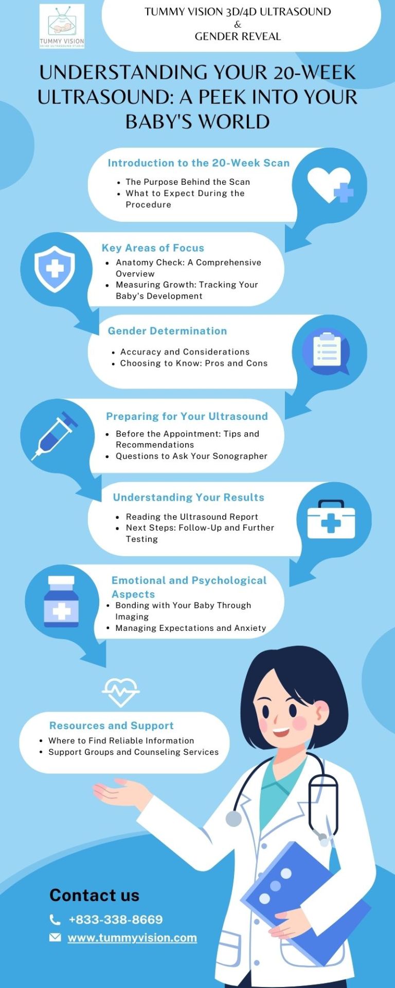
Navigating the results of your 20-week ultrasound can be daunting. This article breaks down common findings, what they mean for your pregnancy, and how to prepare for any possible next steps. Empower yourself with knowledge to understand your baby's health and development.
#20 week ultrasound#14 week 3d ultrasound#6 week ultrasound pictures#3d 4d ultrasound#sneak peek gender test#6 week 3d ultrasound
0 notes
Text
First Digital Atlas of Human Fetal Brain Development Published - Technology Org
New Post has been published on https://thedigitalinsider.com/first-digital-atlas-of-human-fetal-brain-development-published-technology-org/
First Digital Atlas of Human Fetal Brain Development Published - Technology Org
The first digital atlas showing how the human brain develops in the womb has been published by a global research team led by the University of Oxford.
Mother and baby – illustrative photo. The new digital atlas shows the development of the human brain. Image credit: Pixabay, free license
A team of over 200 researchers around the world, involving multiple health and scientific institutions, led by the University of Oxford, has published in the journal Nature, the first digital atlas showing the dynamics of normative maturation of each hemisphere of the fetal brain between 14 and 31 weeks’ gestation – a critical period of human development.
The digital atlas was produced using over 2,500 3-dimensional ultrasound (3D US) brain scans that were acquired serially during pregnancy from 2,194 fetuses in the INTERGROWTH-21st Project, which is a large population-based study of healthy pregnant women living in eight diverse geographical regions of the world (including five in the Global South), whose children had satisfactory growth and neurodevelopment at 2 years of age.
The study involving the creation of the digital atlas for the brain is unique because, for the first time, an international dataset of 3D US scans, collected using standardised methods and equipment, has been analysed with advanced artificial intelligence (AI) and image processing tools to construct a map showing how the fetal brain matures as pregnancy advances.
Demonstrating remarkably similar patterns of fetal brain growth and development across diverse populations represents an important scientific advance in the field of neuroscience. The results are entirely consistent with previously reported findings, from the same INTERGROWTH-21st population, for fetal skeletal growth, newborn size and infant neurocognitive development.
The results of the digital atlas creation also highlight a vitally important public health message: a mother’s health, educational, nutritional and environmental needs must be met to ensure that her child’s body and brain develop healthily.
The findings add to the global impact of the INTERGROWTH-21st Project, which has previously produced international standards for fetal growth, newborn size and the postnatal growth of preterm babies, that are being widely adopted across the world for clinical and research purposes.
Professor Ana Namburete, the first author, whose research group developed the machine learning methods, said: ‘Using AI we enhanced the visibility of brain structures in the 3D US images, and generated an average depiction of the brain at each week of pregnancy during a critical period of development.
Uniquely, our atlas captured patterns of brain growth from as early as 14 weeks’ gestation – filling a 6-week knowledge gap in our understanding of early fetal brain maturation. We also revealed significant asymmetries in brain maturation: for example, in the region associated with language development, which peaked at 20-26 weeks’ gestation and persisted thereafter without any differences between the sexes.’
Professor Stephen Kennedy, co-Principal Investigator of the INTERGROWTH-21st Project, who jointly led the study, said: ‘The atlas will help scientists answer complex biological questions about the fetal origins of cognitive function in childhood, such as how language is acquired. Using the atlas in combination with the soon to be published international standards describing the complementary growth of the fetal brain will be a valuable clinical tool in specialised, referral centres when brain development appears abnormal on ultrasound.’
Professor José Villar, co-Principal Investigator of the INTERGROWTH-21st Project, who jointly led the study, said: ‘This is the latest step in the systematic study of early human growth and development that confirms, using the most advanced research methodology applied to a large number of fetal brain scans, the similarities of growth and development of humans across the world when health, educational, nutritional and environmental needs are met: humans are very similar in all domains, including their brains, when conditions are adequate.’
Source: University of Oxford
You can offer your link to a page which is relevant to the topic of this post.
#3d#3D scanning#Aging news#ai#artificial#Artificial Intelligence#Babies#baby#Biotechnology news#Brain#brain development#Children#cognitive function#development#domains#dynamics#Environmental#equipment#gap#Global#growth#Health#hemisphere#how#human#humans#images#infant#intelligence#language
0 notes
Text
The Premier Destination for E
youtube
The Premier Destination for Early Pregnancy Scans in Leicester ? Ultrasoundplus
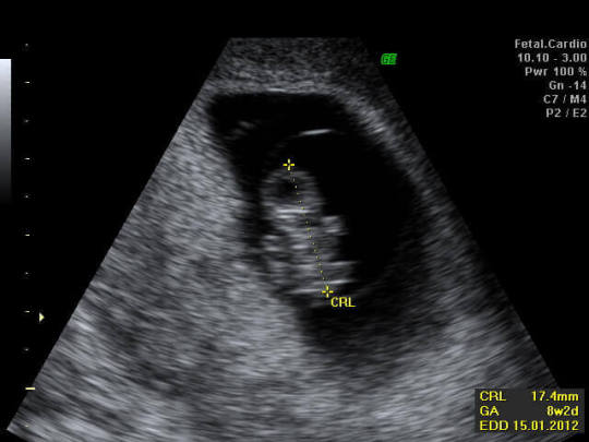
Introduction Your well-behaved Clinic for beforehand Pregnancy Scans in Leicester Are you contemplating an beforehand pregnancy scan in Leicester? The journey of pregnancy is fraught as soon as both anticipation and a plethora of questions. At Ultrasoundplus, located at 75A London Rd, Leicester LE2 0PF, we specialise in alleviating your concerns through a variety of ultrasound scans, including beforehand pregnancy scans, 5-week ultrasound scans, and even more specialised scans for 6 and 10 weeks into your pregnancy journey. The Importance of beforehand Pregnancy Scans Why an beforehand Pregnancy Scan is Crucial An beforehand pregnancy scan serves as a cornerstone for establishing not just fetal move forward but furthermore peace of mind for expecting parents. Here are several reasons why you might opt for one: Confirmation of Pregnancy To begin with, an beforehand pregnancy scan confirms the pregnancy and provides assurance that whatever is progressing as it should be. Accurate Dating One of the primary advantages of beforehand scans is the perfect aspiration of your due date, often a cause of significant speculation in the course of parents-to-be. Fetal Health Monitoring At Ultrasoundplus Leicester, you can opt for an ultrasound scan at 5 weeks to get insights into the beforehand stages of fetal development, thereby ensuring that your journey ahead is as mild as possible. What to Expect During Your Scan Preparing for Your Pregnancy Scan at 5 Weeks, 6 Weeks, or 10 Weeks The preparatory steps for an ultrasound scan at 5 weeks aren't overly intricate. A full bladder is not necessary at this stage, allowing you to be as good as possible during the process. At 5 Weeks During a pregnancy scan at 5 weeks, one can usually see a gestational sac, which serves as an beforehand indicator that your pregnancy is developing as expected. At 6 Weeks When opting for a pregnancy scan at 6 weeks, you'll get more total insights, including the detection of a fetal heartbeata monumental milestone in any pregnancy journey. At 10 Weeks A pregnancy scan at 10 weeks offers even more detailed information, enabling you to witness limb formation and extra necessary developmental stages. Why choose Ultrasoundplus Leicester Your Prime substitute for beforehand Pregnancy Scans in Leicester Our clinic, located at 75A London Rd, Leicester LE2 0PF, not and no-one else provides the latest ultrasound technology but furthermore a team of deeply proficient sonographers who prioritise your comfort and well-being. State-of-the-Art Equipment At Ultrasoundplus Leicester, our equipment is perpetually updated to find the money for the most accurate results possible. Experienced Team Our sonographers bring years of execution and a deep bargain of the emotional nuances that accompany pregnancy scans, ensuring you get the utmost care. https://thepremierdestinationforearly207.blogspot.com/2023/10/the-premier-destination-for-early.html private scan for pregnancy 3d ultrasound leicester 10 weeks ultrasound leicester https://thepremierdestinationforearly263.blogspot.com/ https://thepremierdestinationforearly263.blogspot.com/2023/10/the-premier-destination-for-early.html https://www.tumblr.com/everything-we-all-should-do/732083136558743552 https://a2zhealthmassageschoolslosang273.blogspot.com/2023/10/sacred-geometry-healing-west-palm-beach.html https://www.tumblr.com/rizzocharles74/732078642513969152
0 notes
Text
The Premier Destination for E
youtube
The Premier Destination for Early Pregnancy Scans in Leicester ? Ultrasoundplus
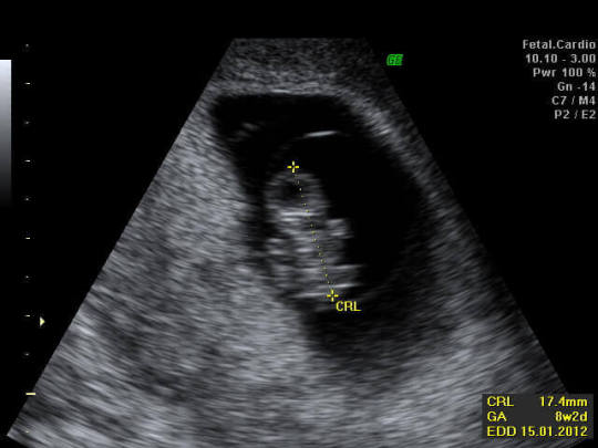
Introduction Your trustworthy Clinic for in front Pregnancy Scans in Leicester Are you contemplating an in front pregnancy scan in Leicester? The journey of pregnancy is fraught subsequently both anticipation and a plethora of questions. At Ultrasoundplus, located at 75A London Rd, Leicester LE2 0PF, we specialise in alleviating your concerns through a variety of ultrasound scans, including in front pregnancy scans, 5-week ultrasound scans, and even more specialised scans for 6 and 10 weeks into your pregnancy journey. The Importance of in front Pregnancy Scans Why an in front Pregnancy Scan is Crucial An in front pregnancy scan serves as a cornerstone for establishing not just fetal development but also good relations of mind for expecting parents. Here are several reasons why you might opt for one: Confirmation of Pregnancy To start with, an in front pregnancy scan confirms the pregnancy and provides assurance that whatever is progressing as it should be. Accurate Dating One of the primary advantages of in front scans is the perfect desire of your due date, often a cause of significant speculation accompanied by parents-to-be. Fetal Health Monitoring At Ultrasoundplus Leicester, you can opt for an ultrasound scan at 5 weeks to get insights into the in front stages of fetal development, thereby ensuring that your journey ahead is as serene as possible. What to Expect During Your Scan Preparing for Your Pregnancy Scan at 5 Weeks, 6 Weeks, or 10 Weeks The preparatory steps for an ultrasound scan at 5 weeks aren't overly intricate. A full bladder is not essential at this stage, allowing you to be as good as possible during the process. At 5 Weeks During a pregnancy scan at 5 weeks, one can usually see a gestational sac, which serves as an in front indicator that your pregnancy is developing as expected. At 6 Weeks When opting for a pregnancy scan at 6 weeks, you'll get more comprehensive insights, including the detection of a fetal heartbeata monumental milestone in any pregnancy journey. At 10 Weeks A pregnancy scan at 10 weeks offers even more detailed information, enabling you to witness limb formation and other essential developmental stages. Why pick Ultrasoundplus Leicester Your Prime unusual for in front Pregnancy Scans in Leicester Our clinic, located at 75A London Rd, Leicester LE2 0PF, not solitary provides the latest ultrasound technology but also a team of extremely intelligent sonographers who prioritise your comfort and well-being. State-of-the-Art Equipment At Ultrasoundplus Leicester, our equipment is perpetually updated to provide the most accurate results possible. Experienced Team Our sonographers bring years of endowment and a deep harmony of the emotional nuances that accompany pregnancy scans, ensuring you get the utmost care. https://thepremierdestinationforearly793.blogspot.com/2023/10/the-premier-destination-for-early.html private scan for pregnancy 3d ultrasound leicester scan at 5 weeks leicester https://www.tumblr.com/easynewsuk/731684882159452160 https://thepremierdestinationforearly557.blogspot.com/ https://thepremierdestinationforearly557.blogspot.com/2023/10/the-premier-destination-for-early.html https://www.tumblr.com/allaboutthenews/731685525650571264 https://spinrewriteraireview602.blogspot.com/
0 notes
Text
Choosing the Best Time for an Elective Ultrasound: A Guide for Expecting Parents

When it comes to experiencing the joys of pregnancy, one of the most exciting moments for expecting parents is seeing their baby during an ultrasound. For many, elective ultrasounds are more than just a medical procedure—they are a keepsake memory. Choosing the right time for an elective ultrasound, whether it's a standard 2D, or a more advanced 3D/4D scan, is essential to getting the best possible experience and images. In this guide, we will explore the best stages of pregnancy for different types of ultrasounds and how this timing plays a crucial role in the elective ultrasound business. The Stages of Pregnancy and Ultrasound Options One of the first things to consider when scheduling an elective ultrasound is understanding the various stages of pregnancy and how they align with different ultrasound types. Whether you're in the first trimester and want a peek at your tiny miracle or you’re later in the pregnancy and hoping to capture detailed facial features in 3D or 4D, timing is key. First Trimester: Weeks 6-12 – Early Sneak Peek The first trimester is a delicate time for both the mother and baby. For those seeking early glimpses, an elective ultrasound can be performed as early as six weeks. While this isn’t the time for detailed images, it can be a significant bonding moment for parents who want to hear their baby’s heartbeat for the first time. Early elective ultrasounds are often 2D and focus primarily on confirming the pregnancy, checking the heartbeat, and verifying the baby’s position in the uterus. If you're thinking about starting an ultrasound business, offering early 2D scans can cater to a market of eager parents who want reassurance and connection during those initial weeks. However, these ultrasounds are best used as a complement to medical check-ups, and you should emphasize that elective scans do not replace medical advice. Second Trimester: Weeks 18-24 – Perfect for Gender Reveal and Clarity The second trimester is when elective ultrasounds truly shine. By week 18, most expecting parents are eager to find out the gender of their baby. Gender reveal scans are popular during this stage, and they provide a perfect opportunity for ultrasound franchise owners to tap into the trend of gender reveal parties. Offering a memorable experience, complete with 2D or 3D imaging, can make your business a go-to choice for keepsake ultrasounds. At this point in the pregnancy, the baby has grown significantly, and you can get clearer images of their entire body. This is the ideal time for parents to schedule a 3D ultrasound if they want to see their baby’s facial features with some clarity but without the fine details that are better captured later in pregnancy. If you’re looking into how to open a 3D ultrasound studio, focusing on second-trimester appointments for gender reveals and basic 3D imaging is a smart business move. Third Trimester: Weeks 28-34 – The Golden Window for 3D/4D Imaging The third trimester is when the magic happens for 3D and 4D ultrasounds. Between weeks 28 and 34, your baby has developed enough fat to have those chubby cheeks and fully formed facial features, making this the best time for capturing keepsake baby ultrasound images. During this golden window, parents can not only see their baby’s face in 3D but also watch them yawn, stretch, and even suck their thumb in real-time 4D. This is also a wonderful opportunity for elective ultrasound business owners to showcase the most advanced technology they have. It’s during this stage that you can truly elevate your service offering, providing an emotional and unforgettable experience for parents. Many parents seek out businesses specifically offering 3D/4D ultrasounds during these weeks, making it an excellent time for targeted ultrasound business marketing tips. However, it’s important to inform clients that after 34 weeks, the baby becomes more cramped in the womb, making it harder to get clear images. Encouraging clients to book their 3D/4D sessions early in the third trimester ensures the best results. Choosing the Right Ultrasound Business Model If you're considering starting an ultrasound business or opening a 3D ultrasound studio, understanding the demand for elective ultrasounds during these key stages of pregnancy is essential. There are several factors to consider when planning your business model: Elective Ultrasound Training and Equipment When starting a 3D/4D ultrasound business, the quality of your equipment and the training of your staff are paramount. Investing in top-tier ultrasound machines and sending your staff to elective ultrasound training programs ensures that your clients receive the best images possible. High-quality imaging is what sets keepsake ultrasounds apart from standard medical ones, and it can help you build a reputation for excellence in the elective ultrasound business. Marketing Your Ultrasound Business Ultrasound business marketing tips often focus on social media and word-of-mouth recommendations, which can be particularly effective when your services include highly shareable moments like gender reveals and 3D/4D images. Consider offering package deals for parents who want multiple scans throughout their pregnancy, such as an early heartbeat scan, a gender reveal, and a 3D/4D ultrasound later on. This creates repeat business and encourages clients to build a relationship with your studio. Additionally, creating partnerships with other pregnancy-related businesses—like maternity boutiques, prenatal yoga studios, and even local hospitals—can help increase your visibility. A comprehensive marketing strategy that includes these partnerships, along with targeted online ads and engaging social media content, can be the key to success. Costs and Profitability The cost of starting an ultrasound business varies depending on factors such as equipment, location, and staffing. However, many businesses find that once they establish a loyal customer base, the return on investment can be substantial. Offering a variety of services, from basic 2D ultrasounds to advanced 4D sessions, can cater to a wider audience and help maximize profitability. Final Thoughts: Timing is Everything Elective ultrasounds offer expecting parents the chance to bond with their baby and create cherished memories. Whether it’s hearing that first heartbeat, discovering the gender, or seeing a baby’s face in 3D for the first time, each stage of pregnancy provides a unique opportunity. As an elective ultrasound business owner, understanding the best timing for these different types of scans is critical to providing a great experience for your clients. Are you considering starting your own 3D/4D ultrasound business? What stage of pregnancy do you think offers the most exciting ultrasound experience? Share your thoughts in the comments below and let’s keep the conversation going! If you found this post helpful, don’t forget to share it with your network. Call-to-Action:Have you started your journey in the ultrasound business? Share your experiences in the comments below and let us know your thoughts! Don’t forget to share this post with your network if you found it helpful. Read the full article
0 notes
Text
The Premier Destination for E
youtube
The Premier Destination for Early Pregnancy Scans in Leicester ? Ultrasoundplus
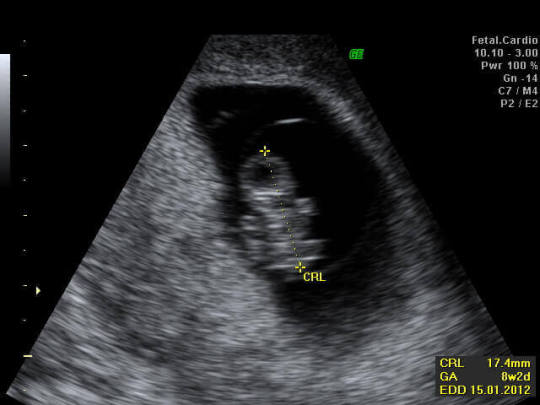
Introduction Your reliable Clinic for early Pregnancy Scans in Leicester Are you contemplating an early pregnancy scan in Leicester? The journey of pregnancy is fraught in the manner of both anticipation and a plethora of questions. At Ultrasoundplus, located at 75A London Rd, Leicester LE2 0PF, we specialise in alleviating your concerns through a variety of ultrasound scans, including early pregnancy scans, 5-week ultrasound scans, and even more specialised scans for 6 and 10 weeks into your pregnancy journey. The Importance of early Pregnancy Scans Why an early Pregnancy Scan is Crucial An early pregnancy scan serves as a cornerstone for establishing not just fetal momentum but afterward friendship of mind for expecting parents. Here are several reasons why you might opt for one: Confirmation of Pregnancy To begin with, an early pregnancy scan confirms the pregnancy and provides assurance that everything is progressing as it should be. Accurate Dating One of the primary advantages of early scans is the correct objective of your due date, often a cause of significant speculation in the midst of parents-to-be. Fetal Health Monitoring At Ultrasoundplus Leicester, you can opt for an ultrasound scan at 5 weeks to gain insights into the early stages of fetal development, thereby ensuring that your journey ahead is as smooth as possible. What to Expect During Your Scan Preparing for Your Pregnancy Scan at 5 Weeks, 6 Weeks, or 10 Weeks The preparatory steps for an ultrasound scan at 5 weeks aren't overly intricate. A full bladder is not valuable at this stage, allowing you to be as satisfying as attainable during the process. At 5 Weeks During a pregnancy scan at 5 weeks, one can usually look a gestational sac, which serves as an early indicator that your pregnancy is developing as expected. At 6 Weeks When opting for a pregnancy scan at 6 weeks, you'll gain more cumulative insights, including the detection of a fetal heartbeata monumental milestone in any pregnancy journey. At 10 Weeks A pregnancy scan at 10 weeks offers even more detailed information, enabling you to witness limb formation and extra valuable developmental stages. Why pick Ultrasoundplus Leicester Your Prime marginal for early Pregnancy Scans in Leicester Our clinic, located at 75A London Rd, Leicester LE2 0PF, not by yourself provides the latest ultrasound technology but afterward a team of highly clever sonographers who prioritise your comfort and well-being. State-of-the-Art Equipment At Ultrasoundplus Leicester, our equipment is perpetually updated to meet the expense of the most accurate results possible. Experienced Team Our sonographers bring years of finishing and a deep promise of the emotional nuances that accompany pregnancy scans, ensuring you receive the utmost care. https://thepremierdestinationforearly897.blogspot.com/2023/10/the-premier-destination-for-early.html private scan for pregnancy 3d ultrasound leicester 10 weeks ultrasound leicester https://thepremierdestinationforearly515.blogspot.com/ https://thepremierdestinationforearly515.blogspot.com/2023/10/the-premier-destination-for-early.html https://www.tumblr.com/things-you-should-know-uk/731175649347272704 https://www.tumblr.com/hispanic-advertising-miami/731023732257243136 https://thepremierdestinationforearly271.blogspot.com/
0 notes
Text
The Premier Destination for E
youtube
The Premier Destination for Early Pregnancy Scans in Leicester ? Ultrasoundplus
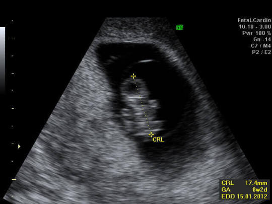
Introduction Your well-behaved Clinic for to the lead Pregnancy Scans in Leicester Are you contemplating an to the lead pregnancy scan in Leicester? The journey of pregnancy is fraught in the manner of both anticipation and a plethora of questions. At Ultrasoundplus, located at 75A London Rd, Leicester LE2 0PF, we specialise in alleviating your concerns through a variety of ultrasound scans, including to the lead pregnancy scans, 5-week ultrasound scans, and even more specialised scans for 6 and 10 weeks into your pregnancy journey. The Importance of to the lead Pregnancy Scans Why an to the lead Pregnancy Scan is Crucial An to the lead pregnancy scan serves as a cornerstone for establishing not just fetal press on but moreover friendship of mind for expecting parents. Here are several reasons why you might opt for one: Confirmation of Pregnancy To start with, an to the lead pregnancy scan confirms the pregnancy and provides assurance that everything is progressing as it should be. Accurate Dating One of the primary advantages of to the lead scans is the perfect goal of your due date, often a cause of significant speculation in the course of parents-to-be. Fetal Health Monitoring At Ultrasoundplus Leicester, you can opt for an ultrasound scan at 5 weeks to gain insights into the to the lead stages of fetal development, thereby ensuring that your journey ahead is as serene as possible. What to Expect During Your Scan Preparing for Your Pregnancy Scan at 5 Weeks, 6 Weeks, or 10 Weeks The preparatory steps for an ultrasound scan at 5 weeks aren't overly intricate. A full bladder is not vital at this stage, allowing you to be as to your liking as realistic during the process. At 5 Weeks During a pregnancy scan at 5 weeks, one can usually look a gestational sac, which serves as an to the lead indicator that your pregnancy is developing as expected. At 6 Weeks When opting for a pregnancy scan at 6 weeks, you'll gain more total insights, including the detection of a fetal heartbeata monumental milestone in any pregnancy journey. At 10 Weeks A pregnancy scan at 10 weeks offers even more detailed information, enabling you to witness limb formation and supplementary vital developmental stages. Why choose Ultrasoundplus Leicester Your Prime unusual for to the lead Pregnancy Scans in Leicester Our clinic, located at 75A London Rd, Leicester LE2 0PF, not lonesome provides the latest ultrasound technology but moreover a team of very skilled sonographers who prioritise your comfort and well-being. State-of-the-Art Equipment At Ultrasoundplus Leicester, our equipment is perpetually updated to provide the most accurate results possible. Experienced Team Our sonographers bring years of carrying out and a deep concord of the emotional nuances that accompany pregnancy scans, ensuring you receive the utmost care. https://thepremierdestinationforearly271.blogspot.com/2023/10/the-premier-destination-for-early.html private scan for pregnancy 3d ultrasound leicester 10 weeks ultrasound leicester https://thepremierdestinationforearly460.blogspot.com/ https://thepremierdestinationforearly460.blogspot.com/2023/10/the-premier-destination-for-early.html https://metropolitanlocalonlinemarket237.blogspot.com/ https://metropolitanlocalonlinemarket237.blogspot.com/2023/10/metropolitan-local-online-marketing.html https://www.tumblr.com/easynewsuk/731173720164581376
0 notes