#vetlabdiagnostic
Explore tagged Tumblr posts
Photo
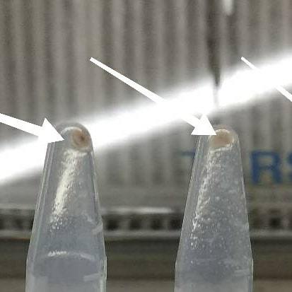
Happy #dnaday to all. A few images and work of my past lab life. Really missing those hard molecular work days... 1 and 2 : DNA extraction and precipitation 3. Electrophoresis 4 and 5. Gel documentation #lablife #vetpath #dnaisolation #dnawork #molecularpathology #vetclinicalpathology #dna #molecularlaboratory #moleculartestinglabs #pcr #vetlabdiagnostic #ethidiumbromide #uvflourescence #dnaladder #deoxyribonucleicacid #researchwork #vetstudies #vetstudent https://www.instagram.com/drdashvetpath/p/Bwt6gdkhLRV/?utm_source=ig_tumblr_share&igshid=1ldf0jbpduy37
#dnaday#lablife#vetpath#dnaisolation#dnawork#molecularpathology#vetclinicalpathology#dna#molecularlaboratory#moleculartestinglabs#pcr#vetlabdiagnostic#ethidiumbromide#uvflourescence#dnaladder#deoxyribonucleicacid#researchwork#vetstudies#vetstudent
0 notes
Photo
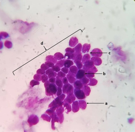
Basal cells: Location: Basal layer of all epithelia. Characteristics and function: Stem cell regenerative compartment for epithelial structures. Shape and size: Small round cells with a high N:C ratio. Nucleus: Round (a) to ovoid (b) centrally placed with compact chromatin. Cytoplasm: Scant with mild basophilia (c). Cytoarchitecture: Pavement (d) or columnar. Ddx: Matured lymphocytes. #vetpath #vetpathology #vetpathologist #vetcytology #vetcyto #veterinaryclinicalpathology #vetclinpath #veterinarycytology #veterinarycytopathology #veterinarymedicine #vetlabdiagnostic #vetcases #vetlife #normalcaninecell #basalcell #vetlablife #veterinary_medicine #vetmed https://www.instagram.com/drdashvetpath/p/BweUHo2Bi7J/?utm_source=ig_tumblr_share&igshid=1hu6g0s917um0
#vetpath#vetpathology#vetpathologist#vetcytology#vetcyto#veterinaryclinicalpathology#vetclinpath#veterinarycytology#veterinarycytopathology#veterinarymedicine#vetlabdiagnostic#vetcases#vetlife#normalcaninecell#basalcell#vetlablife#veterinary_medicine#vetmed
0 notes
Photo

Fleas Ctenocephalides Felis: Most commonly seen fleas in household cats and dogs. Principle host of the cats but also seen infesting dogs and other mammals. Fleas go through 4 life cycle phases egg, larva, pupa and adult. It's is estimated that a female lays somewhere between 5000 to 8000 eggs during her life. The fleas need to feed on a blood meal before they start producing eggs. The eggs develop to larvae in around three to six weeks. The larvae are negatively phototrophic and hide from the light unto the ground substrates. They again moult to pupae by spinning a Coccon and waiting for suitable environmental conditions and for a suitable host. The adult fleas are capable of detecting the CO2 and temperature from the hosts and this helps in lodging of the fleas to the hosts body. Clinical signs and symptoms develop due to allergic reactions towards the substances present in the fleas saliva (Flea allergy dermatitis - FAD). Morphological differentiation between C. felis and C. canis: a. The first spine of genal comb of C. felis is almost the same length as the second, in C. canis is almost half the length. b. Head of C. felis is more rounder and longer than that of C. canis. c. C. felis - tibiae of all 6 legs have 7 to 8 bristles C. canis - tibiae of all 6 legs have 4 to 5 bristles. To effectively control fleas, both the fleas on the animalss as well as the fleas and eggs in the surroundings have to treated. Adulticides along with insect development inhibitors (Lufeneuron) and insect growth regulators (Methoprene) have been used effectively to control flea population. Spot on (Imidacloprid, fipronil, Methoprene) solutions are more effective alternative in pets rather than the commercial available sprays and dips. #vetpath #vetpathology #vetclinpath #vetclinicalpathology #vetlabdiagnostic #veterinarypath #veterinaryclinicalpathology #veterinarypathology #veterinaryparapathologist #vetdermatology #veterinarydermatology #veterinaryentomology #vetlaboratory #vetdiagnostics #vetparasitology #veterinaryparasitology #vetparasite #flea #catflea #dogflea #ctenocephalidesfelis #externalparasites #fleaallergy #fleaallergydermatitis #wetmount #parasites https://www.instagram.com/drdashvetpath/p/BvyuuHyhN9f/?utm_source=ig_tumblr_share&igshid=mnofp94i184l
#vetpath#vetpathology#vetclinpath#vetclinicalpathology#vetlabdiagnostic#veterinarypath#veterinaryclinicalpathology#veterinarypathology#veterinaryparapathologist#vetdermatology#veterinarydermatology#veterinaryentomology#vetlaboratory#vetdiagnostics#vetparasitology#veterinaryparasitology#vetparasite#flea#catflea#dogflea#ctenocephalidesfelis#externalparasites#fleaallergy#fleaallergydermatitis#wetmount#parasites
0 notes
Photo

And again!!!! Lipoma in a dog: Seen are plump adipocytes with optically empty cytoplasm due to the stored fats/lipids in the cells. The nucleus is pushed to the periphery to accomodate the maximum available space for storage of fats/lipids. Adipose tissue are usually found intertwined with collagenous fibres and blood vessels forming a 3D cytoarchitecture. #vetpath #vetpathology #cytopathology #vetdiagnosis #vetdiagnostics #fna #fnac #cytology #cytologyvet #veterinarycytopathology #vetlabdiagnostic #fineneedleaspirationcytology #fineneedleaspirate #lipoma #subcutaneous #veterinaryclinicalpathology #cytologicalexamination #aspirationcytology https://www.instagram.com/drdashvetpath/p/Bvenfy5hfrW/?utm_source=ig_tumblr_share&igshid=306j3bci9mbr
#vetpath#vetpathology#cytopathology#vetdiagnosis#vetdiagnostics#fna#fnac#cytology#cytologyvet#veterinarycytopathology#vetlabdiagnostic#fineneedleaspirationcytology#fineneedleaspirate#lipoma#subcutaneous#veterinaryclinicalpathology#cytologicalexamination#aspirationcytology
0 notes
Photo
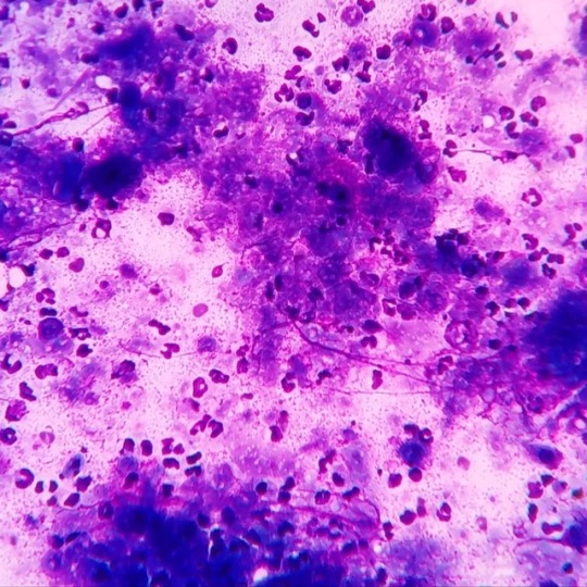
Epidermal cyst with pyogranulomatous inflammation in a dog: As seen in the images many polymorphonuclear cells are seen surrounding the irregular dark basophilic material. Multinucleated foreign body giant cells along with variable number of macrophages can also be observed in aspirates. The dark basophilic material seen are aggregates of keratinized materials which have been ruptured from the cyst into the surrounding tissue. A inflammatory response is set by the host to clear out the debris along with keratinized materials out of the involved tissue. #vetpath #vetpathology #veterinarypathology #veterinarypath #vetclinpath #veterinaryclinpath #veterinaryclinicalpathology #vetclinicalpathology #cytology #vetcytology #veterinarycytology #aspirate #aspirationcytology #aspirate #vetlabdiagnostic #veterinarylabdiagnosis #cytologicalexamination #cytologylab #cytologyvet #fnac #fna #caninetissue #epidermalcyst #dermatology #vetdermatology #veterinarydermatology https://www.instagram.com/drdashvetpath/p/BvGNW39h3R-/?utm_source=ig_tumblr_share&igshid=16c5bpw8m5clt
#vetpath#vetpathology#veterinarypathology#veterinarypath#vetclinpath#veterinaryclinpath#veterinaryclinicalpathology#vetclinicalpathology#cytology#vetcytology#veterinarycytology#aspirate#aspirationcytology#vetlabdiagnostic#veterinarylabdiagnosis#cytologicalexamination#cytologylab#cytologyvet#fnac#fna#caninetissue#epidermalcyst#dermatology#vetdermatology#veterinarydermatology
0 notes
Photo
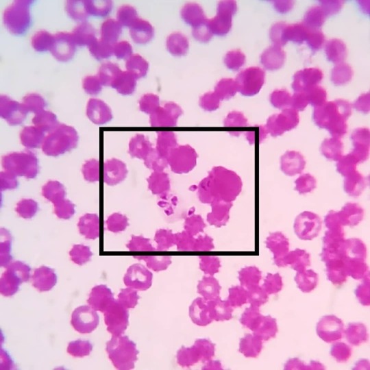
#merozoite 's of #babesiacanis in a #caninebloodsmear: Babesia canis are #intercellular #haemoprotozoa belonging to the family of #piroplasmida e. The babesial organisms are transmitted by the dog #browntick #rhipicephalussanguineus , which are present as #sporozoites in the salivary glands. During their blood meal the sporozoites are passed on to the vertebrate host and attaches itself on the erythrocytic membranes. The parasites enter the RBCs and multiply by asexual reproduction to form merozoites which are seen as leaf like structures. While feeding again the organisms are transferred to the ticks and the organisms enter the gut mucosa and undergo gametogony to form male and female forms which reproduce by sexual and by asexual reproduction to form more sporozoites. And the cycle continues!! Most common babesial organisms seen in canine are Babesia canis and Babesia Gibsoni. Babesia Gibsoni are more smaller, seen as small ring/ circular structures and lack a pyriform shape. Babesia canis on the other hand have large pyriform merozoites and pointed at one end. #vetpathology #vetpath #vetparasitology #veterinaryparasitology #veterinaryparapathologist #bloodparasite #vetcasestudy #vetcases #vetstudies #vetmed #vetmedicine #vetlife #vetknowledge #vetclinicalpathology #vetcliniclife #vetdiagnosis #vetlabdiagnostic https://www.instagram.com/drdashvetpath/p/Bw82BXIh07-/?utm_source=ig_tumblr_share&igshid=15ay9kvz7f12y
#merozoite#babesiacanis#caninebloodsmear#intercellular#haemoprotozoa#piroplasmida#browntick#rhipicephalussanguineus#sporozoites#vetpathology#vetpath#vetparasitology#veterinaryparasitology#veterinaryparapathologist#bloodparasite#vetcasestudy#vetcases#vetstudies#vetmed#vetmedicine#vetlife#vetknowledge#vetclinicalpathology#vetcliniclife#vetdiagnosis#vetlabdiagnostic
0 notes
Photo
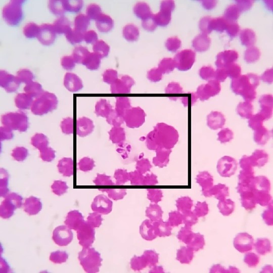
#merozoite 's of #babesiacanis in a #caninebloodsmear: Babesia canis are #intercellular #haemoprotozoa belonging to the family of #piroplasmida e. The babesial organisms are transmitted by the dog #browntick #rhipicephalussanguineus , which are present as #sporozoites in the salivary glands. During their blood meal the sporozoites are passed on to the vertebrate host and attaches itself on the erythrocytic membranes. The parasites enter the RBCs and multiply by asexual reproduction to form merozoites which are seen as leaf like structures. While feeding again the organisms are transferred to the ticks and the organisms enter the gut mucosa and undergo gametogony to form male and female forms which reproduce by sexual and by asexual reproduction to form more sporozoites. And the cycle continues!! Most common babesial organisms seen in canine are Babesia canis and Babesia Gibsoni. Babesia Gibsoni are more smaller, seen as small ring/ circular structures and lack a pyriform shape. Babesia canis on the other hand have large pyriform merozoites and pointed at one end. #vetpathology #vetpath #vetparasitology #veterinaryparasitology #veterinaryparapathologist #bloodparasite #vetcasestudy #vetcases #vetstudies #vetmed #vetmedicine #vetlife #vetknowledge #vetclinicalpathology #vetcliniclife #vetdiagnosis #vetlabdiagnostic https://www.instagram.com/drdashvetpath/p/Bw82BXIh07-/?utm_source=ig_tumblr_share&igshid=1l7cvdwrcv8o9
#merozoite#babesiacanis#caninebloodsmear#intercellular#haemoprotozoa#piroplasmida#browntick#rhipicephalussanguineus#sporozoites#vetpathology#vetpath#vetparasitology#veterinaryparasitology#veterinaryparapathologist#bloodparasite#vetcasestudy#vetcases#vetstudies#vetmed#vetmedicine#vetlife#vetknowledge#vetclinicalpathology#vetcliniclife#vetdiagnosis#vetlabdiagnostic
0 notes