#aspirationcytology
Explore tagged Tumblr posts
Photo

And again!!!! Lipoma in a dog: Seen are plump adipocytes with optically empty cytoplasm due to the stored fats/lipids in the cells. The nucleus is pushed to the periphery to accomodate the maximum available space for storage of fats/lipids. Adipose tissue are usually found intertwined with collagenous fibres and blood vessels forming a 3D cytoarchitecture. #vetpath #vetpathology #cytopathology #vetdiagnosis #vetdiagnostics #fna #fnac #cytology #cytologyvet #veterinarycytopathology #vetlabdiagnostic #fineneedleaspirationcytology #fineneedleaspirate #lipoma #subcutaneous #veterinaryclinicalpathology #cytologicalexamination #aspirationcytology https://www.instagram.com/drdashvetpath/p/Bvenfy5hfrW/?utm_source=ig_tumblr_share&igshid=306j3bci9mbr
#vetpath#vetpathology#cytopathology#vetdiagnosis#vetdiagnostics#fna#fnac#cytology#cytologyvet#veterinarycytopathology#vetlabdiagnostic#fineneedleaspirationcytology#fineneedleaspirate#lipoma#subcutaneous#veterinaryclinicalpathology#cytologicalexamination#aspirationcytology
0 notes
Photo

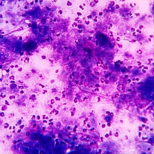
Epidermal cyst with pyogranulomatous inflammation in a dog: As seen in the images many polymorphonuclear cells are seen surrounding the irregular dark basophilic material. Multinucleated foreign body giant cells along with variable number of macrophages can also be observed in aspirates. The dark basophilic material seen are aggregates of keratinized materials which have been ruptured from the cyst into the surrounding tissue. A inflammatory response is set by the host to clear out the debris along with keratinized materials out of the involved tissue. #vetpath #vetpathology #veterinarypathology #veterinarypath #vetclinpath #veterinaryclinpath #veterinaryclinicalpathology #vetclinicalpathology #cytology #vetcytology #veterinarycytology #aspirate #aspirationcytology #aspirate #vetlabdiagnostic #veterinarylabdiagnosis #cytologicalexamination #cytologylab #cytologyvet #fnac #fna #caninetissue #epidermalcyst #dermatology #vetdermatology #veterinarydermatology https://www.instagram.com/drdashvetpath/p/BvGNW39h3R-/?utm_source=ig_tumblr_share&igshid=16c5bpw8m5clt
#vetpath#vetpathology#veterinarypathology#veterinarypath#vetclinpath#veterinaryclinpath#veterinaryclinicalpathology#vetclinicalpathology#cytology#vetcytology#veterinarycytology#aspirate#aspirationcytology#vetlabdiagnostic#veterinarylabdiagnosis#cytologicalexamination#cytologylab#cytologyvet#fnac#fna#caninetissue#epidermalcyst#dermatology#vetdermatology#veterinarydermatology
0 notes