#liposomal serum
Explore tagged Tumblr posts
Text

"Sesderma Azelac RU Liposomal Serum: Your Solution for Radiant Skin. This serum combines the power of liposomes with the benefits of azelaic acid to reduce hyperpigmentation and even out your skin tone. Experience the ultimate skin transformation as it fades dark spots, while its antioxidant-rich formula safeguards your skin. Make Sesderma Azelac RU Liposomal Serum a part of your daily routine for a brighter, more radiant complexion."
0 notes
Text
#bibakart#face serum#anti aging serum#vitamin c serum#skin lightener gel intense pigmentation#soothing restorative serum#age killer serum#toneright serum#hydrating moisturizing serum#skin boosting super serum#avocado pure oil#exquisite serum#japanese ritual smoothing face serum#reti age liposomal mist#bakuchiol serum#anti wrinkle moisturizing serum
0 notes
Text
K-beauty Recovery Reaction! Concentrated Care Guide
Hello! Hello! Hello!
I'm Kim Daehan, the representative of incellderm , the No.1 cosmetics brand in Korea!
Exciting news! Riman Korea has expanded its reach to the United States and Canada! Wow! Applause!! Now, let me guide you on how to become a "Beauty Planner" in the US and Canada!
🌈 INCELLDERM able&co us. Becoming a Beauty Planner!
[must read]How to Join as a Beauty Planner - Step by Step Guide(Click)
🌈 Expansion of Incellderm to the United States and Canada! We are excited to announce that Incellderm is now available in the United States and Canada! Customers in these regions can now experience the benefits of our premium skincare products. ----------------------------------------------------------------------------
"If you are an Incellderm business owner&beauty planner, one thing you must absolutely master is 'Positive Reaction (Skin Regeneration Reaction).' Isn't it the immediate reaction that stands out? The cause of typical positive reactions and detailed coping methods:
Customers experiencing positive reactions show a variety of individual responses, which is why you need to continuously study. I also realized that my knowledge and coping methods regarding positive reactions are insufficient, which could lead to adverse effects on skin management.
Positive Reaction Q&A:
When using Incellderm, various positive reactions appear.
What is a Positive Reaction? Similar to traditional Korean medicine's 'Myeonghyeonbanhaeng' (明顯班現), it refers to a phenomenon where the skin temporarily worsens before improving. Understanding the principle behind positive reactions allows you to enjoy the process as the accumulated epidermal toxins are expelled, and the dermal layer gets replenished. Familiarize yourself with the typical symptoms of positive reactions, their underlying principles, and appropriate coping strategies."
🌈 Itching
Q: Some people experience itching after using Incellderm. Why does this happen?
A: Itching is a clear sign that the dermal layer is undergoing some changes. Incellderm is formulated using the drug delivery system called 'liposome.' The active ingredients of the multi-layered liposome penetrate the skin rapidly, stimulating the capillaries and causing the sensation of itching. This is because new blood vessels are formed, and new skin is generated as the wound heals and the scab falls off. The itching usually occurs in the initial stages but does not last long. However, if the itching persists, it may be necessary to investigate other causes.
Mild Irritation: Itchy sensation accompanied by a feeling like environmental factors are causing it, similar to having hair or insects stuck to the face. ▶ In the basic skincare routine, apply calming gel and serum generously. (Apply the calming gel first and then the serum on top of it.)
Itching Accompanied by Warmth: Caused by strong penetration. ▶ Adjust the application order - Cream → Serum (As the amino acid components in the booster may cause warmth and tingling, reduce the amount of booster used.)
Itching in Trouble or Wounded Areas: Caused by tiny wounds on the skin, often seen in sensitive skin. ▶ Adjust the application area (avoid using on troubled or wounded areas), and to prevent post-inflammatory hyperpigmentation, apply sun gel frequently when going out. Also, maintain excellent hygiene.
Severe Itching: If you experience intense itching that does not subside even when scratched, you should discontinue use. Be cautious about secondary infections caused by scratching with your hands. (Avoid scratching or irritating the skin with your hands as much as possible.) If you have atopic dermatitis, you should seek medical treatment. Please note that the translation provided here is for informational purposes, and it is essential to consult with a healthcare professional or an expert for personalized advice and solutions related to any skin issues.
🌈 Dead Skin Cells (Keratinization)
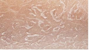
Q: I used to have few dead skin cells, but after using Incellderm, I am experiencing an increase in dead skin cells. Why is this happening?
A: When using Incellderm, the 'keratinization cycle (the cycle of skin cells being generated and shed)' accelerates. In healthy individuals, the keratinization cycle typically takes around 28 days. However, as one ages and undergoes the process of aging, the keratinization cycle also lengthens. For instance, in their 40s, it could be around 40 days, in their 50s, it could be around 50 days, and so on.
Incellderm activates the cells in the dermal layer, pushing accumulated dead skin cells quickly to the surface and normalizing the keratinization cycle. Therefore, you may notice more dead skin cells than usual.
Additionally, dead skin cells are a process of expelling remnants of synthetic surfactants accumulated in the body. As epidermal toxins and impurities are eliminated, the skin tone becomes clearer and brighter.
Please note that you should distinguish dead skin cells from 'psoriasis.' Psoriasis is a condition where unshed dead skin cells accumulate and form a thick armor-like barrier on the skin, causing it to become hard and rigid. For psoriasis, it is essential to seek medical treatment from a dermatologist.
Dry Dead Skin Cells: This refers to fine, dandruff-like flaking, especially around the jawline. This is caused by nutritional deficiency between dead skin cells.
▶ Avoid artificial peeling using washcloths or exfoliating products. Instead, opt for gentle cleansing and moisturizing, allowing dead skin cells to shed naturally. (Drink plenty of water.)
Oily Dead Skin Cells: This type of dead skin cells appear in the form of clumps due to the combination of sebum and dead skin cells, resembling dirt.
▶ Oily dead skin cells can block pores and cause skin troubles, so make sure to perform deep cleansing frequently.
※ Deep Cleansing Method: Create a rich foam with an active powder cleanser using a foaming tool and apply it to the entire face except the eye area, leaving it on for 2-3 minutes. After some time, when the foam penetrates into the pores, rinse with lukewarm water.
If you have attempted exfoliation:
▶ Refrain from additional peeling as it can worsen the condition and cause further irritation. Instead, opt for regular moisturization using calming gel and stay hydrated.
🌈 Facial Redness, Tingling Sensation
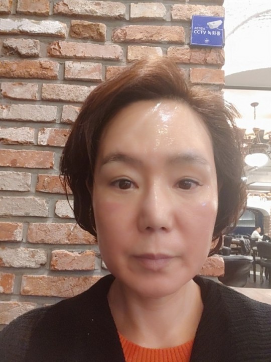
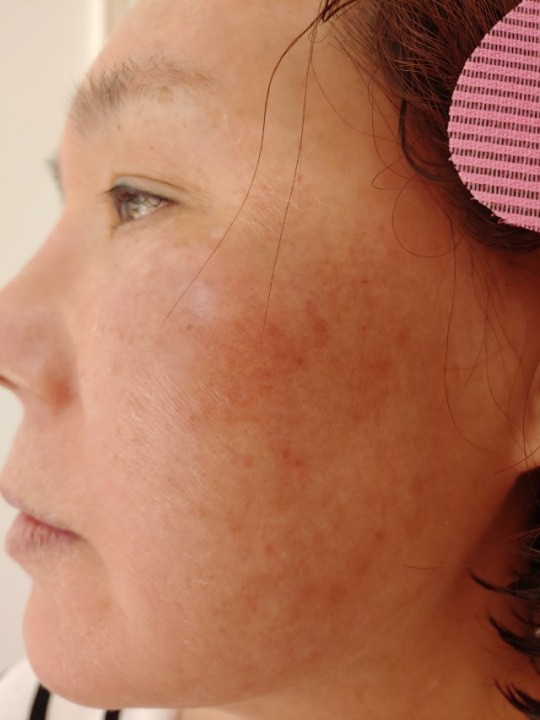
Q: Some people experience redness and occasional tingling sensations on their faces.
A: The development of facial redness and increased redness is often due to skin dehydration. The same applies to tingling sensations. When the skin is dry, especially when it is extremely dry, it means that under a microscope, the lipids between the keratin cells have collapsed, resulting in cracked and flaky skin. In this state, with the lipid layer being disrupted, moisture can easily penetrate, leading to tingling sensations.
This sensation is similar to how steam rises and heat is generated when water is poured onto a dry rice paddy. Usually, tingling sensations and warmth go hand in hand.
Warmth is a typical symptom of the abundant penetration of amino acids. The main ingredients in boosters and serums, amino acids are quickly absorbed into the dermis, causing this sensation. Adjusting the application order is recommended for better results. (Use in the order of sensitivity: Cream -> Serum -> Booster.)
🌈 Red Spots
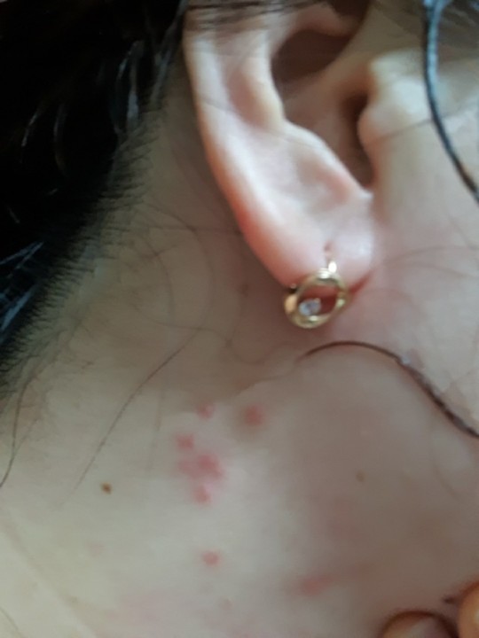
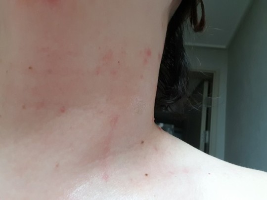
Q: How should I deal with the appearance of red spots?
A: Red spots often occur due to external factors. If they come and go like hives (urticaria), it might be a positive reaction. However, if a specific area continues to worsen, it is more likely to be dermatitis, and you should seek advice from a dermatologist.
If accompanied by itching: The possibility of contact dermatitis is high (e.g., touching your face with dirty hands). Itching is often caused by skin dehydration.
▶ Avoid dry environments and ensure proper hydration. In the basic skincare routine, using oil mist and calming gel can be helpful. Applying sun gel regularly will provide moisture and soothing benefits to the skin.
Fever and rash: Some people frequently wash their face to cool down, but excessive washing can actually dry out the skin.
▶ Reduce the frequency of washing and focus on moisturizing and soothing. Differentiate between hives and dermatitis, and if it's dermatitis, discontinue the use of products.
Occurrence in specific areas (around the eyes, nose, and mouth): Increased activity of sebaceous glands is the main cause. This symptom appears due to an imbalance in the oil and moisture levels.
▶ Perform deep cleansing and restore the oil-moisture balance in the basic skincare routine, especially using oil mist. For the specific area with red spots, take a break from using boosters, serums, and creams, and instead, apply calming gel to prevent enlarged pores.
(Dab calming gel on a cotton pad and apply it to the specific area.)
🌈 Facial Swelling
Q: My face looks swollen, as if it's puffy. Is this a side effect?
A: It is not a side effect. Incellderm is designed to be absorbed into the skin. However, if a customer using the product has poor peripheral circulation due to diabetes, hypertension, hyperlipidemia, or other reasons, the absorbed cosmetics may become stagnant in the skin, leading to swelling.
If you have been taking medication for diabetes, hypertension, hyperlipidemia, or other conditions for a long time, it is advisable to give yourself a period of one or two months to adapt to the product.
Excessive Use: Swelling can occur due to excessive protein intake.
▶ In particular, avoid excessively using cream, as it can cause issues. (Reduce the amount used.)
Swelling Accompanied by Warmth and Itching: This is common among those who frequent saunas or steam rooms.
▶ Avoid saunas and ensure consistent soothing using cold packs or cold compresses.
Switching from Other Products: Accumulated toxins in the skin might be the cause. Chemical cosmetics often contain harmful substances that can accumulate in the skin. When switching to Incellderm from other cosmetics, these toxins may try to be expelled but get stuck, leading to swelling.
▶ To aid in the smooth elimination of toxins, perform lymph node massage. After showering, mix booster and serum in a ratio of 7:3 and apply it to areas such as behind the ears, armpits, and groin, where lymph nodes are located.
Uneven Swelling: This is also often caused by excessive usage. Excessive cream application can lead to mucous edema.
▶ Maintain consistent soothing while adjusting the amount of product used.
🌈 Hyperpigmentation, Freckles, Skin Tone Changes
Q: After using Incellderm, I've noticed an increase in freckles. Also, why does my skin tone keep fluctuating between looking good and bad?
A: It usually takes about 3 to 6 months for melanin spots that were in the dermis to rise to the epidermis. So, it's not possible for freckles to appear immediately after using Incellderm.
The melanocytes that create freckles are located in the innermost layer of the epidermis called the basal layer. What we see as freckles is actually melanocytes filled up to the stratum corneum layer. When you start using Incellderm, the skin condition improves rapidly, and the melanocytes in the basal layer are pushed up to the stratum corneum, so it may seem like freckles are increasing for a while.
However, with continuous use of the product, freckles will gradually become lighter and clearer.
During the initial 7 to 15 days of product usage, the skin tone becomes clearer due to various beneficial cell growth factors, and after about 15 days, the skin tone may fluctuate between good and bad.
This is because the skin, as a tissue composed of cells, promotes cell division when stimulated by various growth factors, much like when it was young. The promotion of cell division means that what was inside the skin is pushed up, which also means that more dead skin cells are produced.
Cells born in the skin rise to the surface and stay there in the form of dead skin cells until they eventually fall off. This process is called turnover, and the normal turnover cycle of healthy skin is about 28 days. Incellderm promotes this skin regeneration process, and the time it becomes visible to the naked eye is around 7 to 15 days after using the product. This is when you might notice your skin tone fluctuating between looking good and bad.
External factors such as pregnancy, genetics, medication use, hormonal changes, and weakened immunity can also cause these phenomena.
If you have undergone treatments for removing freckles or freckle-like pigmentation, the condition you're experiencing might be due to dermal freckles surfacing, and they can reappear at any time.
Neglecting sun protection: Many people tend to focus only on using boosters, serums, and creams and overlook the use of sun gel when going outside (e.g., applying only cream before going out). Proper sun protection is crucial to prevent hyperpigmentation and freckles.
Temporary darkening or dullness of the skin: This is a natural fluctuation in the brightness of the skin and will improve on its own over time.
🌈 Tightness, Dryness
Q: When I use the product, I feel tightness and dryness. What is the reason behind this?
A: The skin is a very thin tissue and cannot distinguish well between the sensation of collagen being formed, which causes tightness, and the dry feeling on the surface due to the dry stratum corneum layer.
When spider-web-like collagen is produced and released, it resists gravity and lifts the skin back up, resulting in a feeling of tightness and increased skin elasticity.
In the case of Incellderm, both of these effects occur:
Inner Tightness: Enjoy this sensation as it is the lifting effect due to collagen production. However, be cautious not to use the product excessively due to misunderstanding.
Surface Dryness (Epidermal Dryness): Proper cleansing and sufficient moisturization are necessary. If the tightness is severe, applying a generous amount of calming gel like a pack before going to bed can help alleviate the sensation.
Insufficient Moisturization: Surprisingly, many people don't drink enough water during the day. Make sure to drink around 2 liters of water daily. Sufficient moisturization enhances the absorption of boosters, serums, and creams. You can think of it like a dry sponge that repels water versus a slightly damp sponge that absorbs more water. Improving lifestyle habits is also important. Consuming salty and carbonated foods, as well as coffee, can deplete the body's moisture.
Dermis Cracking: Although rare, if your skin cracks and bleeding occurs, immediately discontinue the use of the product and seek advice from a dermatologist.
Remember that finding the right balance in product usage and maintaining good skincare habits can help address the tightness and dryness issues effectively.
🌈 Expression Lines
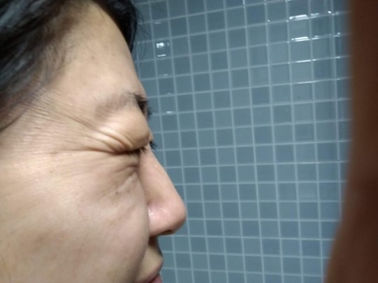
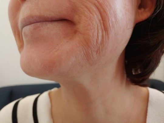
Q: The product I'm using has dual functionality for whitening and wrinkle improvement, but why do I feel like I have more wrinkles now?
A: The cause of wrinkles is the aging of collagen tissues in the dermis, which makes the boundary between the epidermis and dermis uneven. When collagen in the dermis starts to be produced and released like spider webs, the boundary between the epidermis and dermis becomes uneven for a while, resulting in the appearance of wrinkles that weren't there before. However, the wrinkles caused by the product's use do not last long. As wrinkles appear and disappear repeatedly, the dermis gradually fills up, and the boundary between the epidermis and dermis becomes flatter. As a result, the skin's surface becomes smoother, leading to the ultimate wrinkle improvement effect.
Expression lines are wrinkles that occur when unabsorbed ceramides (*ceramides are ingredients most similar to skin cells, and Incellderm products are manufactured using the method of encapsulating active ingredients with ceramides to deliver them to the dermis). This phenomenon can appear as if a vinyl wrap is applied to the skin. This occurs when the product is used excessively, so reducing the amount used can help.
Especially when you apply too much cream around the eye area, it can irritate the delicate skin around the eyes and lead to the appearance of expression lines. To address these expression lines, reduce the amount used and make sure to thoroughly moisturize the area.
By maintaining the proper usage of the product and paying attention to how much is applied, you can achieve the desired results without experiencing any unwanted effects on your skin.
🌈 Troubles (Skin Issues)
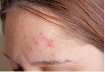
Q: I have developed pimples. What should I do?
A: Pimples and other skin troubles can occur from time to time. In particular, women may experience recurring pimples in relation to their menstrual cycles.
One of the causes of skin troubles is the accumulation of epidermal toxins. Conventional cosmetics use a mixture of water and oil with synthetic surfactants, which may contain harmful epidermal toxins. People who have used chemical cosmetics in the past may have a higher likelihood of having epidermal toxins accumulated in their skin. When using Incellderm products, as the overall condition of the skin improves and the epidermal toxins are eliminated from the skin, a reaction like the improvement of pimples can occur. To minimize such reactions, it is advisable to start using the product with a small amount and gradually increase it to allow for an adaptation period.
Pustules (Pus-filled pimples): They can occur from overusing the product. This is caused by insufficient absorption of protein, leading to the formation of skin troubles. Adjust the usage amount and pay attention to moisturizing. The primary cause of pustules is lack of moisture. When there is not enough moisture, pimples are more likely to form. If pimples have already appeared, apply a calming gel pack to the affected area. (Dip a cotton pad in calming gel and place it on the affected area.)
Comedones (Blackheads and whiteheads): These occur when sebum becomes trapped under the layer of dead skin cells in the pores. Use deep cleansing at least once or twice a week to remove dead skin cells.
Vesicles (Inflammatory bumps): If skin troubles are accompanied by vesicles, it is not a reaction caused by using Incellderm products. These are caused by internal factors such as herpes virus or shingles. In such cases, immediately stop using the product and seek medical treatment.
Prolonged use of a specific brand: Skin troubles can occur due to the residual synthetic surfactants from the prolonged use of conventional chemical cosmetics.
🌈 Other Improvement Reactions
Q: Could it be flat warts?
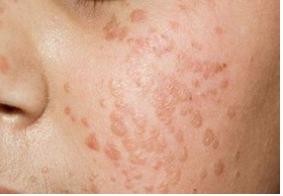
A: It is easy to confuse flat warts with comedones (blackheads and whiteheads). Flat warts are caused by human papillomavirus (HPV) and are often seen when the immune system is weakened. Flat warts are highly contagious, so avoid squeezing them and seek dermatological treatment promptly.
Q: My eyes feel irritated, teary, and uncomfortable, as if there's something in my eyes.
A: Incellderm products are designed to penetrate deep into the skin, so as you age, around your 40s to 50s, your eyes may feel uncomfortable for a while. People with dry eye syndrome may experience this discomfort. The beneficial ingredients in the product help improve the function of the tear glands, leading to increased tear production. With continuous use and adaptation, your eyes should no longer feel uncomfortable.
Q: I haven't lost weight, but people say my face looks smaller, as if I've lost weight.
A: As you age, the skin inside the face may become less dense, and due to gravity, the skin can start to sag. However, with Incellderm products, the skin inside becomes gradually filled, and with consistent use for about 6 months to 1 year, the skin is lifted, resulting in a balanced facial line and making your face appear smaller.
🌈 Expansion of Incellderm to the United States and Canada!
We are excited to announce that Incellderm is now available in the United States and Canada! Customers in these regions can now experience the benefits of our premium skincare products. 🌈 INCELLDERM able&co us. Becoming a Beauty Planner!
[must read]How to Join as a Beauty Planner - Step by Step Guide(Click) For any necessary guides or support in the North American market, I'll update you through Discord! Check out the link tree for community updates! (CLICK)
Referring Beauty Planner ID: 2046299575 * "KIM DAEHAN" Global REFERRAL: 09443 * "안정수"
#cosmetics#kbeauty#beauty#daily#selfie#ootd#trouble#skin aging#young skin#incellderm#botalab#sgmgroup#sgm#rimankorea#kcosmetics#koreanbeauty#koreancosmetics#korean#cosmeticsexport#incelldermusa#incellderus#usa#canada#riman#signup
3 notes
·
View notes
Video
youtube
Liposome Advanced Repair Eye Serum: Interview with 张艺兴 DECORTÉ Global Skincare Ambassador
4 notes
·
View notes
Text
Vitamin C The Essential Pillar of Health and Beauty: Discover how this versatile nutrient transforms your health, skin, and overall well-being.

Introduction
Vitamin C, or ascorbic acid, is far more than a simple cold remedy. This essential nutrient, which the human body cannot produce on its own, plays a critical role in cellular protection, collagen synthesis, and immune support. Historically used to combat scurvy, it is now a cornerstone of modern health and beauty routines. In this article, we explore its scientifically proven benefits, optimal sources, and practical tips to integrate it into your daily life.
Scientifically Proven Benefits of Vitamin C:
1. Powerful Antioxidant: Shield Against Oxidative Stress
Vitamin C neutralizes free radicals — unstable molecules linked to premature aging and chronic diseases (e.g., diabetes, cancer).
Example: A study in The American Journal of Clinical Nutrition found that a daily 500 mg dose boosts blood antioxidant levels by 30%.
Tip: Pair it with vitamin E-rich foods (nuts, avocados) for a synergistic effect.
2. Blood Pressure Regulation
It relaxes blood vessels, reducing systolic blood pressure by an average of 3.8 mmHg.
Note: Not a replacement for hypertension medication.
3. Heart Health Protection
Consuming 700 mg/day lowers heart disease risk by 25%.
Tip: Prioritize dietary sources (bell peppers, citrus fruits) over supplements for this benefit.
4. Gout Prevention
A 30-day supplementation reduces blood uric acid levels by 0.3 mg/dL.
5. Iron Absorption Boost
100 mg of vitamin C increases non-heme iron absorption (from plant sources like lentils) by 67% — ideal for vegetarians!
6. Immune System Support
Stimulates white blood cell production and strengthens skin’s barrier against pathogens.
Example: A University of Helsinki study shows it shortens cold duration by 8% in adults.
7. Cognitive Health
Low blood levels of vitamin C are linked to a higher risk of dementia.
Tip: Kiwis and strawberries are excellent sources for brain health.
8. Skin Rejuvenation
Reduces wrinkles: Boosts collagen production, improving skin elasticity.
Brightens complexion: Fades dark spots and hyperpigmentation.
Sun protection: Enhances sunscreen efficacy against UV damage.
Vitamin C Sources: Food vs. Supplements:
1. Foods Rich in Vitamin C
Vitamin C is abundant in many fruits and vegetables. Here are the top dietary sources and their approximate vitamin C content per serving:
Raw yellow bell pepper: 183 mg per 100 grams (1 small pepper).
Kiwi: 64 mg per medium-sized fruit.
Orange: 53 mg per medium fruit.
Cooked broccoli: 65 mg per 100 grams (about 1 cup).
Strawberries: 89 mg per 150 grams (roughly 1 cup).
Cooking Tip: To retain maximum vitamin C, steam vegetables instead of boiling them. Steaming preserves up to 90% of the nutrient, while boiling can reduce it by 25–50%.
2. Dietary Supplements
For those struggling to meet daily needs through diet alone, supplements are a convenient option:
Forms Available:Tablets/Capsules: Standard options like Nature’s Bounty Vitamin C (500–1,000 mg per serving).Gummies: Tasty alternatives like Nordic Naturals Vitamin C Gummies (250 mg per gummy).Powders: Versatile for mixing into drinks (e.g., NOW Foods Vitamin C Powder).Liposomal Vitamin C: Enhanced absorption formulas like LivOn Labs Lypo-Spheric.
Dosage Guidelines:Adults: 75–90 mg/day (RDA).Upper Limit: 2,000 mg/day to avoid side effects like diarrhea.
Recommendations:Choose buffered forms (e.g., calcium ascorbate or Ester-C) if you have a sensitive stomach.Pair supplements with meals to improve absorption and reduce stomach irritation.
Vitamin C in Skincare: A Practical Guide:
1. Choosing the Right Product
Stable formulas: Look for L-ascorbic acid (10–20%) combined with vitamin E and ferulic acid (SkinCeuticals C E Ferulic).
Packaging: Opaque, airtight bottles to prevent oxidation.
2. Optimal Skincare Routine
Cleanse: Use a gentle cleanser.
Serum: Apply 2–3 drops to dry skin.
Moisturizer: Wait 5 minutes before layering.
SPF: Mandatory in the morning for UV protection.
Avoid: Combining with retinol at night (use vitamin C in the morning).
3. Visible Results
4 weeks: Brighter complexion.
12 weeks: Reduced wrinkles and pigmentation.
Myths vs. Facts About Vitamin C:
Myth 1: “Vitamin C Prevents Colds”
Fact: While vitamin C can reduce the severity and duration of colds by 8–14% in adults, it does not prevent them entirely. Regular supplementation may slightly shorten recovery time but won’t stop you from getting sick.
Myth 2: “Supplements Protect Against Cancer”
Fact: No conclusive evidence links vitamin C supplements to cancer prevention. However, diets rich in vitamin C (from fruits and vegetables) are associated with a lower risk of certain cancers due to their antioxidant and anti-inflammatory properties.
Myth 3: “More Vitamin C = Better Health”
Fact: Excess vitamin C (over 2,000 mg/day) is excreted in urine and can cause side effects like diarrhea, nausea, or stomach cramps. Stick to recommended doses unless advised otherwise by a healthcare provider.
Myth 4: “All Vitamin C Supplements Are the Same”
Fact: Supplements vary in quality and absorption. For example, liposomal or buffered vitamin C (e.g., Ester-C) is gentler on the stomach and better absorbed than standard ascorbic acid tablets.
Myth 5: “Topical Vitamin C Doesn’t Work”
Fact: When formulated correctly (stable, pH-balanced), vitamin C serums do improve skin texture, reduce hyperpigmentation, and boost collagen. Look for products with L-ascorbic acid and airtight packaging.
Myth 6: “Vitamin C Replaces Sunscreen”
Fact: Vitamin C enhances sunscreen’s UV protection but cannot replace it. Always pair vitamin C serums with broad-spectrum SPF 30+ for optimal skin defense.
Myth 7: “Natural Sources Are Always Better Than Supplements”
Fact: While whole foods provide additional nutrients (like fiber and bioflavonoids), supplements are useful for people with dietary restrictions, malabsorption issues, or higher needs (e.g., smokers).
Conclusion
Vitamin C is a non-negotiable ally for robust health and radiant skin. Whether sourced from a strawberry smoothie, a high-end anti-aging serum, or a bioavailable supplement, its potential is limitless. For best results, combine dietary sources, stable topical products, and professional advice.
1 note
·
View note
Text
Amphotericin B Market Overview: Trends, Challenges, and Forecast 2023–2030

The Amphotericin B Market sector is undergoing rapid transformation, with significant growth and innovations expected by 2030. In-depth market research offers a thorough analysis of market size, share, and emerging trends, providing essential insights into its expansion potential. The report explores market segmentation and definitions, emphasizing key components and growth drivers. Through the use of SWOT and PESTEL analyses, it evaluates the sector’s strengths, weaknesses, opportunities, and threats, while considering political, economic, social, technological, environmental, and legal influences. Expert evaluations of competitor strategies and recent developments shed light on geographical trends and forecast the market’s future direction, creating a solid framework for strategic planning and investment decisions.
Brief Overview of the Amphotericin B Market:
The global Amphotericin B Market is expected to experience substantial growth between 2024 and 2031. Starting from a steady growth rate in 2023, the market is anticipated to accelerate due to increasing strategic initiatives by key market players throughout the forecast period.
Get a Sample PDF of Report - https://www.databridgemarketresearch.com/request-a-sample/?dbmr=global-amphotericin-b-market
Which are the top companies operating in the Amphotericin B Market?
The report profiles noticeable organizations working in the water purifier showcase and the triumphant methodologies received by them. It likewise reveals insights about the share held by each organization and their contribution to the market's extension. This Global Amphotericin B Market report provides the information of the Top Companies in Amphotericin B Market in the market their business strategy, financial situation etc.
Bristol-Myers Squibb Company (US), MATINAS BIOPHARMA HOLDINGS, INC. (US), Nano-X Imaging LTD. (Israel), Nanomerics (UK), DNDi (Geneva), Abzena Ltd (UK), Bharat Serums and Vaccines Limited (BSV) (India), Astellas Pharma Inc.(Japan), Leadiant Biosciences (Italy), Inc., Lilly (US), InterMune (US), Jina Pharmaceuticals (US), SteriMax (India), and XGen Pharmaceuticals DJB, Inc. (US) among other
Report Scope and Market Segmentation
Which are the driving factors of the Amphotericin B Market?
The driving factors of the Amphotericin B Market are multifaceted and crucial for its growth and development. Technological advancements play a significant role by enhancing product efficiency, reducing costs, and introducing innovative features that cater to evolving consumer demands. Rising consumer interest and demand for keyword-related products and services further fuel market expansion. Favorable economic conditions, including increased disposable incomes, enable higher consumer spending, which benefits the market. Supportive regulatory environments, with policies that provide incentives and subsidies, also encourage growth, while globalization opens new opportunities by expanding market reach and international trade.
Amphotericin B Market - Competitive and Segmentation Analysis:
**Segments**
- Based on product type, the amphotericin B market can be segmented into conventional amphotericin B and liposomal amphotericin B. The liposomal amphotericin B segment is expected to witness significant growth due to its improved safety profile and efficacy compared to conventional amphotericin B. - By application, the market can be categorized into fungal infections, leishmaniasis, trypanosomiasis, and others. The fungal infections segment is anticipated to dominate the market in 2030, attributed to the high prevalence of fungal infections globally. - On the basis of end-users, the market for amphotericin B is divided into hospitals, specialty clinics, and others. The hospitals segment is projected to hold a substantial market share by 2030, driven by the rising number of patients seeking treatment for various infectious diseases in hospital settings.
**Market Players**
- Gilead Sciences, Inc. - Bristol-Myers Squibb Company - Sigma-Aldrich Co. (Merck KGaA) - Enzon Pharmaceuticals, Inc. - Albemarle Corporation - Xian Wison Biological Technology Co., Ltd. - Orient Europharma Co. Ltd. - Sigmapharm Laboratories LLC - Ciron Drugs & Pharmaceuticals Pvt. Ltd. - Provizer Pharma - Synthetic Biologics, Inc.
The global amphotericin B market is expected to witness substantial growth by 2030, driven by factors such as the increasing prevalence of fungal infections, the development of advanced formulations such as liposomal amphotericin B, and growing investments in healthcare infrastructure. The market players mentioned above are actively involved in strategic initiatives such as product launches, collaborations, and acquisitions to strengthen their market presence and expand their product portfolio. With the rising demand for effective antifungal agents, the amphotericin B market is poised for significant growth in the coming years, especially in regions with high incidThe global amphotericin B market is undergoing significant transformation, driven by various factors that are shaping the industry landscape. As the prevalence of fungal infections continues to rise globally, the demand for effective antifungal agents such as amphotericin B is expected to increase substantially. The market is witnessing a shift towards advanced formulations like liposomal amphotericin B, which offer improved safety and efficacy profiles compared to conventional formulations. This trend is likely to drive the growth of the liposomal amphotericin B segment in the market.
In terms of applications, the fungal infections segment is poised to dominate the market in the forecast period. The high burden of fungal infections worldwide, coupled with the emergence of drug-resistant strains, is driving the demand for antifungal agents such as amphotericin B. Additionally, the increasing awareness about the importance of early diagnosis and treatment of fungal infections is expected to further boost the growth of this segment.
The end-users segment is also playing a crucial role in shaping the amphotericin B market dynamics. Hospitals are expected to hold a significant market share by 2030, driven by the increasing number of patients seeking treatment for various infectious diseases in hospital settings. The availability of advanced healthcare infrastructure and skilled healthcare professionals in hospitals make them key contributors to the growth of the market.
In terms of market players, companies such as Gilead Sciences, Inc., Bristol-Myers Squibb Company, and Merck KGaA are at the forefront of the competitive landscape. These players are actively engaged in strategic initiatives such as product launches, collaborations, and acquisitions to strengthen their market presence and expand their product portfolio. The competitive environment in the amphotericin B market is intense, with companies focusing on research and development activities to introduce novel formulations and expand their market reach.
The amphotericin B market is expected to witness significant growth in the coming years, driven by the increasing incidence of fungal infections and the development of advanced formulations. The market players**Market Players**
- Bristol-Myers Squibb Company (US) - MATINAS BIOPHARMA HOLDINGS, INC. (US) - Nano-X Imaging LTD. (Israel) - Nanomerics (UK) - DNDi (Geneva) - Abzena Ltd (UK) - Bharat Serums and Vaccines Limited (BSV) (India) - Astellas Pharma Inc. (Japan) - Leadiant Biosciences (Italy) - Lilly (US) - InterMune (US) - Jina Pharmaceuticals (US) - SteriMax (India) - XGen Pharmaceuticals DJB, Inc. (US)
**Market Players**
The global amphotericin B market is witnessing significant growth due to various factors like increasing prevalence of fungal infections, the development of advanced formulations such as liposomal amphotericin B, and investments in healthcare infrastructure. Market players such as Gilead Sciences, Inc., Bristol-Myers Squibb Company, and Merck KGaA are leading the market with strategic initiatives like product launches and collaborations. However, competition is intense, with companies focusing on R&D to introduce novel formulations and expand their market reach. The market dynamics are shifting towards advanced formulations like liposomal amphotericin B, driving market growth in the forecast period.
In the landscape of amphotericin B market players, the emergence of companies like Nano-X Imaging LTD., Nanomerics, and Ast
North America, particularly the United States, will continue to exert significant influence that cannot be overlooked. Any shifts in the United States could impact the development trajectory of the Amphotericin B Market. The North American market is poised for substantial growth over the forecast period. The region benefits from widespread adoption of advanced technologies and the presence of major industry players, creating abundant growth opportunities.
Similarly, Europe plays a crucial role in the global Amphotericin B Market, expected to exhibit impressive growth in CAGR from 2024 to 2030.
Explore Further Details about This Research Amphotericin B Market Report https://www.databridgemarketresearch.com/reports/global-amphotericin-b-market
Key Benefits for Industry Participants and Stakeholders: –
Industry drivers, trends, restraints, and opportunities are covered in the study.
Neutral perspective on the Amphotericin B Market scenario
Recent industry growth and new developments
Competitive landscape and strategies of key companies
The Historical, current, and estimated Amphotericin B Market size in terms of value and size
In-depth, comprehensive analysis and forecasting of the Amphotericin B Market
Geographically, the detailed analysis of consumption, revenue, market share and growth rate, historical data and forecast (2024-2031) of the following regions are covered in Chapters
The countries covered in the Amphotericin B Market report are U.S., Canada and Mexico in North America, Brazil, Argentina and Rest of South America as part of South America, Germany, Italy, U.K., France, Spain, Netherlands, Belgium, Switzerland, Turkey, Russia, Rest of Europe in Europe, Japan, China, India, South Korea, Australia, Singapore, Malaysia, Thailand, Indonesia, Philippines, Rest of Asia-Pacific (APAC) in the Asia-Pacific (APAC), Saudi Arabia, U.A.E, South Africa, Egypt, Israel, Rest of Middle East and Africa (MEA) as a part of Middle East and Africa (MEA
Detailed TOC of Amphotericin B Market Insights and Forecast to 2030
Part 01: Executive Summary
Part 02: Scope Of The Report
Part 03: Research Methodology
Part 04: Amphotericin B Market Landscape
Part 05: Pipeline Analysis
Part 06: Amphotericin B Market Sizing
Part 07: Five Forces Analysis
Part 08: Amphotericin B Market Segmentation
Part 09: Customer Landscape
Part 10: Regional Landscape
Part 11: Decision Framework
Part 12: Drivers And Challenges
Part 13: Amphotericin B Market Trends
Part 14: Vendor Landscape
Part 15: Vendor Analysis
Part 16: Appendix
Browse More Reports:
Japan: https://www.databridgemarketresearch.com/jp/reports/global-amphotericin-b-market
China: https://www.databridgemarketresearch.com/zh/reports/global-amphotericin-b-market
Arabic: https://www.databridgemarketresearch.com/ar/reports/global-amphotericin-b-market
Portuguese: https://www.databridgemarketresearch.com/pt/reports/global-amphotericin-b-market
German: https://www.databridgemarketresearch.com/de/reports/global-amphotericin-b-market
French: https://www.databridgemarketresearch.com/fr/reports/global-amphotericin-b-market
Spanish: https://www.databridgemarketresearch.com/es/reports/global-amphotericin-b-market
Korean: https://www.databridgemarketresearch.com/ko/reports/global-amphotericin-b-market
Russian: https://www.databridgemarketresearch.com/ru/reports/global-amphotericin-b-market
Data Bridge Market Research:
Today's trends are a great way to predict future events!
Data Bridge Market Research is a market research and consulting company that stands out for its innovative and distinctive approach, as well as its unmatched resilience and integrated methods. We are dedicated to identifying the best market opportunities, and providing insightful information that will help your business thrive in the marketplace. Data Bridge offers tailored solutions to complex business challenges. This facilitates a smooth decision-making process. Data Bridge was founded in Pune in 2015. It is the product of deep wisdom and experience.
Contact Us:
Data Bridge Market Research
US: +1 614 591 3140
UK: +44 845 154 9652
APAC: +653 1251 1551
Email:- [email protected]
0 notes
Text
Solution for post-inflammatory coloration
Many people have uneven skin color on their faces, including the face as a whole, which is much darker than the neck and arms
Both are related to pigmentation after inflammation, especially if the skin has had acne for a long time, or is often damaged and peeled
In addition to acne marks after inflammation, the place after peeling red will also be more likely to turn black
There are also some laser projects will cause color problems, some can recover, and some can not recover for a long time
If you feel like you're having a hard time turning white, your face looks dirty, and you've used a lot of whitening serums that don't really work
It is likely that the inflammation caused by the color of your skin is particularly uneven, or large areas of the face black
Since seven years ago, I have been researching and sharing various over-the-counter methods to improve post-inflammatory coloration
This year can finally do a perfect version of the summary, I want to share all the content below, in addition to my own and many friends of the hands-on feedback, there is a new release of this year's expert consensus to support.
All the methods I recommend to you follow two principles: 1, suitable for long-term use, high safety and gentleness 2, the recommendation level is strong recommendation
[Outer coating] It is highly recommended that the skin is prone to acne, black after allergy damage, or you already have these color problems, adhere to the use of transaminic acid, azelaic acid, niacinamide, arbutin ingredients
Specific when buying skin care products should be based on their own tolerance, for example, if you tolerate niacinamide, then you can directly buy the formula of Chuanming acid + niacinamide, Chuanming acid is recommended to use 3-5% concentration
If the skin is strongly dry and damaged, it is not necessary to add azelaic acid in a hurry. If the skin is not red at all and the pure color is dark (black), azelaic acid can be applied thinly. High concentration azelaic acid is recommended to be used with face cream in autumn and winter.
Azelaic acid + fruit acid formula can accelerate the peeling, the effect of black acne mark is better, but also to prevent closure, but it is recommended to buy complex acid formula directly, do not use it on your own, the concentration of fruit acid is not too high, and do not use it in the morning.
I do not recommend applying acid A or more intense alcohols to deal with the problem of sinking color.
You may see A variety of teaching people to apply VA freckle, buy A few dollars an acid ointment, low-cost whitening methods, VA class ingredients (mainly refers to A acid in the expert consensus is also weak recommendation), I do not recommend the reason is very simple: VA is needed to establish tolerance, and in the consensus it is classified as: promote skin renewal class.
To put it simply: many people use VA if they are not tolerant, but will peel again, redness, accentuation, heavy color,
[Internal administration] It is recommended to add oral administration if there is enough budget or if the color of inflammation is particularly strong, or if the area is large (such as all blushing black and the pimple mark is particularly large).
Oral administration needs to be adhered to for a long time, and it must be combined with external application at the same time.
There are only two ingredients in the strong oral recommendation: tranexamic acid and glutathione. The former must be prescribed and has many side effects, so I will not recommend it to everyone. If necessary, be sure to consult your doctor; Glutathione I recommend you choose a reliable liposome form, the consensus recommendation is 400mg each time, 3 times a day, I have been eating the amount is not so high, a total of about 500mg a day.
There are also two weak recommended ingredients, namely VC and VE, VC guidelines recommend 200mg×3 times a day, I choose to eat 500mg of liposomal VC every day, but not necessarily every day; The recommended amount of VE is 100mg per day, the amount of general multivitamins + food intake is basically enough, VE is a fat-soluble vitamin, must not hear what VE can help light spots on a one-time eat too much.
If you are the uneven skin color caused by inflammation and want to improve it orally, look for these ingredients to buy regular health products than whitening pills with unknown ways, which are much more reliable.
Whitening pill formulations are often not fully disclosed, either ineffective or very likely to add some endocrine disrupting things.
[Prevention] After the inflammation of the color fade, there are still repeated problems, especially in your face before the repeated color of the place, melanin is not so easy to be completely controlled.
The suggestions put forward by the consensus on the method of preventing PIH are basically the same as the ideas I have been providing to you in previous years. Here, I will pick out the most important points and summarize them for you:
1. Try to control yourself to have less acne and less sensitivity
In particular, excessive skin care, excessive medical beauty repeatedly broken face this matter, can happen less, I have been in contact with and help you successfully become better red and black face cases in recent years, half are excessive skin care (VA+ acid) last year began to excessive medical beauty (improper water light + laser) PIH also began to increase
2. Avoid irritation + pay attention to sun protection
In the first ten years, everyone noticed that if you want to whiten, you must have sun protection
But what everyone has noticed in the last decade is that inflammation can also make you black, and black over and over again
So! When your sunscreen makes your face sensitive, or makes your face red every time you take it off, well! You'll still get dark, which is when you must, immediately, change your sun protection method!
3. Stick to antioxidants
This is really, really important! I always tell you that antioxidants are the most beneficial of all skin care methods, but you have to choose the right antioxidant for you, not everyone has to use Prototype C.
At present, there are not enough easy to use antioxidant products on the market (especially domestic).
0 notes
Photo

The best anti-aging creams: What are they good for? Absolutely loads, it turns out. While you should be wearing one of the best SPF moisturizers (in the streets) and one of the best night creams (in the sheets), incorporating an anti-aging (variously also euphemized as “firming”, “correcting” and “renewing”) cream into your routine will, when used with discipline, transform the look of your complexion.Sorting between them, though, is no easy feat; the category holds a veritable medicine cabinet full of products, ingredients, and market names, all of which mean—and do—different things. Thankfully, the basics aren't too hard to grasp.The key question: which ingredients are actually anti-aging? Here's a quick rundown: squalane and/or hyaluronic acid (HA) plump as well as hydrate, vitamin C protects against environmental stressors, peptides tighten, and retinol (also found in AHAs like glycolic and lactic acid) reduces the appearance of fine lines. On the latter, Harley Street face expert Yannis Alexandrides MD told us “topical retinol leads to a decrease in the appearance of wrinkles… and is invaluable when it comes to improving your skin’s overall texture as you age.” Got it? Good. Below, our grooming expert and product tester Adrian Clark's recommendations.The Best Anti-Aging Creams, At a GlanceBest Anti-Aging Cream Overall: Horace Face Firming GelFormulated with 95.2% ingredients of natural origin, Horace’s cooling and firming face gel helps strengthen skin and reduce the appearance of sagging through peptides. A vegan formula with a distinctive woody and amber scent, it uses two forms of hyaluronic acid that moisturize to help improve skin tone and wrinkles, while protecting your skin from environmental aggressors with the use of antioxidants.Best Budget Anti-Aging Cream: Q+A Ceramide Face CreamAggressive exfoliation, UV exposure, pollution, and stress-inducing harsh ingredients all take a toll on our skin barriers, which in turn contributes to the aging process. Repairing and future-proofing the skin barrier doesn’t, however, need to be complicated or expensive. This daily moisturizer effectively strengthens and supports a compromised skin barrier (even sensitive ones) with its fragrance-free formula that's made with 97.9% naturally derived ingredients. Due to its sensorial rewarding whipped soufflé-like texture, I found myself slathering this on both morning and night. I found it helped reduce dryness (something I have ongoing issues with) and also dialed down redness and protected my skin that's prone to flare-ups and breakouts.Best Vegan Anti-Aging Cream: Eve Lom Radiance Repair Retinol SerumEve LomRadiance Repair Retinol SerumBoasting an all-in-one anti-aging formula, Eve Lom’s Radiance Repair illuminates, hydrates, firms, and smooths the skin. A vegan solution to be used both morning and night, it hydrates skin for up to 72 hours after use. Dermatologically tested, this serum is infused with a cocktail of potent and advanced actives, including liposome-encapsulated retinol, a more stable, gentler, non-irritating form of the active which stimulates collagen production.Best Anti-Aging Cream for Dry Skin: ESPA Men Age-Rebel MoisturizerESPAMen Age-Rebel MoisturiserAge-Rebel is your first line of defense for the early stages of the aging process, like dryness or fine lines. The product is particularly beneficial for dehydrated skin—a single swipe deeply nourishes, energizes, and revitalizes skin. It’s formulated with a blend of acai, velvet horn, and black oat for superior moisture; golden seaweed and sea fennel to wake tired skin up; and beta-glucan and chitin to calm discomfort or irritation. An effective safeguard to help prevent visible signs of premature aging.Best Anti-Aging Cream for Fine Lines: Murad Targeted Wrinkle CorrectorMuradTargeted Wrinkle CorrectorDr. Howard Murad’s clinically proven wrinkle cream helps ease deep-set and fine lines via a powerful peptide-meets-hyaluronic acid-meets squalane treatment (use it sparingly as a little goes a very long way). The triple-action formula targets problem areas where needed, like smoothing forehead creases, and fine lines around the mouth and crow’s feet, helping skin bounce back from lines triggered by repeated facial expressions, and boosting skin’s hydration levels to discourage future wrinkles from forming.Best Anti-Aging Cream for Sensitive Skin: Fresh Black Tea Advanced Age Renewal CreamFreshBlack Tea Advanced Age Renewal CreamFresh's first naturally-derived ingredient blend with a scientifically proven retinol-like performance, Black Tea Advanced Age Renewal Cream works to support lost skin structure due to a natural decline in collagen. Gentle enough to be used twice a day without the known discomfort typical of many retinol alternatives, this dermatologist-tested, lightweight treatment is packed with advanced active ingredients. Suitable for most skin types, including sensitive ones, it is a very effective moisturizer that visibly reduces wrinkles and helps skin regain elasticity and firmness.Best Multi-Purpose Anti-Aging Cream: Kiehl's Age Defender Cream MoisturizerKiehl'sAge Defender Cream MoisturizerThis dual-action daily moisturizer visibly lifts and firms while minimizing the appearance of lines and stubborn wrinkles. Specifically formulated to meet the needs of men’s skin—which tends to be thicker—it effectively helps to strengthen and improve elasticity and texture. On first impression, I thought that the rich and buttery texture of this face cream would be too heavy for my sensitive complexion, but was pleasantly surprised to find that it absorbs quickly and doesn’t leave any greasy residue. Despite being a powerful treatment, it didn’t irritate me in any way; instead, it left my skin feeling revitalized and looking healthier.Best Anti-Aging Serum for Combination Skin: Clarins Double SerumAfter winning 450 global awards, Clarins’ Double Serum needs little introduction. Now in its ninth generation, the dual-system-serum's most powerful formulation to date is the first to target aging from epigenetic changes caused by unbalanced lifestyle choices. I'm a big advocate of the previous generation but found this one even more satisfying. It still targets chronological aging, but also helps combat self-induced damage from exercise, diet, and stress, as well as the environment. I also found it kept my skin hydrated for longer than its predecessor.Best Anti-Aging Serum: Dr. Barbara Sturm Super Anti-Aging Dual SerumDr. Barbara SturmSuper Anti-Aging Dual SerumThis two-in-one dual-phase serum features Dr. Sturm’s renowned hyaluronic acid-based Super Anti-Aging Serum alongside a potent, ceramide-peptide complex that renews the skin barrier and boosts the skin’s natural collagen and elastin reserves. When applied to my skin, I noticed the two blend seamlessly, visibly reducing my fine lines and even some of the tough-to-fix wrinkles. A shortcut to visible age reversal, I found it to be particularly well-suited to dry, dehydrated, and dull complexions.Best Anti-Aging Cream for Plumping: Origins Youthtopia Peptide Plumping Apple CreamYouthtopiaPeptide Plumping Apple CreamThe scientists at the Origins Biotech Labs have taken the humble apple and unlocked its remarkable anti-aging powers. A new era in skincare innovation, the Youthtopia collection harnesses the genius of science and nature. At the core of the collection, the standout Peptide Plumping Apple Cream has been designed to help you hold on to a more youthful-looking complexion. Better suited to those experiencing the early signs of aging, it serves as a deterrent to help future-proof your skin, more than a fixer of already established damage. The first thing I noticed when taking this moisturizer for a spin was the very noticeable and almost instantaneous plumping effect it had and its refreshing boost of hydration.Best Anti-Aging Cream with SPF: Nivea Men Anti-Age Power Moisturizer SPF 30NiveaMen Anti-Age Power Moisturizer SPF 30One of the first brands to introduce male-grooming products specifically formulated to tackle hyperpigmentation, this gem from Nivea Men’s anti-aging line helps prevent the appearance and development of dark spots, uneven skin tone, and sun-induced skin aging. Ultra-light, fast-absorbing, and non-sticky, it uses a combination of powerful and exclusive ingredients that provide the complexion with an instantly refreshed look and a visibly youthful and smoother appearance. It also features an SPF 30 to help protect from any further dark spot-triggering UV damage.Best Anti-Aging Cream for Smoothing: Neal's Yard Remedies Frankincense Intense Age-Defying CreamNeal's YardFrankincense Intense Age-Defying CreamNeal’s Yard Remedies has been at the forefront of wellbeing for more than 40 years and is one of the OG’s of natural and organic skincare. Recognizing advancements in green technology and active organic ingredients, the new and improved Frankincense Intense line features products specifically designed to lift or offer multi-pronged age-defense. I tried out the Age-Defying Cream (which is now also vegan) for several weeks and found it effectively smoothed the texture of my skin and helped plump my fine lines and wrinkles; I also found the signature frankincense fragrance very calming.Best Anti-Aging Cream with Peptides: Ole Henriksen Strength Trainer Peptide Boost MoisturizerOleHenriksenStrength Trainer Peptide Boost MoisturizerThis universal daily face cream fortifies fragile skin barrier which is meant to keep the good stuff (moisture, electrolytes) in and the bad stuff (pollution, free radicals, UV) out. Like a personal trainer for your face, it's infused with a powerful complex of ingredients formulated at highly concentrated levels and aids recovery for skin that is looking a bit lackluster and is showing an onset of fine lines. More of a pre-emptive treatment than a cure, I found it intensely hydrating, improving my skin’s ability to hold onto moisture throughout the day, a problem I struggle with as my skin is often dehydrated and dry.Best Anti-Aging Serum for Volumizing: RéVive Intensité Volumizing SerumRéViveIntensité Volumizing Serum Ultime Targeted Skin FillerAs early as your mid-20s, collagen production begins to slow down, enhancing the appearance of fine lines and folds. Now some good news: this powerful targeted skin filler plumps and firms from within, providing a no-needles-necessary alternative to injectable facial fillers. Powered by RéVive’s signature bio-renewal peptide, bio-volumizing peptide, and painless plumping actives (to help restore the appearance of facial volume), it supports the skin’s production of natural collagen and elastin. Promoting healthier-looking, smoother, and softer skin in just four weeks, the high-intensity formula is particularly good at shifting furrow lines, crow’s feet, nasal-labial folds and is non-comedogenic, meaning it won’t block or inhibit the pores. Apply morning and night and follow with your regular moisturizer to contour, uplift, and restore volume with rapid and transformative results.Best Anti-Aging Cream with Retinol: Indeed Labs Retinol Reface Skin Resurfacer and Intensive Wrinkle Repair SerumIndeed LabsRetinol Reface Skin Wrinkle Repair SerumA specialist in skincare solutions founded on real science, real claims, and real results, Indeed Labs offers budget-friendly and high-performance grooming solutions. Its three-in-one skin-resurfacing retinol targets the signs of aging while you sleep; encouraging the shedding of dead skin to smooth the look of wrinkles, fine lines, and pigmentation, while helping rebuild lost collagen. By using a unique combination of two different types of retinol—a slow-release formula that gently resurfaces the complexion and a multi-faceted and anti-aging ‘retinol-like’ peptide that speeds up cell-turnover—the formula is more effective, faster, and gentler on skin. The addition of bakuchiol, a plant-based retinol alternative, helps alleviate rough texture and dryness. Only use this serum at night, and follow it up with SPF the next morning.Best Drugstore Anti-Aging Cream: RoC Derm Correxion Fill + Treat SerumRoCDerm Correxion Fill + Treat SerumDeveloped in partnership with a panel of expert dermatologists and plastic surgeons, this innovative, non-invasive wrinkle filler targets the appearance of stubborn lines both instantly and over time, with no injections required. I couldn’t wait to get my hands on this targeted treatment to see whether its bold claims were on the level, and not only did I find that it visibly filled the fine lines on my face within minutes of the first application, but I also noticed an improvement in my harder-to-treat wrinkles after a few weeks of continual use both morning and night.Best Anti-Aging Cream for Firming: Caudalie Resveratrol Firming Cashmere CreamCaudalieResveratrol Firming Cashmere CreamOur natural production of collagen reduces over time (by up to 30% as you near the age of 50!) resulting in a loss of elasticity and firmness for the skin. With that in mind, Caudalie has innovated and redesigned its iconic anti-aging line with a new generation of collagen stimulators (the actual collagen molecule is too big to penetrate skin) that uses sustainable and vegan revolutionary green technology. Of the many products I tried and tested that claim to lift, this day cream achieved (with regular use over a three-week period) some of the most transformative re-sculpting results. The first time Caudalie has taken inspiration from cutting-edge technology from the medical world and utilized plant-based ingredients, it offers exceptional anti-aging benefits.Best Derm-Office Anti-Aging Cream: PCA Skin Pro-Max Age Renewal SerumPCA SkinPro-Max Age Renewal SerumClinically proven to visibly lift and firm the appearance of the skin by 60%, this next-level age-renewal serum makes some pretty bold claims—claims that I can substantiate. Only two weeks into using it, my best friend asked if I had indulged in any cosmetic injectables; something he had never asked me before. Supporting collagen production, it dramatically reduced the appearance of sagging, loss of volume, lack of firmness, and hard-to-shift wrinkles: in short, it worked miracles. Using a patent-pending Micro Growth Factor Technology (MGF) that penetrates deeper into the skin, it creates multiple modes of action to help support the anti-inflammatory response. Suitable for all skin types, it can also be used to complement cosmetic injectables, which, incidentally, I still haven’t had.Best Anti-Aging Night Cream: Bynacht Nocturnal Signature Anti-Age CreamBynachtNocturnal Signature Anti-Age CreamResearch and scientific studies have shown us that skin renewal and cellular regeneration happen almost exclusively at night. To take full advantage of this down-time, Bynacht has created a line of über-efficient nocturnal skincare formulas that are complemented with sleep-promoting aromatherapeutic balms and oils that help you effortlessly drift into the land of nod. I found the Nocturnal Signature Anti-Age Cream delivered transformative results in helping diminish the appearance of fine lines and wrinkles, after using it for only four weeks. I also woke up feeling refreshed, with my complexion feeling hydrated and ready to take on the day.What are the best anti-aging ingredients?As far as key ingredients go, you're going to want to look out for a certain top three. Moisturizers with ceramides, squalane and/or hyaluronic acid-based formulas have all been designed to keep skin looking plump and hydrated. Next up, antioxidants such as vitamin C help to protect against environmental damage, which is a main skin concern.Resurfacing ingredients are a little different and are the solution for fading dark spots and fine lines while stimulating cell renewal. Ingredients with these properties include AHAs (Alpha Hydroxy Acids) like glycolic and lactic acid. They also include that perennial grooming buzzword retinol. Ultimately, the best defense against premature aging is to look after your skin, utilizing a proper moisturizer routine, using sunscreen when you head out into the scorching summer heat, and thoroughly taking advantage of our pick of the best age-reversing creams to make your skin look and feel younger and stronger.What about retinol?A potent de-ager that encourages cell production, retinol is a craze in grooming circles right now, but it has been around since the 1970s. Dr Alexandrides explains: “Part of the vitamin A family, retinol works by increasing cell turnover speed to diminish the appearance of fine lines, hyperpigmentation, and uneven skin tone."“Topical retinol leads to a decrease in the appearance of wrinkles and increased production of glycosaminoglycans and collagen," says Dr. Alexandrides. "It's also invaluable when it comes to minimizing the appearance of pores and improving your skin’s overall texture as you age.”Should older men use face cream?These matters are rarely a case of “should”, but if you care about the way your face will look in another five, ten, and even 20 years, then you certainly could consider an anti-aging cream—whatever age you are. At the very least you should be using a daily SPF/sunscreen, but you could incorporate one of the above creams or serums in order to hydrate and plump skin, reduce fine lines, and improve the overall texture of your skin.How we test anti-aging creamsHere at GQ, we're all for staving off the ravages of Father Time. Not with hopes, prayers, and weird tricks we learned off TikTok, but with cold hard science. So out of pure self-interest as much as anything else, we've tested a hell of a lot of anti-aging creams and serums over the years in order that we might remain our most handsome selves for as long as humanely possible. Who did we rope in to complete this task of rolling back the years? Adrian Clark, has been trying and testing all of the best skincare products for British GQ and other publications for over two decades, honing his craft as an expert on staying young and keeping healthy. Each product on this list has been tested and appraised by him over a period of at least two weeks.This piece originally appeared on GQ UK. Source link
0 notes
Photo

The best anti-aging creams: What are they good for? Absolutely loads, it turns out. While you should be wearing one of the best SPF moisturizers (in the streets) and one of the best night creams (in the sheets), incorporating an anti-aging (variously also euphemized as “firming”, “correcting” and “renewing”) cream into your routine will, when used with discipline, transform the look of your complexion.Sorting between them, though, is no easy feat; the category holds a veritable medicine cabinet full of products, ingredients, and market names, all of which mean—and do—different things. Thankfully, the basics aren't too hard to grasp.The key question: which ingredients are actually anti-aging? Here's a quick rundown: squalane and/or hyaluronic acid (HA) plump as well as hydrate, vitamin C protects against environmental stressors, peptides tighten, and retinol (also found in AHAs like glycolic and lactic acid) reduces the appearance of fine lines. On the latter, Harley Street face expert Yannis Alexandrides MD told us “topical retinol leads to a decrease in the appearance of wrinkles… and is invaluable when it comes to improving your skin’s overall texture as you age.” Got it? Good. Below, our grooming expert and product tester Adrian Clark's recommendations.The Best Anti-Aging Creams, At a GlanceBest Anti-Aging Cream Overall: Horace Face Firming GelFormulated with 95.2% ingredients of natural origin, Horace’s cooling and firming face gel helps strengthen skin and reduce the appearance of sagging through peptides. A vegan formula with a distinctive woody and amber scent, it uses two forms of hyaluronic acid that moisturize to help improve skin tone and wrinkles, while protecting your skin from environmental aggressors with the use of antioxidants.Best Budget Anti-Aging Cream: Q+A Ceramide Face CreamAggressive exfoliation, UV exposure, pollution, and stress-inducing harsh ingredients all take a toll on our skin barriers, which in turn contributes to the aging process. Repairing and future-proofing the skin barrier doesn’t, however, need to be complicated or expensive. This daily moisturizer effectively strengthens and supports a compromised skin barrier (even sensitive ones) with its fragrance-free formula that's made with 97.9% naturally derived ingredients. Due to its sensorial rewarding whipped soufflé-like texture, I found myself slathering this on both morning and night. I found it helped reduce dryness (something I have ongoing issues with) and also dialed down redness and protected my skin that's prone to flare-ups and breakouts.Best Vegan Anti-Aging Cream: Eve Lom Radiance Repair Retinol SerumEve LomRadiance Repair Retinol SerumBoasting an all-in-one anti-aging formula, Eve Lom’s Radiance Repair illuminates, hydrates, firms, and smooths the skin. A vegan solution to be used both morning and night, it hydrates skin for up to 72 hours after use. Dermatologically tested, this serum is infused with a cocktail of potent and advanced actives, including liposome-encapsulated retinol, a more stable, gentler, non-irritating form of the active which stimulates collagen production.Best Anti-Aging Cream for Dry Skin: ESPA Men Age-Rebel MoisturizerESPAMen Age-Rebel MoisturiserAge-Rebel is your first line of defense for the early stages of the aging process, like dryness or fine lines. The product is particularly beneficial for dehydrated skin—a single swipe deeply nourishes, energizes, and revitalizes skin. It’s formulated with a blend of acai, velvet horn, and black oat for superior moisture; golden seaweed and sea fennel to wake tired skin up; and beta-glucan and chitin to calm discomfort or irritation. An effective safeguard to help prevent visible signs of premature aging.Best Anti-Aging Cream for Fine Lines: Murad Targeted Wrinkle CorrectorMuradTargeted Wrinkle CorrectorDr. Howard Murad’s clinically proven wrinkle cream helps ease deep-set and fine lines via a powerful peptide-meets-hyaluronic acid-meets squalane treatment (use it sparingly as a little goes a very long way). The triple-action formula targets problem areas where needed, like smoothing forehead creases, and fine lines around the mouth and crow’s feet, helping skin bounce back from lines triggered by repeated facial expressions, and boosting skin’s hydration levels to discourage future wrinkles from forming.Best Anti-Aging Cream for Sensitive Skin: Fresh Black Tea Advanced Age Renewal CreamFreshBlack Tea Advanced Age Renewal CreamFresh's first naturally-derived ingredient blend with a scientifically proven retinol-like performance, Black Tea Advanced Age Renewal Cream works to support lost skin structure due to a natural decline in collagen. Gentle enough to be used twice a day without the known discomfort typical of many retinol alternatives, this dermatologist-tested, lightweight treatment is packed with advanced active ingredients. Suitable for most skin types, including sensitive ones, it is a very effective moisturizer that visibly reduces wrinkles and helps skin regain elasticity and firmness.Best Multi-Purpose Anti-Aging Cream: Kiehl's Age Defender Cream MoisturizerKiehl'sAge Defender Cream MoisturizerThis dual-action daily moisturizer visibly lifts and firms while minimizing the appearance of lines and stubborn wrinkles. Specifically formulated to meet the needs of men’s skin—which tends to be thicker—it effectively helps to strengthen and improve elasticity and texture. On first impression, I thought that the rich and buttery texture of this face cream would be too heavy for my sensitive complexion, but was pleasantly surprised to find that it absorbs quickly and doesn’t leave any greasy residue. Despite being a powerful treatment, it didn’t irritate me in any way; instead, it left my skin feeling revitalized and looking healthier.Best Anti-Aging Serum for Combination Skin: Clarins Double SerumAfter winning 450 global awards, Clarins’ Double Serum needs little introduction. Now in its ninth generation, the dual-system-serum's most powerful formulation to date is the first to target aging from epigenetic changes caused by unbalanced lifestyle choices. I'm a big advocate of the previous generation but found this one even more satisfying. It still targets chronological aging, but also helps combat self-induced damage from exercise, diet, and stress, as well as the environment. I also found it kept my skin hydrated for longer than its predecessor.Best Anti-Aging Serum: Dr. Barbara Sturm Super Anti-Aging Dual SerumDr. Barbara SturmSuper Anti-Aging Dual SerumThis two-in-one dual-phase serum features Dr. Sturm’s renowned hyaluronic acid-based Super Anti-Aging Serum alongside a potent, ceramide-peptide complex that renews the skin barrier and boosts the skin’s natural collagen and elastin reserves. When applied to my skin, I noticed the two blend seamlessly, visibly reducing my fine lines and even some of the tough-to-fix wrinkles. A shortcut to visible age reversal, I found it to be particularly well-suited to dry, dehydrated, and dull complexions.Best Anti-Aging Cream for Plumping: Origins Youthtopia Peptide Plumping Apple CreamYouthtopiaPeptide Plumping Apple CreamThe scientists at the Origins Biotech Labs have taken the humble apple and unlocked its remarkable anti-aging powers. A new era in skincare innovation, the Youthtopia collection harnesses the genius of science and nature. At the core of the collection, the standout Peptide Plumping Apple Cream has been designed to help you hold on to a more youthful-looking complexion. Better suited to those experiencing the early signs of aging, it serves as a deterrent to help future-proof your skin, more than a fixer of already established damage. The first thing I noticed when taking this moisturizer for a spin was the very noticeable and almost instantaneous plumping effect it had and its refreshing boost of hydration.Best Anti-Aging Cream with SPF: Nivea Men Anti-Age Power Moisturizer SPF 30NiveaMen Anti-Age Power Moisturizer SPF 30One of the first brands to introduce male-grooming products specifically formulated to tackle hyperpigmentation, this gem from Nivea Men’s anti-aging line helps prevent the appearance and development of dark spots, uneven skin tone, and sun-induced skin aging. Ultra-light, fast-absorbing, and non-sticky, it uses a combination of powerful and exclusive ingredients that provide the complexion with an instantly refreshed look and a visibly youthful and smoother appearance. It also features an SPF 30 to help protect from any further dark spot-triggering UV damage.Best Anti-Aging Cream for Smoothing: Neal's Yard Remedies Frankincense Intense Age-Defying CreamNeal's YardFrankincense Intense Age-Defying CreamNeal’s Yard Remedies has been at the forefront of wellbeing for more than 40 years and is one of the OG’s of natural and organic skincare. Recognizing advancements in green technology and active organic ingredients, the new and improved Frankincense Intense line features products specifically designed to lift or offer multi-pronged age-defense. I tried out the Age-Defying Cream (which is now also vegan) for several weeks and found it effectively smoothed the texture of my skin and helped plump my fine lines and wrinkles; I also found the signature frankincense fragrance very calming.Best Anti-Aging Cream with Peptides: Ole Henriksen Strength Trainer Peptide Boost MoisturizerOleHenriksenStrength Trainer Peptide Boost MoisturizerThis universal daily face cream fortifies fragile skin barrier which is meant to keep the good stuff (moisture, electrolytes) in and the bad stuff (pollution, free radicals, UV) out. Like a personal trainer for your face, it's infused with a powerful complex of ingredients formulated at highly concentrated levels and aids recovery for skin that is looking a bit lackluster and is showing an onset of fine lines. More of a pre-emptive treatment than a cure, I found it intensely hydrating, improving my skin’s ability to hold onto moisture throughout the day, a problem I struggle with as my skin is often dehydrated and dry.Best Anti-Aging Serum for Volumizing: RéVive Intensité Volumizing SerumRéViveIntensité Volumizing Serum Ultime Targeted Skin FillerAs early as your mid-20s, collagen production begins to slow down, enhancing the appearance of fine lines and folds. Now some good news: this powerful targeted skin filler plumps and firms from within, providing a no-needles-necessary alternative to injectable facial fillers. Powered by RéVive’s signature bio-renewal peptide, bio-volumizing peptide, and painless plumping actives (to help restore the appearance of facial volume), it supports the skin’s production of natural collagen and elastin. Promoting healthier-looking, smoother, and softer skin in just four weeks, the high-intensity formula is particularly good at shifting furrow lines, crow’s feet, nasal-labial folds and is non-comedogenic, meaning it won’t block or inhibit the pores. Apply morning and night and follow with your regular moisturizer to contour, uplift, and restore volume with rapid and transformative results.Best Anti-Aging Cream with Retinol: Indeed Labs Retinol Reface Skin Resurfacer and Intensive Wrinkle Repair SerumIndeed LabsRetinol Reface Skin Wrinkle Repair SerumA specialist in skincare solutions founded on real science, real claims, and real results, Indeed Labs offers budget-friendly and high-performance grooming solutions. Its three-in-one skin-resurfacing retinol targets the signs of aging while you sleep; encouraging the shedding of dead skin to smooth the look of wrinkles, fine lines, and pigmentation, while helping rebuild lost collagen. By using a unique combination of two different types of retinol—a slow-release formula that gently resurfaces the complexion and a multi-faceted and anti-aging ‘retinol-like’ peptide that speeds up cell-turnover—the formula is more effective, faster, and gentler on skin. The addition of bakuchiol, a plant-based retinol alternative, helps alleviate rough texture and dryness. Only use this serum at night, and follow it up with SPF the next morning.Best Drugstore Anti-Aging Cream: RoC Derm Correxion Fill + Treat SerumRoCDerm Correxion Fill + Treat SerumDeveloped in partnership with a panel of expert dermatologists and plastic surgeons, this innovative, non-invasive wrinkle filler targets the appearance of stubborn lines both instantly and over time, with no injections required. I couldn’t wait to get my hands on this targeted treatment to see whether its bold claims were on the level, and not only did I find that it visibly filled the fine lines on my face within minutes of the first application, but I also noticed an improvement in my harder-to-treat wrinkles after a few weeks of continual use both morning and night.Best Anti-Aging Cream for Firming: Caudalie Resveratrol Firming Cashmere CreamCaudalieResveratrol Firming Cashmere CreamOur natural production of collagen reduces over time (by up to 30% as you near the age of 50!) resulting in a loss of elasticity and firmness for the skin. With that in mind, Caudalie has innovated and redesigned its iconic anti-aging line with a new generation of collagen stimulators (the actual collagen molecule is too big to penetrate skin) that uses sustainable and vegan revolutionary green technology. Of the many products I tried and tested that claim to lift, this day cream achieved (with regular use over a three-week period) some of the most transformative re-sculpting results. The first time Caudalie has taken inspiration from cutting-edge technology from the medical world and utilized plant-based ingredients, it offers exceptional anti-aging benefits.Best Derm-Office Anti-Aging Cream: PCA Skin Pro-Max Age Renewal SerumPCA SkinPro-Max Age Renewal SerumClinically proven to visibly lift and firm the appearance of the skin by 60%, this next-level age-renewal serum makes some pretty bold claims—claims that I can substantiate. Only two weeks into using it, my best friend asked if I had indulged in any cosmetic injectables; something he had never asked me before. Supporting collagen production, it dramatically reduced the appearance of sagging, loss of volume, lack of firmness, and hard-to-shift wrinkles: in short, it worked miracles. Using a patent-pending Micro Growth Factor Technology (MGF) that penetrates deeper into the skin, it creates multiple modes of action to help support the anti-inflammatory response. Suitable for all skin types, it can also be used to complement cosmetic injectables, which, incidentally, I still haven’t had.Best Anti-Aging Night Cream: Bynacht Nocturnal Signature Anti-Age CreamBynachtNocturnal Signature Anti-Age CreamResearch and scientific studies have shown us that skin renewal and cellular regeneration happen almost exclusively at night. To take full advantage of this down-time, Bynacht has created a line of über-efficient nocturnal skincare formulas that are complemented with sleep-promoting aromatherapeutic balms and oils that help you effortlessly drift into the land of nod. I found the Nocturnal Signature Anti-Age Cream delivered transformative results in helping diminish the appearance of fine lines and wrinkles, after using it for only four weeks. I also woke up feeling refreshed, with my complexion feeling hydrated and ready to take on the day.What are the best anti-aging ingredients?As far as key ingredients go, you're going to want to look out for a certain top three. Moisturizers with ceramides, squalane and/or hyaluronic acid-based formulas have all been designed to keep skin looking plump and hydrated. Next up, antioxidants such as vitamin C help to protect against environmental damage, which is a main skin concern.Resurfacing ingredients are a little different and are the solution for fading dark spots and fine lines while stimulating cell renewal. Ingredients with these properties include AHAs (Alpha Hydroxy Acids) like glycolic and lactic acid. They also include that perennial grooming buzzword retinol. Ultimately, the best defense against premature aging is to look after your skin, utilizing a proper moisturizer routine, using sunscreen when you head out into the scorching summer heat, and thoroughly taking advantage of our pick of the best age-reversing creams to make your skin look and feel younger and stronger.What about retinol?A potent de-ager that encourages cell production, retinol is a craze in grooming circles right now, but it has been around since the 1970s. Dr Alexandrides explains: “Part of the vitamin A family, retinol works by increasing cell turnover speed to diminish the appearance of fine lines, hyperpigmentation, and uneven skin tone."“Topical retinol leads to a decrease in the appearance of wrinkles and increased production of glycosaminoglycans and collagen," says Dr. Alexandrides. "It's also invaluable when it comes to minimizing the appearance of pores and improving your skin’s overall texture as you age.”Should older men use face cream?These matters are rarely a case of “should”, but if you care about the way your face will look in another five, ten, and even 20 years, then you certainly could consider an anti-aging cream—whatever age you are. At the very least you should be using a daily SPF/sunscreen, but you could incorporate one of the above creams or serums in order to hydrate and plump skin, reduce fine lines, and improve the overall texture of your skin.How we test anti-aging creamsHere at GQ, we're all for staving off the ravages of Father Time. Not with hopes, prayers, and weird tricks we learned off TikTok, but with cold hard science. So out of pure self-interest as much as anything else, we've tested a hell of a lot of anti-aging creams and serums over the years in order that we might remain our most handsome selves for as long as humanely possible. Who did we rope in to complete this task of rolling back the years? Adrian Clark, has been trying and testing all of the best skincare products for British GQ and other publications for over two decades, honing his craft as an expert on staying young and keeping healthy. Each product on this list has been tested and appraised by him over a period of at least two weeks.This piece originally appeared on GQ UK. Source link
0 notes
Photo

The best anti-aging creams: What are they good for? Absolutely loads, it turns out. While you should be wearing one of the best SPF moisturizers (in the streets) and one of the best night creams (in the sheets), incorporating an anti-aging (variously also euphemized as “firming”, “correcting” and “renewing”) cream into your routine will, when used with discipline, transform the look of your complexion.Sorting between them, though, is no easy feat; the category holds a veritable medicine cabinet full of products, ingredients, and market names, all of which mean—and do—different things. Thankfully, the basics aren't too hard to grasp.The key question: which ingredients are actually anti-aging? Here's a quick rundown: squalane and/or hyaluronic acid (HA) plump as well as hydrate, vitamin C protects against environmental stressors, peptides tighten, and retinol (also found in AHAs like glycolic and lactic acid) reduces the appearance of fine lines. On the latter, Harley Street face expert Yannis Alexandrides MD told us “topical retinol leads to a decrease in the appearance of wrinkles… and is invaluable when it comes to improving your skin’s overall texture as you age.” Got it? Good. Below, our grooming expert and product tester Adrian Clark's recommendations.The Best Anti-Aging Creams, At a GlanceBest Anti-Aging Cream Overall: Horace Face Firming GelFormulated with 95.2% ingredients of natural origin, Horace’s cooling and firming face gel helps strengthen skin and reduce the appearance of sagging through peptides. A vegan formula with a distinctive woody and amber scent, it uses two forms of hyaluronic acid that moisturize to help improve skin tone and wrinkles, while protecting your skin from environmental aggressors with the use of antioxidants.Best Budget Anti-Aging Cream: Q+A Ceramide Face CreamAggressive exfoliation, UV exposure, pollution, and stress-inducing harsh ingredients all take a toll on our skin barriers, which in turn contributes to the aging process. Repairing and future-proofing the skin barrier doesn’t, however, need to be complicated or expensive. This daily moisturizer effectively strengthens and supports a compromised skin barrier (even sensitive ones) with its fragrance-free formula that's made with 97.9% naturally derived ingredients. Due to its sensorial rewarding whipped soufflé-like texture, I found myself slathering this on both morning and night. I found it helped reduce dryness (something I have ongoing issues with) and also dialed down redness and protected my skin that's prone to flare-ups and breakouts.Best Vegan Anti-Aging Cream: Eve Lom Radiance Repair Retinol SerumEve LomRadiance Repair Retinol SerumBoasting an all-in-one anti-aging formula, Eve Lom’s Radiance Repair illuminates, hydrates, firms, and smooths the skin. A vegan solution to be used both morning and night, it hydrates skin for up to 72 hours after use. Dermatologically tested, this serum is infused with a cocktail of potent and advanced actives, including liposome-encapsulated retinol, a more stable, gentler, non-irritating form of the active which stimulates collagen production.Best Anti-Aging Cream for Dry Skin: ESPA Men Age-Rebel MoisturizerESPAMen Age-Rebel MoisturiserAge-Rebel is your first line of defense for the early stages of the aging process, like dryness or fine lines. The product is particularly beneficial for dehydrated skin—a single swipe deeply nourishes, energizes, and revitalizes skin. It’s formulated with a blend of acai, velvet horn, and black oat for superior moisture; golden seaweed and sea fennel to wake tired skin up; and beta-glucan and chitin to calm discomfort or irritation. An effective safeguard to help prevent visible signs of premature aging.Best Anti-Aging Cream for Fine Lines: Murad Targeted Wrinkle CorrectorMuradTargeted Wrinkle CorrectorDr. Howard Murad’s clinically proven wrinkle cream helps ease deep-set and fine lines via a powerful peptide-meets-hyaluronic acid-meets squalane treatment (use it sparingly as a little goes a very long way). The triple-action formula targets problem areas where needed, like smoothing forehead creases, and fine lines around the mouth and crow’s feet, helping skin bounce back from lines triggered by repeated facial expressions, and boosting skin’s hydration levels to discourage future wrinkles from forming.Best Anti-Aging Cream for Sensitive Skin: Fresh Black Tea Advanced Age Renewal CreamFreshBlack Tea Advanced Age Renewal CreamFresh's first naturally-derived ingredient blend with a scientifically proven retinol-like performance, Black Tea Advanced Age Renewal Cream works to support lost skin structure due to a natural decline in collagen. Gentle enough to be used twice a day without the known discomfort typical of many retinol alternatives, this dermatologist-tested, lightweight treatment is packed with advanced active ingredients. Suitable for most skin types, including sensitive ones, it is a very effective moisturizer that visibly reduces wrinkles and helps skin regain elasticity and firmness.Best Multi-Purpose Anti-Aging Cream: Kiehl's Age Defender Cream MoisturizerKiehl'sAge Defender Cream MoisturizerThis dual-action daily moisturizer visibly lifts and firms while minimizing the appearance of lines and stubborn wrinkles. Specifically formulated to meet the needs of men’s skin—which tends to be thicker—it effectively helps to strengthen and improve elasticity and texture. On first impression, I thought that the rich and buttery texture of this face cream would be too heavy for my sensitive complexion, but was pleasantly surprised to find that it absorbs quickly and doesn’t leave any greasy residue. Despite being a powerful treatment, it didn’t irritate me in any way; instead, it left my skin feeling revitalized and looking healthier.Best Anti-Aging Serum for Combination Skin: Clarins Double SerumAfter winning 450 global awards, Clarins’ Double Serum needs little introduction. Now in its ninth generation, the dual-system-serum's most powerful formulation to date is the first to target aging from epigenetic changes caused by unbalanced lifestyle choices. I'm a big advocate of the previous generation but found this one even more satisfying. It still targets chronological aging, but also helps combat self-induced damage from exercise, diet, and stress, as well as the environment. I also found it kept my skin hydrated for longer than its predecessor.Best Anti-Aging Serum: Dr. Barbara Sturm Super Anti-Aging Dual SerumDr. Barbara SturmSuper Anti-Aging Dual SerumThis two-in-one dual-phase serum features Dr. Sturm’s renowned hyaluronic acid-based Super Anti-Aging Serum alongside a potent, ceramide-peptide complex that renews the skin barrier and boosts the skin’s natural collagen and elastin reserves. When applied to my skin, I noticed the two blend seamlessly, visibly reducing my fine lines and even some of the tough-to-fix wrinkles. A shortcut to visible age reversal, I found it to be particularly well-suited to dry, dehydrated, and dull complexions.Best Anti-Aging Cream for Plumping: Origins Youthtopia Peptide Plumping Apple CreamYouthtopiaPeptide Plumping Apple CreamThe scientists at the Origins Biotech Labs have taken the humble apple and unlocked its remarkable anti-aging powers. A new era in skincare innovation, the Youthtopia collection harnesses the genius of science and nature. At the core of the collection, the standout Peptide Plumping Apple Cream has been designed to help you hold on to a more youthful-looking complexion. Better suited to those experiencing the early signs of aging, it serves as a deterrent to help future-proof your skin, more than a fixer of already established damage. The first thing I noticed when taking this moisturizer for a spin was the very noticeable and almost instantaneous plumping effect it had and its refreshing boost of hydration.Best Anti-Aging Cream with SPF: Nivea Men Anti-Age Power Moisturizer SPF 30NiveaMen Anti-Age Power Moisturizer SPF 30One of the first brands to introduce male-grooming products specifically formulated to tackle hyperpigmentation, this gem from Nivea Men’s anti-aging line helps prevent the appearance and development of dark spots, uneven skin tone, and sun-induced skin aging. Ultra-light, fast-absorbing, and non-sticky, it uses a combination of powerful and exclusive ingredients that provide the complexion with an instantly refreshed look and a visibly youthful and smoother appearance. It also features an SPF 30 to help protect from any further dark spot-triggering UV damage.Best Anti-Aging Cream for Smoothing: Neal's Yard Remedies Frankincense Intense Age-Defying CreamNeal's YardFrankincense Intense Age-Defying CreamNeal’s Yard Remedies has been at the forefront of wellbeing for more than 40 years and is one of the OG’s of natural and organic skincare. Recognizing advancements in green technology and active organic ingredients, the new and improved Frankincense Intense line features products specifically designed to lift or offer multi-pronged age-defense. I tried out the Age-Defying Cream (which is now also vegan) for several weeks and found it effectively smoothed the texture of my skin and helped plump my fine lines and wrinkles; I also found the signature frankincense fragrance very calming.Best Anti-Aging Cream with Peptides: Ole Henriksen Strength Trainer Peptide Boost MoisturizerOleHenriksenStrength Trainer Peptide Boost MoisturizerThis universal daily face cream fortifies fragile skin barrier which is meant to keep the good stuff (moisture, electrolytes) in and the bad stuff (pollution, free radicals, UV) out. Like a personal trainer for your face, it's infused with a powerful complex of ingredients formulated at highly concentrated levels and aids recovery for skin that is looking a bit lackluster and is showing an onset of fine lines. More of a pre-emptive treatment than a cure, I found it intensely hydrating, improving my skin’s ability to hold onto moisture throughout the day, a problem I struggle with as my skin is often dehydrated and dry.Best Anti-Aging Serum for Volumizing: RéVive Intensité Volumizing SerumRéViveIntensité Volumizing Serum Ultime Targeted Skin FillerAs early as your mid-20s, collagen production begins to slow down, enhancing the appearance of fine lines and folds. Now some good news: this powerful targeted skin filler plumps and firms from within, providing a no-needles-necessary alternative to injectable facial fillers. Powered by RéVive’s signature bio-renewal peptide, bio-volumizing peptide, and painless plumping actives (to help restore the appearance of facial volume), it supports the skin’s production of natural collagen and elastin. Promoting healthier-looking, smoother, and softer skin in just four weeks, the high-intensity formula is particularly good at shifting furrow lines, crow’s feet, nasal-labial folds and is non-comedogenic, meaning it won’t block or inhibit the pores. Apply morning and night and follow with your regular moisturizer to contour, uplift, and restore volume with rapid and transformative results.Best Anti-Aging Cream with Retinol: Indeed Labs Retinol Reface Skin Resurfacer and Intensive Wrinkle Repair SerumIndeed LabsRetinol Reface Skin Wrinkle Repair SerumA specialist in skincare solutions founded on real science, real claims, and real results, Indeed Labs offers budget-friendly and high-performance grooming solutions. Its three-in-one skin-resurfacing retinol targets the signs of aging while you sleep; encouraging the shedding of dead skin to smooth the look of wrinkles, fine lines, and pigmentation, while helping rebuild lost collagen. By using a unique combination of two different types of retinol—a slow-release formula that gently resurfaces the complexion and a multi-faceted and anti-aging ‘retinol-like’ peptide that speeds up cell-turnover—the formula is more effective, faster, and gentler on skin. The addition of bakuchiol, a plant-based retinol alternative, helps alleviate rough texture and dryness. Only use this serum at night, and follow it up with SPF the next morning.Best Drugstore Anti-Aging Cream: RoC Derm Correxion Fill + Treat SerumRoCDerm Correxion Fill + Treat SerumDeveloped in partnership with a panel of expert dermatologists and plastic surgeons, this innovative, non-invasive wrinkle filler targets the appearance of stubborn lines both instantly and over time, with no injections required. I couldn’t wait to get my hands on this targeted treatment to see whether its bold claims were on the level, and not only did I find that it visibly filled the fine lines on my face within minutes of the first application, but I also noticed an improvement in my harder-to-treat wrinkles after a few weeks of continual use both morning and night.Best Anti-Aging Cream for Firming: Caudalie Resveratrol Firming Cashmere CreamCaudalieResveratrol Firming Cashmere CreamOur natural production of collagen reduces over time (by up to 30% as you near the age of 50!) resulting in a loss of elasticity and firmness for the skin. With that in mind, Caudalie has innovated and redesigned its iconic anti-aging line with a new generation of collagen stimulators (the actual collagen molecule is too big to penetrate skin) that uses sustainable and vegan revolutionary green technology. Of the many products I tried and tested that claim to lift, this day cream achieved (with regular use over a three-week period) some of the most transformative re-sculpting results. The first time Caudalie has taken inspiration from cutting-edge technology from the medical world and utilized plant-based ingredients, it offers exceptional anti-aging benefits.Best Derm-Office Anti-Aging Cream: PCA Skin Pro-Max Age Renewal SerumPCA SkinPro-Max Age Renewal SerumClinically proven to visibly lift and firm the appearance of the skin by 60%, this next-level age-renewal serum makes some pretty bold claims—claims that I can substantiate. Only two weeks into using it, my best friend asked if I had indulged in any cosmetic injectables; something he had never asked me before. Supporting collagen production, it dramatically reduced the appearance of sagging, loss of volume, lack of firmness, and hard-to-shift wrinkles: in short, it worked miracles. Using a patent-pending Micro Growth Factor Technology (MGF) that penetrates deeper into the skin, it creates multiple modes of action to help support the anti-inflammatory response. Suitable for all skin types, it can also be used to complement cosmetic injectables, which, incidentally, I still haven’t had.Best Anti-Aging Night Cream: Bynacht Nocturnal Signature Anti-Age CreamBynachtNocturnal Signature Anti-Age CreamResearch and scientific studies have shown us that skin renewal and cellular regeneration happen almost exclusively at night. To take full advantage of this down-time, Bynacht has created a line of über-efficient nocturnal skincare formulas that are complemented with sleep-promoting aromatherapeutic balms and oils that help you effortlessly drift into the land of nod. I found the Nocturnal Signature Anti-Age Cream delivered transformative results in helping diminish the appearance of fine lines and wrinkles, after using it for only four weeks. I also woke up feeling refreshed, with my complexion feeling hydrated and ready to take on the day.What are the best anti-aging ingredients?As far as key ingredients go, you're going to want to look out for a certain top three. Moisturizers with ceramides, squalane and/or hyaluronic acid-based formulas have all been designed to keep skin looking plump and hydrated. Next up, antioxidants such as vitamin C help to protect against environmental damage, which is a main skin concern.Resurfacing ingredients are a little different and are the solution for fading dark spots and fine lines while stimulating cell renewal. Ingredients with these properties include AHAs (Alpha Hydroxy Acids) like glycolic and lactic acid. They also include that perennial grooming buzzword retinol. Ultimately, the best defense against premature aging is to look after your skin, utilizing a proper moisturizer routine, using sunscreen when you head out into the scorching summer heat, and thoroughly taking advantage of our pick of the best age-reversing creams to make your skin look and feel younger and stronger.What about retinol?A potent de-ager that encourages cell production, retinol is a craze in grooming circles right now, but it has been around since the 1970s. Dr Alexandrides explains: “Part of the vitamin A family, retinol works by increasing cell turnover speed to diminish the appearance of fine lines, hyperpigmentation, and uneven skin tone."“Topical retinol leads to a decrease in the appearance of wrinkles and increased production of glycosaminoglycans and collagen," says Dr. Alexandrides. "It's also invaluable when it comes to minimizing the appearance of pores and improving your skin’s overall texture as you age.”Should older men use face cream?These matters are rarely a case of “should”, but if you care about the way your face will look in another five, ten, and even 20 years, then you certainly could consider an anti-aging cream—whatever age you are. At the very least you should be using a daily SPF/sunscreen, but you could incorporate one of the above creams or serums in order to hydrate and plump skin, reduce fine lines, and improve the overall texture of your skin.How we test anti-aging creamsHere at GQ, we're all for staving off the ravages of Father Time. Not with hopes, prayers, and weird tricks we learned off TikTok, but with cold hard science. So out of pure self-interest as much as anything else, we've tested a hell of a lot of anti-aging creams and serums over the years in order that we might remain our most handsome selves for as long as humanely possible. Who did we rope in to complete this task of rolling back the years? Adrian Clark, has been trying and testing all of the best skincare products for British GQ and other publications for over two decades, honing his craft as an expert on staying young and keeping healthy. Each product on this list has been tested and appraised by him over a period of at least two weeks.This piece originally appeared on GQ UK. Source link
0 notes
Text

Sesderma C-Vit Liposomal Serum delivers a burst of radiance to your skin. Packed with Vitamin C and liposomes for deep absorption, it brightens, firms, and protects your complexion. Restore your skin's natural glow with this potent serum, and embrace a healthier, more youthful look. Discover the benefits of Sesderma C-Vit today.
0 notes
Photo

The best anti-aging creams: What are they good for? Absolutely loads, it turns out. While you should be wearing one of the best SPF moisturizers (in the streets) and one of the best night creams (in the sheets), incorporating an anti-aging (variously also euphemized as “firming”, “correcting” and “renewing”) cream into your routine will, when used with discipline, transform the look of your complexion.Sorting between them, though, is no easy feat; the category holds a veritable medicine cabinet full of products, ingredients, and market names, all of which mean—and do—different things. Thankfully, the basics aren't too hard to grasp.The key question: which ingredients are actually anti-aging? Here's a quick rundown: squalane and/or hyaluronic acid (HA) plump as well as hydrate, vitamin C protects against environmental stressors, peptides tighten, and retinol (also found in AHAs like glycolic and lactic acid) reduces the appearance of fine lines. On the latter, Harley Street face expert Yannis Alexandrides MD told us “topical retinol leads to a decrease in the appearance of wrinkles… and is invaluable when it comes to improving your skin’s overall texture as you age.” Got it? Good. Below, our grooming expert and product tester Adrian Clark's recommendations.The Best Anti-Aging Creams, At a GlanceBest Anti-Aging Cream Overall: Horace Face Firming GelFormulated with 95.2% ingredients of natural origin, Horace’s cooling and firming face gel helps strengthen skin and reduce the appearance of sagging through peptides. A vegan formula with a distinctive woody and amber scent, it uses two forms of hyaluronic acid that moisturize to help improve skin tone and wrinkles, while protecting your skin from environmental aggressors with the use of antioxidants.Best Budget Anti-Aging Cream: Q+A Ceramide Face CreamAggressive exfoliation, UV exposure, pollution, and stress-inducing harsh ingredients all take a toll on our skin barriers, which in turn contributes to the aging process. Repairing and future-proofing the skin barrier doesn’t, however, need to be complicated or expensive. This daily moisturizer effectively strengthens and supports a compromised skin barrier (even sensitive ones) with its fragrance-free formula that's made with 97.9% naturally derived ingredients. Due to its sensorial rewarding whipped soufflé-like texture, I found myself slathering this on both morning and night. I found it helped reduce dryness (something I have ongoing issues with) and also dialed down redness and protected my skin that's prone to flare-ups and breakouts.Best Vegan Anti-Aging Cream: Eve Lom Radiance Repair Retinol SerumEve LomRadiance Repair Retinol SerumBoasting an all-in-one anti-aging formula, Eve Lom’s Radiance Repair illuminates, hydrates, firms, and smooths the skin. A vegan solution to be used both morning and night, it hydrates skin for up to 72 hours after use. Dermatologically tested, this serum is infused with a cocktail of potent and advanced actives, including liposome-encapsulated retinol, a more stable, gentler, non-irritating form of the active which stimulates collagen production.Best Anti-Aging Cream for Dry Skin: ESPA Men Age-Rebel MoisturizerESPAMen Age-Rebel MoisturiserAge-Rebel is your first line of defense for the early stages of the aging process, like dryness or fine lines. The product is particularly beneficial for dehydrated skin—a single swipe deeply nourishes, energizes, and revitalizes skin. It’s formulated with a blend of acai, velvet horn, and black oat for superior moisture; golden seaweed and sea fennel to wake tired skin up; and beta-glucan and chitin to calm discomfort or irritation. An effective safeguard to help prevent visible signs of premature aging.Best Anti-Aging Cream for Fine Lines: Murad Targeted Wrinkle CorrectorMuradTargeted Wrinkle CorrectorDr. Howard Murad’s clinically proven wrinkle cream helps ease deep-set and fine lines via a powerful peptide-meets-hyaluronic acid-meets squalane treatment (use it sparingly as a little goes a very long way). The triple-action formula targets problem areas where needed, like smoothing forehead creases, and fine lines around the mouth and crow’s feet, helping skin bounce back from lines triggered by repeated facial expressions, and boosting skin’s hydration levels to discourage future wrinkles from forming.Best Anti-Aging Cream for Sensitive Skin: Fresh Black Tea Advanced Age Renewal CreamFreshBlack Tea Advanced Age Renewal CreamFresh's first naturally-derived ingredient blend with a scientifically proven retinol-like performance, Black Tea Advanced Age Renewal Cream works to support lost skin structure due to a natural decline in collagen. Gentle enough to be used twice a day without the known discomfort typical of many retinol alternatives, this dermatologist-tested, lightweight treatment is packed with advanced active ingredients. Suitable for most skin types, including sensitive ones, it is a very effective moisturizer that visibly reduces wrinkles and helps skin regain elasticity and firmness.Best Multi-Purpose Anti-Aging Cream: Kiehl's Age Defender Cream MoisturizerKiehl'sAge Defender Cream MoisturizerThis dual-action daily moisturizer visibly lifts and firms while minimizing the appearance of lines and stubborn wrinkles. Specifically formulated to meet the needs of men’s skin—which tends to be thicker—it effectively helps to strengthen and improve elasticity and texture. On first impression, I thought that the rich and buttery texture of this face cream would be too heavy for my sensitive complexion, but was pleasantly surprised to find that it absorbs quickly and doesn’t leave any greasy residue. Despite being a powerful treatment, it didn’t irritate me in any way; instead, it left my skin feeling revitalized and looking healthier.Best Anti-Aging Serum for Combination Skin: Clarins Double SerumAfter winning 450 global awards, Clarins’ Double Serum needs little introduction. Now in its ninth generation, the dual-system-serum's most powerful formulation to date is the first to target aging from epigenetic changes caused by unbalanced lifestyle choices. I'm a big advocate of the previous generation but found this one even more satisfying. It still targets chronological aging, but also helps combat self-induced damage from exercise, diet, and stress, as well as the environment. I also found it kept my skin hydrated for longer than its predecessor.Best Anti-Aging Serum: Dr. Barbara Sturm Super Anti-Aging Dual SerumDr. Barbara SturmSuper Anti-Aging Dual SerumThis two-in-one dual-phase serum features Dr. Sturm’s renowned hyaluronic acid-based Super Anti-Aging Serum alongside a potent, ceramide-peptide complex that renews the skin barrier and boosts the skin’s natural collagen and elastin reserves. When applied to my skin, I noticed the two blend seamlessly, visibly reducing my fine lines and even some of the tough-to-fix wrinkles. A shortcut to visible age reversal, I found it to be particularly well-suited to dry, dehydrated, and dull complexions.Best Anti-Aging Cream for Plumping: Origins Youthtopia Peptide Plumping Apple CreamYouthtopiaPeptide Plumping Apple CreamThe scientists at the Origins Biotech Labs have taken the humble apple and unlocked its remarkable anti-aging powers. A new era in skincare innovation, the Youthtopia collection harnesses the genius of science and nature. At the core of the collection, the standout Peptide Plumping Apple Cream has been designed to help you hold on to a more youthful-looking complexion. Better suited to those experiencing the early signs of aging, it serves as a deterrent to help future-proof your skin, more than a fixer of already established damage. The first thing I noticed when taking this moisturizer for a spin was the very noticeable and almost instantaneous plumping effect it had and its refreshing boost of hydration.Best Anti-Aging Cream with SPF: Nivea Men Anti-Age Power Moisturizer SPF 30NiveaMen Anti-Age Power Moisturizer SPF 30One of the first brands to introduce male-grooming products specifically formulated to tackle hyperpigmentation, this gem from Nivea Men’s anti-aging line helps prevent the appearance and development of dark spots, uneven skin tone, and sun-induced skin aging. Ultra-light, fast-absorbing, and non-sticky, it uses a combination of powerful and exclusive ingredients that provide the complexion with an instantly refreshed look and a visibly youthful and smoother appearance. It also features an SPF 30 to help protect from any further dark spot-triggering UV damage.Best Anti-Aging Cream for Smoothing: Neal's Yard Remedies Frankincense Intense Age-Defying CreamNeal's YardFrankincense Intense Age-Defying CreamNeal’s Yard Remedies has been at the forefront of wellbeing for more than 40 years and is one of the OG’s of natural and organic skincare. Recognizing advancements in green technology and active organic ingredients, the new and improved Frankincense Intense line features products specifically designed to lift or offer multi-pronged age-defense. I tried out the Age-Defying Cream (which is now also vegan) for several weeks and found it effectively smoothed the texture of my skin and helped plump my fine lines and wrinkles; I also found the signature frankincense fragrance very calming.Best Anti-Aging Cream with Peptides: Ole Henriksen Strength Trainer Peptide Boost MoisturizerOleHenriksenStrength Trainer Peptide Boost MoisturizerThis universal daily face cream fortifies fragile skin barrier which is meant to keep the good stuff (moisture, electrolytes) in and the bad stuff (pollution, free radicals, UV) out. Like a personal trainer for your face, it's infused with a powerful complex of ingredients formulated at highly concentrated levels and aids recovery for skin that is looking a bit lackluster and is showing an onset of fine lines. More of a pre-emptive treatment than a cure, I found it intensely hydrating, improving my skin’s ability to hold onto moisture throughout the day, a problem I struggle with as my skin is often dehydrated and dry.Best Anti-Aging Serum for Volumizing: RéVive Intensité Volumizing SerumRéViveIntensité Volumizing Serum Ultime Targeted Skin FillerAs early as your mid-20s, collagen production begins to slow down, enhancing the appearance of fine lines and folds. Now some good news: this powerful targeted skin filler plumps and firms from within, providing a no-needles-necessary alternative to injectable facial fillers. Powered by RéVive’s signature bio-renewal peptide, bio-volumizing peptide, and painless plumping actives (to help restore the appearance of facial volume), it supports the skin’s production of natural collagen and elastin. Promoting healthier-looking, smoother, and softer skin in just four weeks, the high-intensity formula is particularly good at shifting furrow lines, crow’s feet, nasal-labial folds and is non-comedogenic, meaning it won’t block or inhibit the pores. Apply morning and night and follow with your regular moisturizer to contour, uplift, and restore volume with rapid and transformative results.Best Anti-Aging Cream with Retinol: Indeed Labs Retinol Reface Skin Resurfacer and Intensive Wrinkle Repair SerumIndeed LabsRetinol Reface Skin Wrinkle Repair SerumA specialist in skincare solutions founded on real science, real claims, and real results, Indeed Labs offers budget-friendly and high-performance grooming solutions. Its three-in-one skin-resurfacing retinol targets the signs of aging while you sleep; encouraging the shedding of dead skin to smooth the look of wrinkles, fine lines, and pigmentation, while helping rebuild lost collagen. By using a unique combination of two different types of retinol—a slow-release formula that gently resurfaces the complexion and a multi-faceted and anti-aging ‘retinol-like’ peptide that speeds up cell-turnover—the formula is more effective, faster, and gentler on skin. The addition of bakuchiol, a plant-based retinol alternative, helps alleviate rough texture and dryness. Only use this serum at night, and follow it up with SPF the next morning.Best Drugstore Anti-Aging Cream: RoC Derm Correxion Fill + Treat SerumRoCDerm Correxion Fill + Treat SerumDeveloped in partnership with a panel of expert dermatologists and plastic surgeons, this innovative, non-invasive wrinkle filler targets the appearance of stubborn lines both instantly and over time, with no injections required. I couldn’t wait to get my hands on this targeted treatment to see whether its bold claims were on the level, and not only did I find that it visibly filled the fine lines on my face within minutes of the first application, but I also noticed an improvement in my harder-to-treat wrinkles after a few weeks of continual use both morning and night.Best Anti-Aging Cream for Firming: Caudalie Resveratrol Firming Cashmere CreamCaudalieResveratrol Firming Cashmere CreamOur natural production of collagen reduces over time (by up to 30% as you near the age of 50!) resulting in a loss of elasticity and firmness for the skin. With that in mind, Caudalie has innovated and redesigned its iconic anti-aging line with a new generation of collagen stimulators (the actual collagen molecule is too big to penetrate skin) that uses sustainable and vegan revolutionary green technology. Of the many products I tried and tested that claim to lift, this day cream achieved (with regular use over a three-week period) some of the most transformative re-sculpting results. The first time Caudalie has taken inspiration from cutting-edge technology from the medical world and utilized plant-based ingredients, it offers exceptional anti-aging benefits.Best Derm-Office Anti-Aging Cream: PCA Skin Pro-Max Age Renewal SerumPCA SkinPro-Max Age Renewal SerumClinically proven to visibly lift and firm the appearance of the skin by 60%, this next-level age-renewal serum makes some pretty bold claims—claims that I can substantiate. Only two weeks into using it, my best friend asked if I had indulged in any cosmetic injectables; something he had never asked me before. Supporting collagen production, it dramatically reduced the appearance of sagging, loss of volume, lack of firmness, and hard-to-shift wrinkles: in short, it worked miracles. Using a patent-pending Micro Growth Factor Technology (MGF) that penetrates deeper into the skin, it creates multiple modes of action to help support the anti-inflammatory response. Suitable for all skin types, it can also be used to complement cosmetic injectables, which, incidentally, I still haven’t had.Best Anti-Aging Night Cream: Bynacht Nocturnal Signature Anti-Age CreamBynachtNocturnal Signature Anti-Age CreamResearch and scientific studies have shown us that skin renewal and cellular regeneration happen almost exclusively at night. To take full advantage of this down-time, Bynacht has created a line of über-efficient nocturnal skincare formulas that are complemented with sleep-promoting aromatherapeutic balms and oils that help you effortlessly drift into the land of nod. I found the Nocturnal Signature Anti-Age Cream delivered transformative results in helping diminish the appearance of fine lines and wrinkles, after using it for only four weeks. I also woke up feeling refreshed, with my complexion feeling hydrated and ready to take on the day.What are the best anti-aging ingredients?As far as key ingredients go, you're going to want to look out for a certain top three. Moisturizers with ceramides, squalane and/or hyaluronic acid-based formulas have all been designed to keep skin looking plump and hydrated. Next up, antioxidants such as vitamin C help to protect against environmental damage, which is a main skin concern.Resurfacing ingredients are a little different and are the solution for fading dark spots and fine lines while stimulating cell renewal. Ingredients with these properties include AHAs (Alpha Hydroxy Acids) like glycolic and lactic acid. They also include that perennial grooming buzzword retinol. Ultimately, the best defense against premature aging is to look after your skin, utilizing a proper moisturizer routine, using sunscreen when you head out into the scorching summer heat, and thoroughly taking advantage of our pick of the best age-reversing creams to make your skin look and feel younger and stronger.What about retinol?A potent de-ager that encourages cell production, retinol is a craze in grooming circles right now, but it has been around since the 1970s. Dr Alexandrides explains: “Part of the vitamin A family, retinol works by increasing cell turnover speed to diminish the appearance of fine lines, hyperpigmentation, and uneven skin tone."“Topical retinol leads to a decrease in the appearance of wrinkles and increased production of glycosaminoglycans and collagen," says Dr. Alexandrides. "It's also invaluable when it comes to minimizing the appearance of pores and improving your skin’s overall texture as you age.”Should older men use face cream?These matters are rarely a case of “should”, but if you care about the way your face will look in another five, ten, and even 20 years, then you certainly could consider an anti-aging cream—whatever age you are. At the very least you should be using a daily SPF/sunscreen, but you could incorporate one of the above creams or serums in order to hydrate and plump skin, reduce fine lines, and improve the overall texture of your skin.How we test anti-aging creamsHere at GQ, we're all for staving off the ravages of Father Time. Not with hopes, prayers, and weird tricks we learned off TikTok, but with cold hard science. So out of pure self-interest as much as anything else, we've tested a hell of a lot of anti-aging creams and serums over the years in order that we might remain our most handsome selves for as long as humanely possible. Who did we rope in to complete this task of rolling back the years? Adrian Clark, has been trying and testing all of the best skincare products for British GQ and other publications for over two decades, honing his craft as an expert on staying young and keeping healthy. Each product on this list has been tested and appraised by him over a period of at least two weeks.This piece originally appeared on GQ UK. Source link
0 notes
Photo

The best anti-aging creams: What are they good for? Absolutely loads, it turns out. While you should be wearing one of the best SPF moisturizers (in the streets) and one of the best night creams (in the sheets), incorporating an anti-aging (variously also euphemized as “firming”, “correcting” and “renewing”) cream into your routine will, when used with discipline, transform the look of your complexion.Sorting between them, though, is no easy feat; the category holds a veritable medicine cabinet full of products, ingredients, and market names, all of which mean—and do—different things. Thankfully, the basics aren't too hard to grasp.The key question: which ingredients are actually anti-aging? Here's a quick rundown: squalane and/or hyaluronic acid (HA) plump as well as hydrate, vitamin C protects against environmental stressors, peptides tighten, and retinol (also found in AHAs like glycolic and lactic acid) reduces the appearance of fine lines. On the latter, Harley Street face expert Yannis Alexandrides MD told us “topical retinol leads to a decrease in the appearance of wrinkles… and is invaluable when it comes to improving your skin’s overall texture as you age.” Got it? Good. Below, our grooming expert and product tester Adrian Clark's recommendations.The Best Anti-Aging Creams, At a GlanceBest Anti-Aging Cream Overall: Horace Face Firming GelFormulated with 95.2% ingredients of natural origin, Horace’s cooling and firming face gel helps strengthen skin and reduce the appearance of sagging through peptides. A vegan formula with a distinctive woody and amber scent, it uses two forms of hyaluronic acid that moisturize to help improve skin tone and wrinkles, while protecting your skin from environmental aggressors with the use of antioxidants.Best Budget Anti-Aging Cream: Q+A Ceramide Face CreamAggressive exfoliation, UV exposure, pollution, and stress-inducing harsh ingredients all take a toll on our skin barriers, which in turn contributes to the aging process. Repairing and future-proofing the skin barrier doesn’t, however, need to be complicated or expensive. This daily moisturizer effectively strengthens and supports a compromised skin barrier (even sensitive ones) with its fragrance-free formula that's made with 97.9% naturally derived ingredients. Due to its sensorial rewarding whipped soufflé-like texture, I found myself slathering this on both morning and night. I found it helped reduce dryness (something I have ongoing issues with) and also dialed down redness and protected my skin that's prone to flare-ups and breakouts.Best Vegan Anti-Aging Cream: Eve Lom Radiance Repair Retinol SerumEve LomRadiance Repair Retinol SerumBoasting an all-in-one anti-aging formula, Eve Lom’s Radiance Repair illuminates, hydrates, firms, and smooths the skin. A vegan solution to be used both morning and night, it hydrates skin for up to 72 hours after use. Dermatologically tested, this serum is infused with a cocktail of potent and advanced actives, including liposome-encapsulated retinol, a more stable, gentler, non-irritating form of the active which stimulates collagen production.Best Anti-Aging Cream for Dry Skin: ESPA Men Age-Rebel MoisturizerESPAMen Age-Rebel MoisturiserAge-Rebel is your first line of defense for the early stages of the aging process, like dryness or fine lines. The product is particularly beneficial for dehydrated skin—a single swipe deeply nourishes, energizes, and revitalizes skin. It’s formulated with a blend of acai, velvet horn, and black oat for superior moisture; golden seaweed and sea fennel to wake tired skin up; and beta-glucan and chitin to calm discomfort or irritation. An effective safeguard to help prevent visible signs of premature aging.Best Anti-Aging Cream for Fine Lines: Murad Targeted Wrinkle CorrectorMuradTargeted Wrinkle CorrectorDr. Howard Murad’s clinically proven wrinkle cream helps ease deep-set and fine lines via a powerful peptide-meets-hyaluronic acid-meets squalane treatment (use it sparingly as a little goes a very long way). The triple-action formula targets problem areas where needed, like smoothing forehead creases, and fine lines around the mouth and crow’s feet, helping skin bounce back from lines triggered by repeated facial expressions, and boosting skin’s hydration levels to discourage future wrinkles from forming.Best Anti-Aging Cream for Sensitive Skin: Fresh Black Tea Advanced Age Renewal CreamFreshBlack Tea Advanced Age Renewal CreamFresh's first naturally-derived ingredient blend with a scientifically proven retinol-like performance, Black Tea Advanced Age Renewal Cream works to support lost skin structure due to a natural decline in collagen. Gentle enough to be used twice a day without the known discomfort typical of many retinol alternatives, this dermatologist-tested, lightweight treatment is packed with advanced active ingredients. Suitable for most skin types, including sensitive ones, it is a very effective moisturizer that visibly reduces wrinkles and helps skin regain elasticity and firmness.Best Multi-Purpose Anti-Aging Cream: Kiehl's Age Defender Cream MoisturizerKiehl'sAge Defender Cream MoisturizerThis dual-action daily moisturizer visibly lifts and firms while minimizing the appearance of lines and stubborn wrinkles. Specifically formulated to meet the needs of men’s skin—which tends to be thicker—it effectively helps to strengthen and improve elasticity and texture. On first impression, I thought that the rich and buttery texture of this face cream would be too heavy for my sensitive complexion, but was pleasantly surprised to find that it absorbs quickly and doesn’t leave any greasy residue. Despite being a powerful treatment, it didn’t irritate me in any way; instead, it left my skin feeling revitalized and looking healthier.Best Anti-Aging Serum for Combination Skin: Clarins Double SerumAfter winning 450 global awards, Clarins’ Double Serum needs little introduction. Now in its ninth generation, the dual-system-serum's most powerful formulation to date is the first to target aging from epigenetic changes caused by unbalanced lifestyle choices. I'm a big advocate of the previous generation but found this one even more satisfying. It still targets chronological aging, but also helps combat self-induced damage from exercise, diet, and stress, as well as the environment. I also found it kept my skin hydrated for longer than its predecessor.Best Anti-Aging Serum: Dr. Barbara Sturm Super Anti-Aging Dual SerumDr. Barbara SturmSuper Anti-Aging Dual SerumThis two-in-one dual-phase serum features Dr. Sturm’s renowned hyaluronic acid-based Super Anti-Aging Serum alongside a potent, ceramide-peptide complex that renews the skin barrier and boosts the skin’s natural collagen and elastin reserves. When applied to my skin, I noticed the two blend seamlessly, visibly reducing my fine lines and even some of the tough-to-fix wrinkles. A shortcut to visible age reversal, I found it to be particularly well-suited to dry, dehydrated, and dull complexions.Best Anti-Aging Cream for Plumping: Origins Youthtopia Peptide Plumping Apple CreamYouthtopiaPeptide Plumping Apple CreamThe scientists at the Origins Biotech Labs have taken the humble apple and unlocked its remarkable anti-aging powers. A new era in skincare innovation, the Youthtopia collection harnesses the genius of science and nature. At the core of the collection, the standout Peptide Plumping Apple Cream has been designed to help you hold on to a more youthful-looking complexion. Better suited to those experiencing the early signs of aging, it serves as a deterrent to help future-proof your skin, more than a fixer of already established damage. The first thing I noticed when taking this moisturizer for a spin was the very noticeable and almost instantaneous plumping effect it had and its refreshing boost of hydration.Best Anti-Aging Cream with SPF: Nivea Men Anti-Age Power Moisturizer SPF 30NiveaMen Anti-Age Power Moisturizer SPF 30One of the first brands to introduce male-grooming products specifically formulated to tackle hyperpigmentation, this gem from Nivea Men’s anti-aging line helps prevent the appearance and development of dark spots, uneven skin tone, and sun-induced skin aging. Ultra-light, fast-absorbing, and non-sticky, it uses a combination of powerful and exclusive ingredients that provide the complexion with an instantly refreshed look and a visibly youthful and smoother appearance. It also features an SPF 30 to help protect from any further dark spot-triggering UV damage.Best Anti-Aging Cream for Smoothing: Neal's Yard Remedies Frankincense Intense Age-Defying CreamNeal's YardFrankincense Intense Age-Defying CreamNeal’s Yard Remedies has been at the forefront of wellbeing for more than 40 years and is one of the OG’s of natural and organic skincare. Recognizing advancements in green technology and active organic ingredients, the new and improved Frankincense Intense line features products specifically designed to lift or offer multi-pronged age-defense. I tried out the Age-Defying Cream (which is now also vegan) for several weeks and found it effectively smoothed the texture of my skin and helped plump my fine lines and wrinkles; I also found the signature frankincense fragrance very calming.Best Anti-Aging Cream with Peptides: Ole Henriksen Strength Trainer Peptide Boost MoisturizerOleHenriksenStrength Trainer Peptide Boost MoisturizerThis universal daily face cream fortifies fragile skin barrier which is meant to keep the good stuff (moisture, electrolytes) in and the bad stuff (pollution, free radicals, UV) out. Like a personal trainer for your face, it's infused with a powerful complex of ingredients formulated at highly concentrated levels and aids recovery for skin that is looking a bit lackluster and is showing an onset of fine lines. More of a pre-emptive treatment than a cure, I found it intensely hydrating, improving my skin’s ability to hold onto moisture throughout the day, a problem I struggle with as my skin is often dehydrated and dry.Best Anti-Aging Serum for Volumizing: RéVive Intensité Volumizing SerumRéViveIntensité Volumizing Serum Ultime Targeted Skin FillerAs early as your mid-20s, collagen production begins to slow down, enhancing the appearance of fine lines and folds. Now some good news: this powerful targeted skin filler plumps and firms from within, providing a no-needles-necessary alternative to injectable facial fillers. Powered by RéVive’s signature bio-renewal peptide, bio-volumizing peptide, and painless plumping actives (to help restore the appearance of facial volume), it supports the skin’s production of natural collagen and elastin. Promoting healthier-looking, smoother, and softer skin in just four weeks, the high-intensity formula is particularly good at shifting furrow lines, crow’s feet, nasal-labial folds and is non-comedogenic, meaning it won’t block or inhibit the pores. Apply morning and night and follow with your regular moisturizer to contour, uplift, and restore volume with rapid and transformative results.Best Anti-Aging Cream with Retinol: Indeed Labs Retinol Reface Skin Resurfacer and Intensive Wrinkle Repair SerumIndeed LabsRetinol Reface Skin Wrinkle Repair SerumA specialist in skincare solutions founded on real science, real claims, and real results, Indeed Labs offers budget-friendly and high-performance grooming solutions. Its three-in-one skin-resurfacing retinol targets the signs of aging while you sleep; encouraging the shedding of dead skin to smooth the look of wrinkles, fine lines, and pigmentation, while helping rebuild lost collagen. By using a unique combination of two different types of retinol—a slow-release formula that gently resurfaces the complexion and a multi-faceted and anti-aging ‘retinol-like’ peptide that speeds up cell-turnover—the formula is more effective, faster, and gentler on skin. The addition of bakuchiol, a plant-based retinol alternative, helps alleviate rough texture and dryness. Only use this serum at night, and follow it up with SPF the next morning.Best Drugstore Anti-Aging Cream: RoC Derm Correxion Fill + Treat SerumRoCDerm Correxion Fill + Treat SerumDeveloped in partnership with a panel of expert dermatologists and plastic surgeons, this innovative, non-invasive wrinkle filler targets the appearance of stubborn lines both instantly and over time, with no injections required. I couldn’t wait to get my hands on this targeted treatment to see whether its bold claims were on the level, and not only did I find that it visibly filled the fine lines on my face within minutes of the first application, but I also noticed an improvement in my harder-to-treat wrinkles after a few weeks of continual use both morning and night.Best Anti-Aging Cream for Firming: Caudalie Resveratrol Firming Cashmere CreamCaudalieResveratrol Firming Cashmere CreamOur natural production of collagen reduces over time (by up to 30% as you near the age of 50!) resulting in a loss of elasticity and firmness for the skin. With that in mind, Caudalie has innovated and redesigned its iconic anti-aging line with a new generation of collagen stimulators (the actual collagen molecule is too big to penetrate skin) that uses sustainable and vegan revolutionary green technology. Of the many products I tried and tested that claim to lift, this day cream achieved (with regular use over a three-week period) some of the most transformative re-sculpting results. The first time Caudalie has taken inspiration from cutting-edge technology from the medical world and utilized plant-based ingredients, it offers exceptional anti-aging benefits.Best Derm-Office Anti-Aging Cream: PCA Skin Pro-Max Age Renewal SerumPCA SkinPro-Max Age Renewal SerumClinically proven to visibly lift and firm the appearance of the skin by 60%, this next-level age-renewal serum makes some pretty bold claims—claims that I can substantiate. Only two weeks into using it, my best friend asked if I had indulged in any cosmetic injectables; something he had never asked me before. Supporting collagen production, it dramatically reduced the appearance of sagging, loss of volume, lack of firmness, and hard-to-shift wrinkles: in short, it worked miracles. Using a patent-pending Micro Growth Factor Technology (MGF) that penetrates deeper into the skin, it creates multiple modes of action to help support the anti-inflammatory response. Suitable for all skin types, it can also be used to complement cosmetic injectables, which, incidentally, I still haven’t had.Best Anti-Aging Night Cream: Bynacht Nocturnal Signature Anti-Age CreamBynachtNocturnal Signature Anti-Age CreamResearch and scientific studies have shown us that skin renewal and cellular regeneration happen almost exclusively at night. To take full advantage of this down-time, Bynacht has created a line of über-efficient nocturnal skincare formulas that are complemented with sleep-promoting aromatherapeutic balms and oils that help you effortlessly drift into the land of nod. I found the Nocturnal Signature Anti-Age Cream delivered transformative results in helping diminish the appearance of fine lines and wrinkles, after using it for only four weeks. I also woke up feeling refreshed, with my complexion feeling hydrated and ready to take on the day.What are the best anti-aging ingredients?As far as key ingredients go, you're going to want to look out for a certain top three. Moisturizers with ceramides, squalane and/or hyaluronic acid-based formulas have all been designed to keep skin looking plump and hydrated. Next up, antioxidants such as vitamin C help to protect against environmental damage, which is a main skin concern.Resurfacing ingredients are a little different and are the solution for fading dark spots and fine lines while stimulating cell renewal. Ingredients with these properties include AHAs (Alpha Hydroxy Acids) like glycolic and lactic acid. They also include that perennial grooming buzzword retinol. Ultimately, the best defense against premature aging is to look after your skin, utilizing a proper moisturizer routine, using sunscreen when you head out into the scorching summer heat, and thoroughly taking advantage of our pick of the best age-reversing creams to make your skin look and feel younger and stronger.What about retinol?A potent de-ager that encourages cell production, retinol is a craze in grooming circles right now, but it has been around since the 1970s. Dr Alexandrides explains: “Part of the vitamin A family, retinol works by increasing cell turnover speed to diminish the appearance of fine lines, hyperpigmentation, and uneven skin tone."“Topical retinol leads to a decrease in the appearance of wrinkles and increased production of glycosaminoglycans and collagen," says Dr. Alexandrides. "It's also invaluable when it comes to minimizing the appearance of pores and improving your skin’s overall texture as you age.”Should older men use face cream?These matters are rarely a case of “should”, but if you care about the way your face will look in another five, ten, and even 20 years, then you certainly could consider an anti-aging cream—whatever age you are. At the very least you should be using a daily SPF/sunscreen, but you could incorporate one of the above creams or serums in order to hydrate and plump skin, reduce fine lines, and improve the overall texture of your skin.How we test anti-aging creamsHere at GQ, we're all for staving off the ravages of Father Time. Not with hopes, prayers, and weird tricks we learned off TikTok, but with cold hard science. So out of pure self-interest as much as anything else, we've tested a hell of a lot of anti-aging creams and serums over the years in order that we might remain our most handsome selves for as long as humanely possible. Who did we rope in to complete this task of rolling back the years? Adrian Clark, has been trying and testing all of the best skincare products for British GQ and other publications for over two decades, honing his craft as an expert on staying young and keeping healthy. Each product on this list has been tested and appraised by him over a period of at least two weeks.This piece originally appeared on GQ UK. Source link
0 notes
Video
youtube
Introducing Liposome Advanced Repair Eye Serum
3 notes
·
View notes
Text
Anti-aging EGF market Analysis, Size, Share, Growth, Trends, and Forecasts by 2031

The Anti-aging EGF Market, a dynamic and transformative sector within the beauty and skincare industry, presents a captivating landscape of innovations, trends, and consumer demands. This market revolves around EGF, or Epidermal Growth Factor, a remarkable protein with extraordinary properties that have captured the attention of individuals seeking to reverse the effects of aging.
𝐆𝐞𝐭 𝐚 𝐅𝐫𝐞𝐞 𝐒𝐚𝐦𝐩𝐥𝐞 𝐑𝐞𝐩𝐨𝐫𝐭:https://www.metastatinsight.com/request-sample/2449
Top Companies
BIOEFFECT
LipoTrue S.L.
Skin Actives Scientific LLC
Spec-Chem Industry Inc.
TUNEMAKERS
DHC Corporation
C. Biotec Inc.
Kate Ryan Skincare. LLC
Bio-Vitae
Overt Skincare
Gold Biotechnology, Inc. (GoldBio)
Cell Guidance Systems
Peptide Institute, Inc.
Thermo Fisher Scientific Inc.
Prospec-Tany Technogene Ltd.
EGF, discovered by renowned scientists Stanley Cohen and Rita Levi-Montalcini in the 1980s, plays a central role in regulating cell growth, differentiation, and tissue repair, making it a powerful ally in the quest for youthful, rejuvenated skin.
𝐄𝐱𝐩𝐥𝐨𝐫𝐞 𝐭𝐡𝐞 𝐅𝐮𝐥𝐥 𝐑𝐞𝐩𝐨𝐫𝐭:@https://www.metastatinsight.com/report/anti-aging-egf-market/2449
At the core of the Anti-aging EGF Market lies a multifaceted array of products, each designed to harness the potential of EGF and deliver its benefits to consumers. From serums and creams to specialized treatments, the market offers a diverse range of options for those looking to enhance the vitality and elasticity of their skin. These products often boast scientific breakthroughs and state-of-the-art formulations, promising visible results in reducing wrinkles, improving skin texture, and restoring a youthful complexion.
Furthermore, the Anti-aging EGF Market is deeply intertwined with advancements in biotechnology and skincare research. The development of advanced delivery systems, such as nanotechnology and liposomal encapsulation, has enabled EGF to penetrate the skin more effectively, maximizing its regenerative effects. This synergy between science and beauty has become a driving force behind the market's growth.
In recent years, the demand for natural and sustainable beauty products has also permeated the Anti-aging EGF Market. As consumers increasingly seek eco-friendly and cruelty-free options, manufacturers have responded with EGF-based products that align with these values. This shift reflects the broader trend in the beauty industry toward a more ethical and environmentally responsible approach.
In addition to technological advancements and ethical considerations, the Anti-aging EGF Market is characterized by a constant pursuit of personalization and individualization. As each person's skin is unique, a one-size-fits-all solution is no longer sufficient. The market now offers personalized skincare regimens, often incorporating EGF, tailored to an individual's specific needs and concerns. This customization enables consumers to address their unique aging challenges effectively.
Global Anti-aging EGF market is estimated to reach $199.0 Million by 2030; growing at a CAGR of 5.9% from 2023 to 2030.
Contact Us:
+1 214 613 5758
#Anti-agingEGF#Anti-agingEGFmarket#Anti-agingEGFindustry#marketsize#marketgrowth#marketforecast#marketanalysis#marketdemand#marketreport#marketresearch
0 notes
Text
Therapeutic Effect of Phycocyanin on Skin Injury in Mice
Abstract: In order to investigate the therapeutic effect of CPC on skin injury in mice, ICR mice were divided into five treatment groups, namely, normal control group (Con), model group (Mod), blank dressing group (BD), alginate dressing group (CPC TDD), and alginate dressing + alginate intraperitoneal injection group (CPC + ip.), and then scalded in a scald test. The results showed that the CPC TDD group and CPC+ip. group had the shortest wound healing time, reduced the serum malondialdehyde (MDA) content, increased the superoxide dismutase (SOD) content, and lowered the serum levels of inflammatory factors TNF-α and IL-6. Phycocyanin enhances the antioxidant capacity of the body and reduces the level of inflammatory factors, thus promoting the healing of skin injury.
With the increasing frequency of China's marine activities and the complexity of the surrounding marine situation, the incidence of burns combined with marine accidents is increasing [1, 2]. Seawater has special physicochemical properties such as hypertonicity, alkalinity, low temperature, and bacteria, etc. Burns combined with seawater immersion will cause hypertonicity, electrolyte disorders, hemodynamic disorders, and microcirculatory disorders of traumatic tissues; and the secondary injuries are more serious, which can easily lead to multiple organ failures, such as acute lung injuries [3].
C-phycocyanin (CPC) is a blue powder that is soluble in water and insoluble in lipids. C-phycocyanin is a blue powder, water-soluble and insoluble in lipids. The molecule consists of an open-chain tetrapyrrole compound and a deaccessorized protein bound by a thioether bond [4]. Spirulina has a relatively high phycocyanin production rate and phycocyanin content per unit of biomass [5]. In pharmacological studies, CPC has been used in the treatment of oxidative stress-induced diseases and as a powerful anti-inflammatory agent [6-8] and antioxidant [9-11]. In recent years, studies on CPC have mainly focused on the pigment phycocyanin, fluorescence of CPC, antioxidant and anticancer activities of CPC, as well as the modification and stability of CPC [12]. At present, it has been reported at home and abroad that phycocyanin has a strong wound healing effect with a high safety factor.
Madhyastha et al [13] demonstrated that CPC accelerated wound healing by inducing fibroblast proliferation through the cell cycle-dependent kinases CDK1 and CDK2 on the one hand, and promoting cell migration to the wound on the other, and subsequently prepared biodegradable CPC scaffolds [14], which were applied to accelerate the first phase of wound healing in mice with skin injuries.In a study by Manconi et al [15], conventional phospholipid microcapsules were found to increase the anti-inflammatory activity of CPC in mice in a dose-dependent relationship. Manconi et al. [15] found that conventional phospholipid microcapsules were able to increase the anti-inflammatory activity of CPC in mice in a dose-dependent manner, whereas liposomes produced the same anti-inflammatory response as the free protein at only 1/2 the dose of CPC; Madhyastha et al. [16] synthesized silver nanoparticles (AgCPCNPs), a low toxicity dermal wound repair material, by using alginate cyanoblue (CPC) as a raw material, which has the advantage of CPC as a wound repair material, and has the advantage of CPC as a wound repair material. Madhyastha et al. [16] synthesized silver nanoparticles (AgCPCNPs), a less toxic material for dermal wound repair using alginin (CPC) as a raw material, which has the dual advantages of CPC as a wound repair agent and Ag nanometal as an antibacterial agent.
In the present study, alginate was prepared as a dressing and its therapeutic effects, such as anti-inflammatory, antioxidant, and accelerated wound healing, were investigated in a mouse skin injury test.
1 Materials and Methods
1.1 Materials
1.1.1 Laboratory animals: 90 male ICR mice (Certificate No. 20201130Abzz0105999214 SPF grade) with intact skin and weighing 20-25 g. The 60Co sterilized feed and sterilized bedding used to feed the mice were supplied by the Zhejiang Laboratory Animal Center.
1.1.2 Instruments YP1002N electronic balance, BT125D analytical balance (Shanghai Minqiao Precision Scientific Instrument Co., Ltd.), UPT-II-60L floor-standing ultrapure water machine (Shanghai Sike Instrument Co., Ltd.), CF16RN high-speed tabletop freezing centrifuge (HITA ⁃ CHI Company, Japan), DK-S24 electrothermal thermostatic water bath (Shanghai Senxin Laboratory Instrument Co., Ltd.), DHG-9140A blast drying oven (Shanghai Jinghong Experimental Equipment Co., Ltd.). Ltd.), DHG-9140A blower drying oven (Shanghai Jinghong Experimental Equipment Co., Ltd.).
1.1.3 Reagents
PVA-124, CPC, ethanol, ethyl hydroxyphenyl ester, propylene glycol, nitrogen ketone, chloral hydrate, sodium sulfide, sodium chloride, magnesium chloride, magnesium sulfate, calcium chloride, sodium bromide, sodium bicarbonate and potassium chloride were purchased from Sinopharm Chemical Reagent Corporation; superoxide dismutase (SOD) and malondialdehyde (MDA) kits were purchased from Shanghai Biyuntian Bio-Technology Co; Interleukin-6 (IL-6) kit and tumor necrosis factor (TNF-α) kit were purchased from Abbott (Shanghai) Trading Co.
1.2 Methodology
1.2.1 Preparation of alginate dressing Weigh 0.9 g PVA-124 and 0.03 g hydroxyphenethyl ester added to pure water, placed in a 90 ℃ water bath to dissolve for 24 h, colloid cooling standby . Weigh 0.03 g CPC to join the colloid dissolved, and then add 5 mL of anhydrous ethanol, 0.3 mL of 1% propylene glycol solution and 0.3 mL of 1% azetidone solution mixed homogeneously, add ddH2O volumizing to 30 mL standby.
1.2.2 Sea water conditions
The seawater used was prepared according to the formula of the Third Institute of Oceanography of the State Oceanic Administration (Table 1), and the main components and contents were close to those of the seawater off the southeast coast of China, with a pH of 8.2, a relative density of 1.05, an osmotic pressure of 1,300 mmol/L, and a 0.22 μm filtration membrane, and then placed in 4 ℃ for storage.
Table 1 Seawater formulations
name (of a thing)
Content//g/L
NaCl
26.518
MgCl2
2.447
MgSO4
3.305
CaCl2
1.141
NaBr
0.083
NaHCO3
0.202
KCl
0.725
1.2.3 Preparation of animal models for grouping and drug administration
(1) Grouping of mice and administration of drugs. Mice were randomly divided into Control group (Con), Model group (Mod), Blank dressing group (BD), C-phycocyanin transdermal drug delivery group (CPC TDD), CPC transdermal drug delivery+CPC in⁃ traperit injection group (CPC TDD), and CPC transdermal drug delivery+CPC in⁃ traperit injection group (CPC TDD). BD, Blank dressing group (BD), C-phycocyanin transdermal drug delivery group (CPC TDD), CPC transdermal drug delivery + CPC in⁃ traperitoneal injection group (CPC+ip.). The drug delivery methods are shown in Table 2.
Table 2 Grouping of animals and administration of drugs
Grouping Administration
Con Mod BD
CPC TDD
CPC+ip.
leave sth. untouched
Apply sterile saline evenly to the wound, 12 h 1 time
A blank dressing was evenly applied to the wound, with a drug thickness of 0.1 cm, once every 12 h.
Apply a prescription phycocyanin dressing to the wound uniformly, with a drug thickness of 0.1 cm.
12 h 1 time
A phycocyanin dressing was applied uniformly to the wound with a drug thickness of 0.1 cm.
12 h once; intraperitoneal injection of phycocyanin at a concentration of 200 mg/kg, 24 h once
Scald test .
Thirty mice were fed and watered ad libitum and housed under standard environmental conditions (24±2°C, 40%~60% humidity, and a 12-h light-dark cycle) for 1 week after acclimatization.
②Dehairing: 8 g NaS and 100 mL ddH2O were used to make dehairing agent, mice were dehaired and left for 24 h to recover naturally. (iii) Modeling: Each mouse was anesthetized by intraperitoneal injection of 10% chloral hydrate solution (0.04 mL/10 g). Then a preheated round weight [(95±2)°C] with a diameter of 1.5 cm was placed on the hair removal area on the back of the mice with a camera clamped to cause scalding for 10 s, resulting in scalding of the skin of the mice, and the scalding area was kept consistent in each mouse.
Post-scald immersion in seawater. The scald test was repeated, and the scalded mice were immediately tied and immersed in the prepared seawater for 1 h. After the mice were recovered, the wounds were rinsed with saline.
4) Post-traumatic seawater immersion test . The dehairing area of mice was sterilized with 75% ethanol solution, and a 1 cm × 1 cm square wound was cut into the dorsal spine, deep enough to reach the mucous layer of the skin. The mice were immersed in seawater for 1 h after traumatization, and after the mice were recovered from the water, the seawater on the body surface was wiped dry, and the wounds were rinsed with physiological saline.
1.2.4 Indicator testing
1) Body weight . The body weight of the mice was examined during the wound healing process, and the trend of body weight change was observed.
2) Wound healing time. Record the wound healing time of each group of mice from the time of wound infliction to the time when the wound surface starts to crust and the wound is basically healed.
(3) Changes in wound area and wound healing rate . The wound area was recorded on days 3, 7, and 14, and the wound healing rate was calculated. Wound healing rate = [(initial wound area - area on the day of measurement)/initial wound area] × 100%.
(4) Histological analysis. On the 3rd, 7th and 14th days after wounding, mice were anesthetized, and tissue specimens of 1 cm×1 cm in size and not more than 3 mm in thickness were cut from the traumatized surfaces, and HE staining was used to observe the histological changes of the skin and evaluate the pathological changes.
5) Determine the serum levels of inflammatory factors and antioxidant factors . TNF-α, IL-6, MDA and SOD levels in serum were determined.
1.2.5 Data analysis
SPSS13.0 statistical analysis software was used to analyze the data of each group statistically, and the data were expressed as mean ± standard deviation (x ± S). The t-test was used for multiple comparisons within groups, and then the Newman-Coyles test was used. p<0.05, the difference between the comparison groups was significant.
2 Results and analysis
2.1 Scald test results
2. 1. 1 Molding effect The molding effect is shown in Figure 1.
2. 1.2 Effects of alginate on body weight of mice
As shown in Fig. 2, the weight of mice in the Con group increased steadily, and except for the Con group, the weight of mice in other treatment groups had a tendency of decreasing and then increasing at the beginning of the treatment period; the weight of the CPC+ip. group recovered the most quickly, followed by the CPC TDD group; the recovery of the CPC+ip. group was close to that of the Con group at the end of the scalded period. The results showed that phycocyanin treatment had the effect of promoting the recovery of body weight in mice due to the symptoms of dehydration, decreased appetite and inflammation at the initial stage of scald injury.
2. 1.3 Effects of algal blue proteins on changes in wound area
From Figures 3 and 4, it can be seen that the Mod group had the slowest wound healing speed, and the wound was still not completely healed at day 14, while the BD group was similar to the Mod group. Compared with the Mod group, the CPC TDD group had the fastest healing speed and the wound was basically healed at day 14. Compared with the other groups, the CPC+ip. group had the fastest wound healing rate, and the wound was completely healed and the body hair began to recover at day 14. The results showed that after the scalded back of mice, the tissue fluid exuded a lot, and then dried and hardened to form a scab. Blank dressings had little effect on skin healing, while phycocyanin promoted skin healing and was more effective when supplemented with intraperitoneal injections.
1.4 Results of histological changes
As shown in Figure 5, during the healing process, a large number of platelets aggregated early to form clots, which gradually hardened to form scabs. The capillary permeability of the wound edge increased, and a large number of inflammatory cells (lymphocytes and mononuclear macrophages) exuded, mainly concentrated around the wound edge. 3 d, the epithelial cells of each group were obviously destroyed, and most of the epithelial tissue was detached; 7 d, there were already more healed and about to be healed parts in the CPC TDD group and the CPC+ip. group; 14 d, the epithelial cells were arranged neatly and continuously, with good coloration of the epithelial cells in the CPC TDD and the CPC+ip. group, and the dermal tissues and accessory structures were clear. In the CPC TDD and CPC+ip. groups, at 14 d, the epidermal cells were arranged neatly and continuously, the epithelial cells were well colored, the dermal tissues and appendages were clear and intact, and the collagen cells of the dermis were arranged neatly. The results showed that phycocyanin could promote the structural repair of scalded wounds and protect the integrity of skin tissues and cell structures.
2. 1.5 Effect of algin on serum MDA and SOD levels in scalded mice
As shown in Figure 6, compared with the Mod group, the serum MDA content of the CPC+ip. group decreased significantly and the SOD content increased significantly (P<0.05), while the serum MDA content of the CPC TDD group also decreased and the SOD content increased, but not significantly. The results showed that phycocyanin could promote the expression of SOD and reduce the content of MDA to improve the antioxidant ability of mice, thus promoting wound healing.
1.6 Effect of algin on TNF-α and IL-6 in the serum of scalded mice
As shown in Figure 7, the serum levels of TNF-α and IL-6 were increased in the Mod group compared to the Con group. Compared with the Con group, the serum levels of TNF-α and IL-6 in the CPC+ip. group were significantly reduced (P<0.05). Thus, phycocyanin could reduce the inflammatory response of scalded mice by down-regulating the expression of inflammatory factors.
2.2 Results of seawater immersion test after burns
2.2.1 Modeling Effect The modeling effect is shown in Figure 8.
2.2.2 Effects of alginate on body weight of mice
As shown in Fig. 9, the body weight of Con group basically showed a stable growth trend, the body weight of Mod, BD and CPC TDD groups firstly decreased and then increased, and the body weight of CPC+ip. group showed an increasing trend, which was obviously higher than that of other treatment groups. The results showed that phycocyanin could promote the recovery of body weight in mice, and the effect was better when supplemented with intraperitoneal injection.
2.2.3 Effect of alginate on changes in wound area
As shown in Figures 10 and 11, the wounds of the Mod and BD groups were still not healed at 14 d. The wound healing rate was lower than that of the other two groups, and there were still scabs. CPC TDD and CPC+ip. mice took less time to lose scabs than the Mod and Con groups, and the CPC+ip. group took the shortest time, and the CPC+ip. group had almost completely healed their wounds on the 14th day. The results showed that phycocyanin could promote the healing of injured skin in mice, and the best effect was achieved by intraperitoneal injection.
2.2.4 Results of histologic changes
As shown in Figure 12, 3 days after the injury, the traumatic tissues of mice in all treatment groups showed varying degrees of inflammatory cell infiltration, loose submucosa, and destruction of hair follicles, and the degree of inflammation in the traumatic tissues of the Mod and BD groups was significantly higher than that in the CPC+ip. and CPC TDD groups, with severe detachment of the epithelial cells from the mucous membranes, and the arrangement of the cells was sparse and irregular. On day 7, the degree of inflammation in the CPC+ip. group was significantly reduced, hair follicles increased, and epidermal cells were structurally intact and proliferated vigorously, whereas the epidermal cells in the other treatment groups were still sparse and irregularly arranged. On day 14, the wounds of mice in the CPC+ip. and CPC TDD groups were close to healing, and the cells were structurally intact and well organized. On day 21, the epidermal cells of the wounds of mice in all treatment groups were tightly arranged and well organized.
2.2.5 Effects of alginate on MDA and SOD contents in the blood serum of sea water immersed mice after scald injury As shown in Fig. 13, compared with the Mod group, the SOD contents of the CPC TDD group and the CPC+ip. group increased, the MDA contents decreased, and there was a significant difference in the CPC+ip. group (P<0.05). The results showed that phycocyanin could enhance the antioxidant stress response in vivo, and the effect was better when supplemented with intraperitoneal injection.
2.2.6 Effect of algin on TNF-α and IL-6 in the serum of scalded mice As shown in Fig. 14, compared with the Con group, the serum levels of TNF-α and IL-6 in the Mod group increased. Compared with the Mod group, the serum levels of TNF-α and IL-6 in the CPCTDD and CPC+ip. groups were decreased, and the serum levels of TNF-α and IL-6 in the CPC+ip. group were significantly lower than those in the Mod group (P<0.05). The results indicated that phycocyanin could down-regulate the expression of inflammatory factors.
2.3 Post-traumatic sea water immersion test
2.3.1 Molding effect The molding effect is shown in Fig. 15.
2.3.2 Changes in body weight of mice As shown in Fig. 16, the body weight of the Con group showed a steady increase, while the body weights of mice in the CPC TDD group and the CPC+ip. group showed a plateau within 5 days, after which the body weights began to increase rapidly.
2.3.3 Wound healing time As shown in Figures 17 and 18, the wounds of the Mod and BD groups were still not healed at 14 d. The wound healing rate was lower than that of the other two groups, and the scabs were detached from all groups.The time taken to detach the scabs of the mice in the CPC TDD and CPC+ip. groups was lower than that of the Mod and Con groups, and there was a significant difference between the CPC+ip. group and the Mod group (P<0.05). There was a significant difference between the CPC+ip. group and the Mod group (P<0.05). The wounds of the CPC+ip. group were basically healed at the 14th day. The results indicated that phycocyanin could promote the healing of injured skin in mice.
2.3.4 Histological changes As can be seen in Figure 19, at day 7, the Mod group had oedema, massive inflammatory cell infiltration, necrosis of epidermal cells, and an incomplete traumatic surface, while the CPC TDD and CPC+ip groups had reduced inflammatory cell infiltration and the epidermis was gradually recovered. On the 14th day, in Mod group, part of the wound surface was necrotic, the epidermis was still incomplete, there were scars and neoplastic tissues appeared. in CPC TDD group and CPC+ip. group, the epidermis was thickened and the wound surface was intact, the structure of the skin was clear, and the formation of granulation tissues could be seen.
2.3.5 Effects of alginate on MDA and SOD in the blood serum of sea water immersed mice after scald injury As shown in Fig. 20, compared with the Con group, the serum SOD content of the Mod group decreased and the MDA content increased. Compared with the MOD group, the SOD content in the CPC TDD group and CPC+ip. group increased and the MDA content decreased, with a significant difference in the CPC+ip. group (P < 0.05). The results showed that phycocyanin could enhance the antioxidant stress response in vivo, and the effect of intraperitoneal injection was better.
2.3.6 Effects of algin on the serum levels of TNF-α and IL-6 in scalded mice As shown in Fig. 21, compared with the Con group, the serum levels of TNF-α and IL-6 in the Mod group increased. Compared with the Mod group, the TNF-α and IL-6 levels decreased in the CPC TDD and CPC+ip. groups. The serum levels of TNF-α and IL-6 in the CPC+ip. group were significantly lower than those in the Mod group (P<0.05). The results indicated that phycocyanin could down-regulate the expression of inflammatory factors.
3 Summary and discussion
SOD can effectively scavenge oxidized free radicals, maintain the balance of free radicals in the body, and protect the tissues from oxidative damage. low SOD content will lead to a large accumulation of oxygen free radicals, which will lead to lipid peroxidation, generating a large amount of lipid peroxides (MDA), and damaging the body. Liu Qi et al. [17] found that alginate had a preventive effect on the oxidative system damage model induced by X-ray radiation in mice. Compared with the model group, the serum and liver tissue of the alginate prevention group showed an increase in SOD content, a decrease in MDA content, and an increase in ROS and GSH-PX content in the liver tissue, indicating that alginate can protect the oxidative system in the body and improve the body's antioxidant ability. In the present study, the SOD content increased and MDA content decreased in mice using algin dressing, and the effect of supplementing with intraperitoneal injection of algin was more significant.
TNF-α induces cytokines such as IL-1, IL-10, IFN-γ and other cytokines to participate in the inflammatory response in injured mice.The higher levels of IL-6 and TNF-α indicate a more intense inflammatory response and slower wound healing in mice.Grover et al.[18] found that phycocyanin could inhibit the production of inflammatory cytokines (IL-1α, IL-1β, IL-2, IL-4, IL-6, IL-12, IL-13, IFN-c, TNF-a), thus increasing the immunity of the organism, IL-1α, IL-1β, IL-2, IL-4, IL-6, IL-12, IL-13, IFN-c, TNF-a), thus increasing the immunity of the body. The present study demonstrated that CPC could reduce the synthesis of IL-6 and TNF-a in the serum of mouse skin injury model, and reduce the inflammatory response of mice, thus promoting the healing of mouse skin and achieving therapeutic effects.
Algae blue protein also has functional activities such as anti-tumor, anti-fibrosis, and enhancement of immunity [19]. The study of Sun Yingxin [20] found that algal blue protein could reduce paraquat-induced pulmonary fibrosis in rats, lower the MDA content in serum and lung tissue, increase the content of SOD antioxidant enzyme, inhibit the activity of TNF-α and lower the level of TGF-β1. Zhai Shixiang [21] found that phycocyanin could alleviate LPS-induced hepatic stellate cell fibrosis and CCL4-induced hepatic fibrosis in rats, and found that phycocyanin could increase the abundance of intestinal flora with anti-inflammatory activity in rats. The study of Han Minmin [22] found that phycocyanin significantly inhibited the growth of eight types of tumor cells, including human hepatocellular carcinoma cells and human pancreatic cancer cells, and was able to promote the development of immune organs and enhance immunity in mice. Zhang Xin et al. [23] found that phycocyanin and its α and β subunits significantly inhibited the growth of human lung adenocarcinoma SPC-A-1 cells, and the cell survival rate was inversely proportional to the concentration. Chu et al. [24] found that C-alginin induced apoptosis and inhibited the proliferation of breast cancer cells, and the main mechanism may be related to the activation of p38MAPK and JNK signaling.
References:
[ 1] Xia Zhaofan, Ma Bing. A few thoughts on the treatment of naval war trauma under modern war conditions [J]. PLA Medical Journal, 2016, 41(12):973-976.
[2] Luo Dan, Wang Yina, Wu Jianxin, et al. Mastery of key operational skills for self-rescue and mutual rescue of soldiers with marine injuries[J]. PLA Nursing Journal, 2020, 37(7):66-68.
[3] Ma Jun, Discussion on the mechanism of acute lung injury after burns combined with seawater immersion[D]. Guangzhou: Southern Medical University, 2017.
[4]LIU L N, CHEN X L, ZHANG Y Z, et al. Characterization, struc⁃ ture and function of linker polypeptides in phycobilisomes of cyano ⁃ bacteria and red algae: an overview[J]. : An overview[J]. BBA-bioenergetics, 2005, 1708(2):133-142.
[5] Fang Hui. Rapid Detection of Cyanobacterial Protein Content in Spirulina obtususus based on Spectroscopic Technology [D]. Hangzhou: Zhejiang University, 2020.
[6]OU Y, ZHENG S, LIN L, et al. Protective effect of C-phycocyanin against carbon tetrachloride-induced hepatocyte damage in vitro and in vivo[J]. Chem Biol Interact, 2010, 185(2):94-100.
[7]MANCONIA M, PENDÁS J, LEDÓN N, et al. Phycocyanin lipo⁃ somes for topical anti-inflammatory activity: In-vitro in-vivo studies [J]. Pharm Pharmacol, 2009, 61(4):423-430.
[8]CADDEO C, CHESSA M, VASSALLO A, et al. Extraction, purifi⁃ cation and nanoformulation of natural phycocyanin (from Klamath algae) for dermal and deeper soft tissue delivery[J]. Biomed Nano⁃ technol, 2013, 9(11):1929-1938.
[9] THANGAM R, SURESH V, ASENATH P, et al. C-Phycocyanin from Oscillatoria tenuis exhibited an antioxidant and in vitro antip. roliferative activity through induction of apoptosis and G0/G1 cell cycle arrest [J]. Food Chem, 2013, 140(1-2):262-272.
[10] MIN S K, KWON Y S, CHO M K, et al. Nasal delivery of antioxi⁃ dants by cholesterol-incorporated liposomes extends the neuropro ⁃ tective time window in cerebral ischemia[J]. Current pharmaceuti⁃ cal design, 2017, 23(40):6223-6230.
[11] CASTANGIA I, MANCA M L, CATALÁN-LATORRE A, et al. Phycocyanin-encapsulating hyalurosomes as carrier for skin deliv ⁃ery and protection from oxidative stress damage[J]. Mater Sci Ma⁃ ter Med, 2016, 27(4):75.
[ 12] Shen Xiangyang . Research and application of extraction purification and refining of algal blue protein from Spirulina obtususifera [D]. Nanning: Guangxi University, 2019.
[13]MADHYASTHA H K, RADHA K S, NAKAJIMA Y, et al. uPA dependent and independent mechanisms of wound healing by C- phycocyanin[J]. Cell Mol Med, 2008, 12(6B):2691-2703.
[14] MADHYASTHA H K, MADHYASTHA R, NAKAJIMA Y, et al. Regulation of growth factors-associated cell migration by C-phyco ⁃ cyanin scaffold in dermal wound healing[J]. Clin Exp Pharmacol Physiol, 2012, 39(1):13-29.
[15]MANCONI M, MURA S, MANCA M L, et al. Chitosomes as drug delivery systems for C-phycocyanin: Preparation and characteriza⁃ tion[J]. Int J Pharm, 2010, 392(1-2):92-100.
[16] MADHYASTHA H K, MADHYASTHA R, THAKUR A, et al. c- Phycocyanin primed silver nano conjugates: studies on red blood cell stress resilience mechanism [J J]. Colloids Surf B Biointerfac⁃ es, 2020, 194:111211.
[ 17] Liu Qi, Li Wenjun, Lu Lina, et al. Protective effects of phycocyanin on radiation-induced oxidative damage in mice [J]. Nuclear Technology, 2018, 41(1):31-36.
[18] GROVER P, BHATNAGAR A, KUMARI N, et al. C-Phycocya⁃ nin-a novel protein from Spirulina platensis-In vivo toxicity, anti⁃ oxidant and immunomodulatory studies[J].Saudi J Biol Sci, 2021, 28(3):1853-1859.
[19]Jiang Guoqing, Yan Qiuli, Li Dong, et al. Progress of extraction, purification and stabilization of cyanobacterial proteins in Spirulina[J]. Journal of Food Safety and Quality Testing, 2021, 12(6):2332-2338.
[20] Sun Yingxin. Study on anti-paraquat induced pulmonary fibrillation in rats by Spirogyra albicans blue protein[D]. Beijing: University of Chinese Academy of Sciences, 2012.
[21] Zhai Shixiang. Research on the mechanism of in vitro and in vivo anti-hepatic fibrosis of Spirulina alginata [D]. Bejing: University of Chinese Academy of Sciences, 2020.
[22] Han Min Min. Anti-cellulite effect of Spiroplasma obtusum and its mechanism [D]. Nanning: Guangxi University of Traditional Chinese Medicine, 2021.
[23]Zhang Xin, Li Jianyong, Gong Xingguo. Separation of C-phycocyanin subunits and antitumor activity of Spirulina [J]. Journal of Zhejiang University (Science Edition), 2010, 37(3):319-323.
[24] Chu Jing, Zhou Xiaolan, Wang Lixin, et al. Study on the anticancer effect of C-alginin on breast cancer cells[J]. Genomics and Applied Biology, 2020, 39(2):934-940.
#Phycocyanin #Phycocyaninpowder #Phycocyaninuses #Phycocyaninbenefits #CPhycocyanin
0 notes