#imagingtechnology
Explore tagged Tumblr posts
Text
Global Contrast Media Market Size, Share & Trends Analysis Report by Type (Iodine-Based Contrast Media, Gadolinium-Based Contrast Media (GBCAs),Microbubble Contrast Media, Barium-Based Contrast Media), by Modality (X-ray/CT Contrast Media, MRI Contrast Media, Ultrasound Contrast Media), by Route of Administration (Intravenous (IV),Oral, Rectal), by Indication(Cardiovascular Diseases, Oncology, Neurology, Gastrointestinal Disorders), by End-User(Hospitals & Clinics, Diagnostic Imaging Centres, Research Institutes) and Geography (North America, Europe, Asia-Pacific, Middle East and Africa, and South America) | Forecast [2024-2032]
#ContrastMediaMarket#MedicalImaging#Radiology#DiagnosticImaging#ContrastAgents#HealthcareInnovation#MRIContrast#CTScan#XRayImaging#Radiopharmaceuticals#HealthcareMarket#ImagingTechnology#PatientCare#MarketTrends#MedicalDiagnostics
0 notes
Text
Top MRI and CT Scan Centers in Delhi, NCR: A Comprehensive Guide
When it comes to medical imaging, MRI and CT scans are among the most advanced and widely used diagnostic tools. In the bustling region of Delhi NCR, finding the right center for these scans can be crucial for accurate diagnosis and effective treatment. This comprehensive guide will help you navigate the top MRI and CT scan centers in Delhi NCR, ensuring you receive the best care possible.
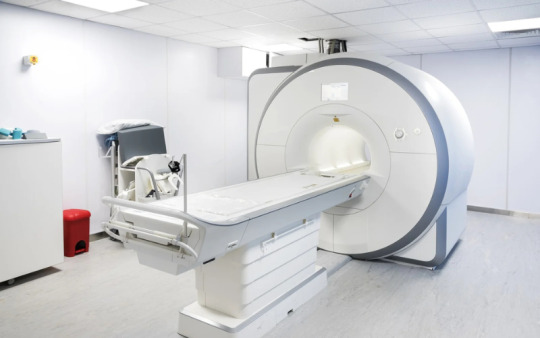
Understanding MRI and CT Scans
MRI (Magnetic Resonance Imaging) and CT (Computed Tomography) scans are both non-invasive imaging techniques used to create detailed images of the inside of the body. While both are essential for diagnosing various conditions, they have distinct differences:
MRI Scans: Use strong magnetic fields and radio waves to produce detailed images of organs and tissues. They are particularly useful for imaging soft tissues, such as the brain, muscles, and ligaments.
CT Scans: Use X-rays to create cross-sectional images of the body. They are excellent for visualizing bone structures, detecting tumors, and identifying internal injuries.
Choosing the Right Imaging Center
When selecting an imaging center, consider the following factors:
Technology and Equipment: Ensure the center is equipped with the latest MRI and CT scan machines.
Expertise: Look for centers with experienced radiologists and technicians.
Accessibility: Choose a center that is conveniently located and offers flexible appointment timings.
Cost: Compare prices and check if the center accepts your insurance.
Top MRI and CT Scan Centers in Delhi, NCR
Here are some of the top centers in Delhi NCR, categorized by their specialties and locations:
MRI Centers
MRI Centre in Maharani Bagh: Known for its state-of-the-art MRI machines, this center offers high-resolution imaging for accurate diagnosis. The facility is easily accessible and provides a comfortable environment for patients.
CT Scan Centre Near Maharani Bagh: This center is equipped with advanced CT scan technology, ensuring quick and precise imaging. It is conveniently located for residents of Maharani Bagh and surrounding areas.
Color Doppler Test Centers
Color Doppler Test Centre Near Apollo Hospital: Specializing in vascular imaging, this center uses color Doppler ultrasound to assess blood flow and detect any abnormalities. Its proximity to Apollo Hospital makes it a preferred choice for many patients.
Color Doppler Centre Test in Okhla: This center offers comprehensive Doppler studies, including arterial and venous Doppler. It is known for its accurate results and experienced staff.
Ultrasound Centers
Ultrasound Centre in Tughlakabad: Providing a range of ultrasound services, this center is equipped with modern machines and skilled technicians. It is a trusted name for prenatal and abdominal ultrasounds.
Ultrasound Centre in South Ex: Located in South Extension, this center offers detailed ultrasound imaging for various medical conditions. It is known for its patient-friendly approach and efficient service.
X-Ray Centers
X-Ray Centre in Govindpuri: This center provides high-quality digital X-rays, ensuring minimal radiation exposure. It is a reliable choice for quick and accurate imaging.
Dental X Ray Centre in Jasola: Specializing in dental X-rays, this center offers panoramic and cephalometric imaging. It is equipped with the latest technology to provide clear and detailed images.
#MRI#CTScan#MedicalImaging#DiagnosticImaging#Radiology#MRIScan#CTScanProcedure#HealthDiagnostics#BodyImaging#MedicalTests#BrainScan#ImagingTechnology
0 notes
Text
Digital Microscope 23.2 mm

Labnic Digital Microscope features an achromatic optical system with a 40x–1000x magnification range, bright field capability, and a binocular head that rotates 360° and inclines 30°. It includes two PL10X/18 mm eyepieces, a 23.2 mm diameter, and a spiral up-and-down adjustment for precise focusing.
0 notes
Text
3D Reconstruction: Capturing the World in Three Dimensions

3D reconstruction refers to the process of capturing the shape and appearance of real objects using specialized scanners and software and converting those scans into digital 3D models. These 3D models allow us to represent real-world objects in virtual 3D space on computers very accurately. The technology behind 3D reconstruction utilizes a variety of different scanning techniques, from laser scanning to photometric stereo, in order to digitally preserve real objects and environments.
History and Early Developments
One of the earliest forms of 3D scanning and reconstruction dates back to the 1960s, when laser range scanning first emerged as a method. Early laser scanners were slow and bulky, but provided a way to accurately capture 3D coordinates from surfaces. In the 1970s and 80s, raster stereo techniques came about, using cameras instead of lasers to capture depth maps of scenes. Over time, scanners got faster, accuracy improved, and multiple scanning technologies started to converge into unified 3D modeling pipelines. By the 1990s, digitizing entire buildings or archeological sites became possible thanks to the increased capabilities of 3D reconstruction tools and hardware.
Photogrammetry and Structure from Motion
One major development that helped accelerate 3D Reconstruction was the emergence of photogrammetry techniques. Photogrammetry uses 2D imagery, often from consumer cameras, to extract 3D geometric data. Structure from motion algorithms allow one to take unordered image collections, determine overlapping features across images, and reconstruct the camera positions and a sparse 3D point cloud. These image-based methods paved the way for reconstructing larger exterior environments accurately and cheaply compared to laser scanners alone. Photogrammetry is now a widely used technique for cultural heritage documentation, architecture projects, and even film/game production.
Integral 3D Scanning Technologies
Today there are many 3D scanning technologies in use for different types of objects and environments. Structured light scanning projects patterns of lines or dots onto surfaces and reads distortions from a camera to calculate depth. It works well for industrial inspection, small objects and scanning interiors with controlled lighting. Laser scanning uses time-of-flight or phase-shift measurements to rapidly capture millions of precise 3D data points. Laser scanners excel at large outdoor environments like cultural heritage sites, construction projects and full building documentation. Multi-view stereo and photometric stereo algorithms fuse together image collections into full and detailed 3D reconstructions even with untextured surfaces.
Applications in Mapping and Modeling
3D reconstruction mapping applications are widespread. Cultural heritage sites are extensively documented with 3D models to make high resolution digital archives and share artifacts globally online. Forensic reconstruction of accident or crime scenes relies on 3D modeling to understand what happened. Engineering companies use 3D scans to digitally inspect parts for quality control. Manufacturers design products in CAD but validate and improve designs with physical prototypes that are 3D scanned. Cities are mapped in 3D with aerial and mobile scanning to plan infrastructure projects or monitor urban development over time. The gaming and animation industries also reconstruct detailed digital environments, objects and characters for immersive 3D experiences. Advancing computing power allows more complex reconstructions than ever before across many industries.
Ongoing Developments and the Future of 3D
There is continued work to improve the speed, resolution, automation and scaling of 3D acquisition techniques. Multi-sensor fusion and simultaneous localization and mapping (SLAM) is leading to capabilities like interior mapping with handheld scanners. Artificial intelligence and deep learning are also entering the field with applications like automatically registering separate scans or recognizing objects in 3D data. On the modeling side, semantics and building information models (BIM) are merging geometric shapes with metadata, properties and relationships. Advances will open the door to dynamic 3D representations that change over time, interaction with virtual objects through augmented reality and further immersive experiences in artificial worlds reconstructed from real environments. The future vision is seamlessly integrating virtual 3D spaces with our daily physical world.
Get more insights on 3D Reconstruction
About Author:
Money Singh is a seasoned content writer with over four years of experience in the market research sector. Her expertise spans various industries, including food and beverages, biotechnology, chemical and materials, defense and aerospace, consumer goods, etc. (https://www.linkedin.com/in/money-singh-590844163)
#3DReconstruction#ComputerVision#ImagingTechnology#SpatialData#DigitalModeling#Photogrammetry#PointClouds#3DScanning
0 notes
Text

#HyperspectralImaging#HyperspectralSensors#RemoteSensing#ImagingTechnology#IndustrialInspection#industrialautomation#SatelliteImaging#SensorTechnology#TechTrends
0 notes
Text
Apply Now!
Launch Your Career in Healthcare with Longowal Polytechnic College!
Paramedical Courses 2024-25 M.Sc & B.Sc in Radiology & Imaging Technology
State-of-the-Art Facilities Expert Faculty Hands-On Training
🔗 Don't miss out on this opportunity! Secure your future in the medical field.
Enroll Today!
𝑃𝑙𝑠 𝐶𝑜𝑛𝑡𝑎𝑐𝑡 𝐿𝑜𝑛𝑔𝑜𝑤𝑎𝑙 𝐺𝑟𝑜𝑢𝑝 𝑜𝑓 𝐶𝑜𝑙𝑙𝑒𝑔𝑒𝑠. 𝐷𝑒𝑟𝑎𝑏𝑎𝑠𝑠𝑖,𝐷𝑖𝑠𝑡𝑟𝑖𝑐𝑡 𝑀𝑜ℎ𝑎𝑙𝑖, 𝑁𝑒𝑎𝑟 𝐶ℎ𝑎𝑛𝑑𝑖𝑔𝑎𝑟ℎ. 𝐶𝑎𝑙𝑙 9915158000 𝑤𝑤𝑤.𝑙𝑔𝑐𝑑𝑒𝑟𝑎𝑏𝑎𝑠𝑠𝑖.𝑜𝑟𝑔
#HealthcareCareer#Radiology#ImagingTechnology#LongowalPolytechnic#AdmissionsOpen#FutureInHealthcare#ApplyNow#MedicalCourses#Admissions2024#ParamedicalStudies#JoinNow#Education#CareerInMedicine
0 notes
Text
Cone Beam Computed Tomography (CBCT) Market Outlook Report 2024-2030: Trends, Strategic Insights, and Growth Opportunities | GQ Research
The Cone Beam Computed Tomography (CBCT) market is set to witness remarkable growth, as indicated by recent market analysis conducted by GQ Research. In 2023, the global Cone Beam Computed Tomography (CBCT) market showcased a significant presence, boasting a valuation of US$ 541.23 Million. This underscores the substantial demand for Cone Beam Computed Tomography (CBCT) technology and its widespread adoption across various industries.
Get Sample of this Report at: https://gqresearch.com/request-sample/global-cone-beam-computed-tomography-cbct-market/
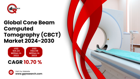
Projected Growth: Projections suggest that the Cone Beam Computed Tomography (CBCT) market will continue its upward trajectory, with a projected value of US$ 1102.59 Million by 2030. This growth is expected to be driven by technological advancements, increasing consumer demand, and expanding application areas.
Compound Annual Growth Rate (CAGR): The forecast period anticipates a Compound Annual Growth Rate (CAGR) of 10.70%, reflecting a steady and robust growth rate for the Cone Beam Computed Tomography (CBCT) market over the coming years.
Technology Adoption:
Cone Beam Computed Tomography (CBCT) has witnessed significant adoption across various industries, including dentistry, orthopedics, and oncology. Its precise imaging capabilities and three-dimensional visualization have revolutionized diagnostic and treatment planning processes. In dentistry, CBCT is increasingly replacing traditional two-dimensional imaging techniques due to its ability to provide detailed images of dental structures, facilitating more accurate diagnoses and treatment interventions. Similarly, in orthopedics and oncology, CBCT enables clinicians to obtain high-resolution images of bones, joints, and soft tissues, aiding in the diagnosis and monitoring of musculoskeletal and oncological conditions.
Application Diversity:
The versatility of CBCT technology extends beyond medical applications. It finds utility in fields such as archaeology, veterinary medicine, and industrial inspection. Archaeologists utilize CBCT to non-destructively examine artifacts and fossils, revealing internal structures and aiding in historical research. In veterinary medicine, CBCT assists veterinarians in diagnosing complex conditions in animals, such as dental abnormalities and musculoskeletal disorders. Furthermore, CBCT is employed in industrial settings for quality control and inspection purposes, facilitating the analysis of internal structures and defects in manufactured components.
Consumer Preferences:
Consumer preferences for CBCT systems are shaped by factors such as image quality, radiation dose, cost-effectiveness, and ease of use. End-users prioritize systems that offer high-resolution images with minimal radiation exposure to patients. Additionally, affordability and compatibility with existing infrastructure influence purchasing decisions, especially for small to medium-sized clinics and imaging centers. User-friendly interfaces and streamlined workflow integration are also key considerations, as they contribute to operational efficiency and staff satisfaction.
Technological Advancements:
The CBCT market is characterized by ongoing technological advancements aimed at enhancing imaging quality, reducing radiation dose, and improving system efficiency. Manufacturers are investing in innovations such as advanced image reconstruction algorithms, iterative reconstruction techniques, and dose optimization software to achieve higher image resolution while minimizing radiation exposure. Additionally, developments in detector technology, such as digital flat-panel detectors and photon-counting detectors, contribute to improved image quality and faster scan times. Integration with artificial intelligence (AI) algorithms for image analysis and interpretation represents another area of innovation, enabling automated detection of anatomical structures and pathology.
Market Competition:
The CBCT market is highly competitive, with numerous players vying for market share through product differentiation, pricing strategies, and technological innovation. Established medical imaging companies, as well as niche players specializing in CBCT systems, compete to meet the diverse needs of end-users across various industries. Key competitive factors include product performance, reliability, after-sales support, and brand reputation. Market consolidation through mergers and acquisitions is also observed as companies seek to expand their product portfolios and global presence.
Environmental Considerations:
Environmental considerations play an increasingly important role in the CBCT market, with a focus on reducing energy consumption, optimizing resource utilization, and minimizing waste generation. Manufacturers are adopting eco-friendly manufacturing processes and materials to mitigate environmental impact throughout the product lifecycle. Efforts to design energy-efficient systems with reduced power consumption contribute to lower operational costs and carbon footprint. Furthermore, initiatives to recycle and responsibly dispose of end-of-life equipment and components support sustainability goals within the healthcare and imaging industry.
Regional Dynamics: Different regions may exhibit varying growth rates and adoption patterns influenced by factors such as consumer preferences, technological infrastructure and regulatory frameworks.
Key players in the industry include:
Carestream Health, Inc.
Dentsply Sirona Inc.
Planmeca Group
Vatech Co., Ltd.
Danaher Corporation (KaVo Dental)
Cefla Medical Equipment
Morita Corporation
PreXion Corporation
CurveBeam LLC
NewTom Srl
The research report provides a comprehensive analysis of the Cone Beam Computed Tomography (CBCT) market, offering insights into current trends, market dynamics and future prospects. It explores key factors driving growth, challenges faced by the industry, and potential opportunities for market players.
For more information and to access a complimentary sample report, visit Link to Sample Report: https://gqresearch.com/request-sample/global-cone-beam-computed-tomography-cbct-market/
About GQ Research:
GQ Research is a company that is creating cutting edge, futuristic and informative reports in many different areas. Some of the most common areas where we generate reports are industry reports, country reports, company reports and everything in between.
Contact:
Jessica Joyal
+1 (614) 602 2897 | +919284395731 Website - https://gqresearch.com/
0 notes
Text
Are you ready to revolutionize your vision? In this comprehensive guide to LIDAR Imaging - Beamagine, we delve deep into the future of imaging technology. LIDAR Imaging offers a whole new dimension to visual perception, and with Beamagine's cutting-edge solutions, we're taking it to the next level. Join us as we dive into the world of 3D visualization and uncover the incredible potential of LIDAR Imaging - Beamagine.
Know more: https://www.4shared.com/s/fFCbWzYXzjq
0 notes
Text
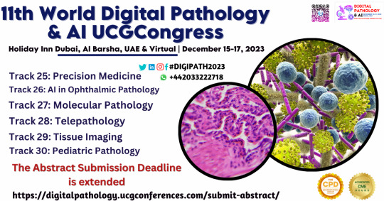
Call for Abstract
Don't miss the opportunity for your research to gain some much-deserved visibility at the CME/CPD accredited 11th World Digital Pathology & AI UCGCongress from December 15–17, 2023, in Holiday Inn Dubai, Al Barsha, UAE & Virtual,
.WhatsApp us: https://wa.me/442033222718?text=
submit your abstract here: https://digitalpathology.ucgconferences.com/submit-abstract/
#CellularAnalysis#TissueAnalysis#ImagingTechnology#MedicalImaging#TissueVisualization#DigitalHistopathology#PathologyImages#ImagingInMedicine#BiologicalSamples#HighResolutionImaging#TissueResearch#Pathology
0 notes
Text
World Radiology Day

Celebrating World Radiography Day! 📷✨
Today, we honor the skilled radiographers who help save lives through diagnostic imaging. Their expertise is a beacon of hope in healthcare. Thank you for your dedication and commitment!
Learn more>> https://www.sriramakrishnahospital.com/
#sriramakrishnahospital#WorldRadiographyDay#healthcareheroes#ImagingTechnology#healthcare#coimbatore#multispecialityhospital#healthandwellness#bookyourappointment
0 notes
Text
Global Contrast Media Market Size, Share & Trends Analysis Report by Type (Iodine-Based Contrast Media, Gadolinium-Based Contrast Media (GBCAs),Microbubble Contrast Media, Barium-Based Contrast Media), by Modality (X-ray/CT Contrast Media, MRI Contrast Media, Ultrasound Contrast Media), by Route of Administration (Intravenous (IV),Oral, Rectal), by Indication(Cardiovascular Diseases, Oncology, Neurology, Gastrointestinal Disorders), by End-User(Hospitals & Clinics, Diagnostic Imaging Centres, Research Institutes) and Geography (North America, Europe, Asia-Pacific, Middle East and Africa, and South America) | Forecast [2024-2032]
#ContrastMediaMarket#MedicalImaging#Radiology#DiagnosticImaging#ContrastAgents#HealthcareInnovation#MRIContrast#CTScan#XRayImaging#Radiopharmaceuticals#HealthcareMarket#ImagingTechnology#PatientCare#MarketTrends#MedicalDiagnostics
0 notes
Text
Cone Beam Computed Tomography Market Strategic Assessment: Market Size, Share, Growth Projections
The global cone beam computed tomography market size is expected to reach USD 1,217.1 million by 2030, registering a CAGR of 9.6% from 2024 to 2030, according to a new report by Grand View Research, Inc. The market is driven by a rise in the volume of digital imaging procedures, especially in the field of dentistry. Dental surgeries and other diagnostic imaging fields are undergoing a digital transformation process with intraoral optical scanners, 3D imaging technologies, and software applications designed for smooth operations of the imaging devices.
Cone Beam Computed Tomography Market Report Highlights
Based on application, the dental implantology segment dominated the market and accounted for a share of 26.8% in 2023 as the implants offer patients feasible options for teeth replacement treatments. With the rising R&D in this field, dental implants are available with better biomaterials and improved designs
The orthodontics segment is expected to register the fastest CAGR during the forecast period from 2024 to 2030 as this segment covers complex dental treatments, including misaligned teeth, smile gaps, and crooked teeth
By patient position, the seated position segment accounted for the largest revenue share in 2023 as patients can be still in a seating position than a standing position. CBCT imaging can be done easily in a seating position
In terms of end-use the dental clinics segment dominated the market in 2023 and is likely to witness the fastest growth over the forecast period. Most of the patients prefer visiting clinics due to the availability of specialists, cost efficiency, and improved technology
The North America cone beam computed tomography market accounted for a revenue share of 47.9% in 2023 owing to the factors such as the presence of independent clinics and rising R&D activities in digital imaging. The Asia Pacific cone beam tomography market is expected to grow significantly in the coming years due to the rising awareness about 3D imaging procedures and expanding healthcare infrastructure in the region
For More Details or Sample Copy please visit link @: Cone Beam Computed Tomography Market Report
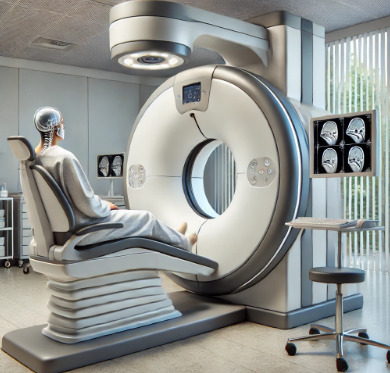
Cone beam CT imaging is performed with the help of a rotating platform with the X-ray source and detector and uses a single flat panel. Cone beam computed tomography (CBCT) produces a cone beam of radiation instead of a fan beam. The entire scanning process of the target area is done in a single rotation, hence reducing the radiation exposure significantly. These highly accurate, low radiation dose, and small footprint CBCT systems have transformed daily clinical practices.
As per the American Cancer Society, about 35,000 cases of mouth, tongue, and throat cancers are diagnosed in the U.S. annually, with the average age of most people diagnosed with these cancers is over 60 years. According to the World Dental Federation, globally, about 30% of people aged 65 to 74 years have no natural tooth, and this burden is expected to increase with the aging of the population. The growing incidence rate of such oral diseases in this age group is likely to increase the demand for digital diagnostic imaging in the coming years.
List of major companies in the Cone Beam Computed Tomography Market
Dentsply Sirona.
J. MORITA MFG. CORP.
VATECH
CurveBeam AI, Ltd.
Carestream Health Inc. (ONEX Corporation)
Danaher Corporation
For Customized reports or Special Pricing please visit @: Cone Beam Computed Tomography Market Analysis Report We have segmented the global cone beam computed tomography market on the basis of on application, patient position, end-use, and region.
#ConeBeamCT#MedicalImaging#CBCTMarket#DentalImaging#RadiologyTech#3DImaging#DiagnosticImaging#HealthcareInnovation#Orthodontics#ImagingTechnology#CBCTScanner#MedicalDevices#RadiologySolutions#DentistryTech#HealthcareEquipment#SurgicalPlanning#ENTImaging#MaxillofacialImaging#AdvancedDiagnostics#RadiologyMarket
0 notes
Text
Exploring IT Technology Trends in Hospital Imaging: Unveiling Insights into Software Use and Medical
The fusion of IT technology and hospital imaging has sparked a revolution in the rapidly advancing healthcare landscape. The combination of software solutions and medical reporting workflows reshapes how hospitals operate and enhances patient care. This blog provides insights into IT technology trends in hospital imaging.
Read Full blog here: https://www.grgonline.com/post/exploring-it-technology-trends-in-hospital-imaging-unveiling-insights-into-software-use-and-medical
The Evolution of Hospital Imaging
Hospital imaging has transcended traditional boundaries, propelled by IT technology. From X-rays to MRIs, digitization has ushered in an era of heightened accuracy, efficiency, and accessibility in diagnosing medical conditions.
Seamless Integration of Software
Cutting-edge software solutions have been seamlessly integrated into medical imaging processes. Radiology information systems (RIS) and picture archiving and communication systems (PACS) are streamlining image storage, retrieval, and distribution, reducing manual efforts and the risk of errors.
Enhancing Medical Reporting Workflow
The flow of medical information is pivotal. Modern software has revolutionized medical reporting, automating the creation, distribution, and archival of reports. This translates to quicker diagnoses and more timely treatment decisions.
AI-Powered Insights
Artificial intelligence (AI) is not science fiction; it's transforming hospital imaging. AI algorithms analyze medical images with unmatched precision, flagging anomalies that might elude human eyes. This assists radiologists and enhances diagnostic accuracy.
Remote Access and Telehealth
The convergence of IT and imaging has transcended physical barriers. Remote access to medical images allows experts to collaborate across distances and offer timely consultations. Telehealth consultations are becoming more seamless and efficient, benefiting both patients and medical professionals.
Data Security and Compliance
As data becomes digital gold, its security becomes paramount. IT solutions enable efficient imaging and ensure compliance with stringent data protection regulations. Patient confidentiality remains a top priority.
Enhanced Patient Engagement
Empowering patients with access to their medical images fosters transparency and patient engagement. Online portals and mobile apps enable patients to view their images and reports, facilitating informed discussions with healthcare providers.
Real-Time Analytics for Strategic Decisions
Modern software solutions offer real-time analytics, providing hospital administrators with insights into imaging trends, usage patterns, and operational efficiencies. These data-driven insights guide strategic decisions for resource allocation and workflow enhancement.
Collaboration Beyond Boundaries
The global healthcare community is benefiting from IT innovations in imaging. Cross-border collaborations allow experts to share knowledge, review cases, and offer opinions, transcending geographical constraints.
Future Prospects
The horizon of IT technology trends in hospital imaging is bright. The integration of virtual reality (VR) and augmented reality (AR) into imaging processes holds promise for enhanced visualization and training.
Conclusion
The combination of IT technology and hospital imaging has birthed a new era in healthcare. From streamlined workflows to enhanced patient care, software solutions are leaving an indelible mark. As we navigate the evolving trends, it's clear that the future of hospital imaging is intertwined with IT innovation.
The journey towards precision, efficiency and patient-centric care continues, powered by the synergy of technology and healthcare.
Visit our website now: https://www.grgonline.com/
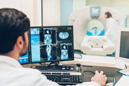
#Healthcare#healthcareanalytics#healthcare it#imagingtechnology#medical software#medical imaging#techtrends#hospitality#diagnosticimaging#digitalhealthcare#marketing
0 notes
Text
Ct Scan Bangalore
Discover advanced CT Scan Bangalore for precise diagnostic imaging. Experience state-of-the-art technology and skilled radiologists delivering accurate and detailed results. Enhance your healthcare journey with reliable CT scan services in Bangalore, ensuring a comprehensive and effective approach to medical diagnostics. Trust in the expertise and quality of CT scans to aid in the accurate diagnosis and treatment planning of various medical conditions.
#CTScan#BangaloreMedicalServices#AdvancedImaging#DiagnosticExcellence#MedicalTechnology#RadiologyServices#HealthcareJourney#AccurateDiagnosis#TreatmentPlanning#MedicalDiagnostics#PrecisionImaging#HealthcareQuality#CTScanBangalore#MedicalExcellence#HealthcareInnovation#ImagingTechnology#MedicalDiagnosis#HealthcareServices#RadiologyExpertise#MedicalImaging
0 notes
Text
Is It Possible For CT Scans To Detect Kidney Stones?
Medical imaging has revolutionized the field of diagnostics, providing physicians with valuable insights into the human body.
When it comes to detecting kidney stones, one of the most common urological conditions, computed tomography (CT) scans have proven to be a highly effective tool.
With their remarkable precision and ability to capture detailed images of the internal structures, CT scans have become a go-to method for diagnosing and visualizing kidney stones.
Kidney stones, medically referred to as renal calculi, are solid formations composed of minerals and salts that develop within the kidneys.
These stones can vary in size and shape, ranging from tiny grains to large obstructions that can cause excruciating pain. Timely and accurate detection of kidney stones is crucial to ensure proper treatment and prevent further complications.
So, how do CT scans play a role in detecting kidney stones? CT scans utilize a combination of X-ray technology and computer processing to create cross-sectional images of the body.
This imaging technique produces highly detailed images that allow healthcare professionals to visualize the kidneys, urinary tract, and any potential abnormalities, such as kidney stones.
When it comes to detecting kidney stones, CT scans are particularly effective due to their ability to capture images in multiple planes.
Unlike traditional X-rays, which provide only two-dimensional images, CT scans can generate three-dimensional representations of the kidneys. This comprehensive view enables radiologists to accurately locate and assess the size, shape, and position of kidney stones.
Furthermore, CT scans can distinguish between different types of kidney stones based on their density. This information is invaluable for determining the most appropriate treatment approach.
For instance, calcium-based stones, the most common type, are easily identified on CT scans as they appear as bright white structures. On the other hand, uric acid stones, which are less dense, may require specialized imaging techniques such as dual-energy CT scans to enhance their visibility.
In addition to their remarkable imaging capabilities, CT scans offer several other advantages when it comes to detecting kidney stones. They are non-invasive, meaning they do not require any surgical intervention or the insertion of instruments into the body. This significantly reduces patient discomfort and eliminates the risk of complications associated with invasive procedures.
Moreover, CT scans are quick and efficient, producing images within a matter of minutes. This rapid turnaround time is particularly crucial in emergency cases where prompt diagnosis is necessary to alleviate severe pain and prevent further complications.
The speed and accuracy of CT scans make them an invaluable tool for healthcare professionals in managing kidney stone cases effectively.
Despite their numerous advantages, CT scans do have some limitations when it comes to detecting kidney stones. One notable concern is the exposure to ionizing radiation. CT scans utilize X-rays, which involve a small amount of radiation.
While the radiation dose is typically low and considered safe, repeated exposure can potentially increase the risk of long-term effects, especially in younger patients or individuals with a history of radiation exposure.
To mitigate this concern, radiologists and healthcare providers strive to minimize radiation exposure during CT scans. They employ various techniques such as low-dose protocols and specialized software algorithms that optimize image quality while reducing radiation levels.
Additionally, alternative imaging modalities, such as ultrasound and magnetic resonance imaging (MRI), may be considered for patients who are more sensitive to radiation or require repeated imaging.
CT scans have proven to be a highly effective method for detecting kidney stones. Their ability to provide detailed, three-dimensional images allows healthcare professionals to accurately locate, evaluate, and classify kidney stones.
CT scans offer a non-invasive, efficient, and reliable diagnostic approach, particularly in emergency situations. However, concerns regarding radiation exposure should be carefully considered and appropriate measures taken to minimize risks.
As medical technology continues to advance, the field of diagnostic imaging will undoubtedly bring forth even more sophisticated tools and techniques to improve the detection and management of kidney stones.
Well, are you interested in discovering a leading diagnostic provider in Australia? Follow this link to explore an esteemed team of radiologists who deliver exceptional care and utilize state-of-the-art technology to ensure the highest quality diagnostics.
0 notes
Text
24/7 radiologist
If you are searching for bespoke 24/7 radiologist services for your hospitals at a moderate price, then contact Radblox, the trusted and globally accepted radiology service provider. At Radblox, we aim to be an extending helping hand for emergency imaging report reading for our clients, physicians and hospitals.
If you need 24/7 radiologist services for your hospitals at an affordable price, Radblox is a great option. With a long-standing history of top-notch service and trustworthiness, Here at Radblox, we strive to be an invaluable resource when it comes to urgent imaging report analysis. We assist our clients, physicians and hospitals by providing reliable services in a timely manner.
#247Radiology#Teleradiology#Imaging#Diagnostics#MedicalImaging#RadiologyServices#RadiologyGroups#RadiologyClinics#Radiologist#ImagingTechnology
0 notes