#gastrul
Explore tagged Tumblr posts
Text
for reasons [did the 'taking classes' year of grad school in 2020/21 and was desperate not to be on zoom anymore] i only took 7/8 required credits for my phd when i was supposed to and now have to take a class. already kind of cringe and undignified given my advanced age in grad school years. but i couldn't even use it to take something like linalg that i actually need training in because it only counts toward my degree if it's a biology class. not only do i absolutely not need anyone to teach any biology topic to me at this point, because my job is to interpret and synthesize primary biology literature, but i also already took every even faintly relevant biology course in college, some of them twice, the majority of them at grad level with graduate students. and they were taught better and more effectively, because my undergraduate institution simply had better pedagogical practices than *** by basically any metric.
all of this has cashed out to me taking the paper-reading course about my field taught by one of my committee members, because it only meets once a week, i've read half the papers before, and it's graded on class participation which is my best stat. unfortunately, because it's taught by one of my committee members, i can't admit that i am taking it literally only for credit and would be capable of teaching the class given a couple of weeks to prep, and instead have to pretend that i am concerned about it meaningfully impairing my career that, while i have an encyclopedic knowledge of early embryonic development in flies and worms, i'm really more familiar with late embryonic development in mammals. this is requiring some contortions.
#the events i study happen post-gastrulation at minimum. i just don't need to worry about whether a mouse embryo is a cup#or what you're suppose to call the wnt-inhibiting organizer region#but i would like to maintain this professional relationship.#box opener#he was like ''oh right you never took this as a first year!''#and i had to try very hard not to be like. yes. because if you look at my college transcript you will note that i took#1. developmental mechanisms 2. stem cells and regen 3. dev genetics of model organisms 4. genetics and development (general)#and 5. plant development#and the syllabi of the first three. contain 90% of the syllabus of this class between them.#unfortunately i'm instead channeling this into being really pedantic about why we're using certain archaic terminology#that i don't believe cleaves reality at the joints.#but that too is a kind of participating.
18 notes
·
View notes
Text

Culture of Continuity
Models of embryo formation called blastoids created from human pluripotent stem cells grown on a 3D matrix bear characteristics of continuous early development from pre-implantation to early gastrulation – the point of multi-layered organisation
Read the published research paper here
Image from work by Rowan M. Karvas and colleagues
Department of Developmental Biology and Center of Regenerative Medicine, Washington University School of Medicine, St. Louis, MO, USA
Image originally published with a Creative Commons Attribution 4.0 International (CC BY 4.0)
Published in Cell Stem Cell, September 2023
You can also follow BPoD on Instagram, Twitter and Facebook
#science#biomedicine#immunofluorescence#biology#embryo development#gastrulation#stem cells#developmental biology
3 notes
·
View notes
Text
developmental biology really just straight up sucks
#I'm sorry mr (redacted)... I like you very much and I know you love spiral cleavage and gastrulation but it just isnt it okay?#op dasloddl
1 note
·
View note
Text

TSRNOSS. Page 174.
#lithium#metallothionein#gastrulation#oxygen consumption rate#oxidative phosphorylation#foetus#spontaneous abortion#spiral#clockwise#alpha particle#sponges#Coriolis force#histone solubility#manuscript#theoretical biology#notebooks#handwriting
0 notes
Text
Reference preserved in our archive (Daily updates!)
Just a 'mild' reversal of a fetus's internal organs...
Context and significance A striking increase in situs inversus cases, diagnosed by ultrasound, was observed several months following removal of zero-COVID-19 policies in China, which coincided with a surge in SARS-CoV-2 infection. The rare clinical evidence presented here reveals a previously unobserved possible fetal consequence of maternal SARS-CoV-2 infection specifically during gestational weeks 4–6. To date, visceral lateralization has never been definitively linked to a specific developmental time in humans due to the rarity of such fetal samples. This study advances our current understanding of gastrulation-stage development and possibly provides the most robust support yet linking an environmental factor to occurrence of situs inversus, opening new research directions into mechanisms of visceral lateralization in humans and consequences of SARS-CoV-2 infection in pregnancy.
Highlights • Situs inversus is associated with SARS-CoV-2 infection at gestational weeks (GWs) 4–6 • Results herein support previously undefined visceral lateralization at GWs 4–6 in humans • SARS-CoV-2 infection at GWs 4–6 is well supported as an environmental risk factor for situs inversus
Summary Background A dramatic increase in fetal situs inversus diagnoses by ultrasound in the months following the severe acute respiratory syndrome coronavirus 2 (SARS-CoV-2) surge of December 2022 in China led us to investigate whether maternal SARS-CoV-2 exposure could be associated with elevated risk of fetal situs inversus.
Methods In this multi-institutional, hospital-based, matched case-control study, we investigated pregnant women who underwent ultrasonographic fetal biometric assessment at gestational weeks 20–24 at our hospitals. Each pregnant woman carrying a situs inversus fetus was randomly matched with four controls based on the date of confinement. Relevant information, including SARS-CoV-2 infection, and other potential risk factors were collected. Conditional logistic regression was used to test possible associations between fetal situs inversus and SARS-CoV-2 infection at different gestational weeks as well as individual risk factors.
Findings A total of 52 pregnant women diagnosed with fetal situs inversus between January 1 and October 31, 2023 and 208 matched controls with normal fetuses were enrolled. We found no association between an increased risk of fetal situs inversus with gestational SARS-CoV-2 infection or with other risk factors. However, fetal situs inversus was significantly associated with SARS-CoV-2 infection specifically in gestational weeks 4–6 (adjusted odds ratio [aOR] 6.54 [95% confidence interval 1.76–24.34]), but not with infection at other gestational ages, after adjusting for covariates.
Conclusions Increased risk of fetal situs inversus is significantly associated with maternal SARS-CoV-2 infection at gestational weeks 4–6, corresponding to the fetal developmental window for visceral lateralization in humans.
#mask up#covid#pandemic#public health#wear a mask#wear a respirator#covid 19#still coviding#coronavirus#sars cov 2
33 notes
·
View notes
Text
embryology turning out to be a soul-sucking subject to study for cause i could not. care less about gastrulation in birds. actively painful to read about i'm gonna be honest
9 notes
·
View notes
Note
Do you know any cool science facts? Bonus points if the inspire awe and wonder!
As a former science educator, it is my moral duty to have and share cool science facts. ;)
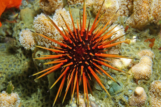
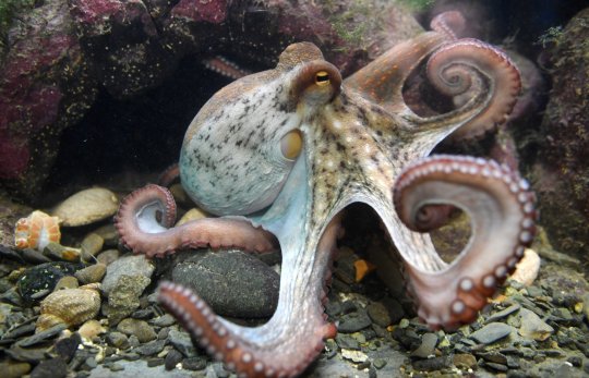
Would you believe that humans are more closely related (evolutionarily speaking) to a sea urchin than to an octopus?


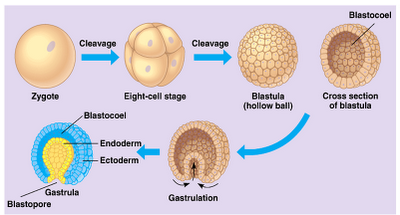
One of our strongest pieces of evidence for this comes from the study of embryonic development, or embryology. Some time after germ cells combine to form a single-celled zygote, the cell divides several times to form a hollow sphere of cells called a blastula. In animals with bilateral body plans (i.e., digestive tract surrounded by a separate internal body cavity, with a mouth at one end and an anus at the other), that digestive tract begins forming as the blastula undergoes gastrulation, a process by which one part of the surface of the sphere turns in on itself.

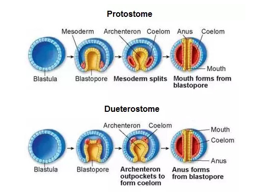
This initial opening into the digestive tract, the blastopore, is either the mouth or the anus. In protostomes ("mouth-first"), the blastopore is the the mouth! As the digestive tract develops, it will eventually form an anus at the other end of the embryo. In deuterstomes ("mouth-second"), the mouth... forms second. The blastopore is the anus.

As it turns out, many worms, slugs, and the like - including molluscs and cephalopods - are protostomes. Deuterostomes then encompass many of the creatures you might name with bilateral body plans (and related, radial body plans like starfish and sea urchins), including arthropods, birds, fish, reptiles, mammals, even tunicates! And humans.
At one point in our development, each and every one of us was nothing more than a butthole. Sometimes it seems some people never progressed past that stage!
8 notes
·
View notes
Text
More "holiday" music -- for biology lovers and nerds:
An a cappella version of the "Hallelujah" Chorus -- with the lyrics changed to celebrate the entire Animal Kingdom (Lyrics in the video, and this post. Eye contact. 3 minutes, 45 seconds. Proper closed captions in Galician, auto-generated captions in Spanish):
youtube
Lyrics:
Animalia! (x10)
Our cells are phospholipid membranous Animalia (x4) But have no form-restrictive containers Animalia (x4) Our high motility is what shapes us For heterotrophy as predators Chordata, annelids and ctenophores Animalia!
The opisthokonta clade Has become The kingdoms of fungi And animals And animals
In the domain of eukaryota ATP made inside mitochondria But we contain no plastids like chloroplasts With blastulae as diplo- or triploblasts
Kingdom of Mollusca Tardigrada Arthropoda Chordata:
The phylum of Larvacia Agnatha Amphibia And Mammalia:
Classis of Rodentia Cetacea Carnivora Primates:
The order of Lemuridae Tarsiidae Atelidae Hominidae:
Family of Gorilla and Pongo Chimpanzee And homo:
The genus of Homo sapiens sapiens
(But we all came from before the Cambrian)
But we all came But we all came But we all came But we all came
From a concestor in the Precambrian
Protostomes (And deuter- ostomia) With desmosomes (Gap junctions, tight junctions) And microtubules nucleated by centrosomes
Coelomates with hollow cores Gastrulate from blastopores And we chordates have tails and gill slits and notochords
Echinoderms (Platyzoa, Nematoda) And Arthropods (Porifera and Cnidaria) Animalia (x4)
ANIMALIA!
#music#a cappella#biology#the Animal Kingdom#Christmas Season music#(even though this was originally composed for Easter)#eye contact#open captions#Youtube
4 notes
·
View notes
Text
finally something that makes sense gastrulation i love you everyone say thank you gastrulation
2 notes
·
View notes
Note
For everyone!! If you could pic another job aside from the mafia...what would you choose?
And to the mod, have a lovely day 💜
Risotto Nero - I'd probably be a teacher, science in particular
Pros - I'd be an architect, almost went to university for it before things went south
Pesci - I honestly don't know...
Maggi - A vet or someone who works at an animal shelter I think, it would be very fulfilling
LuLu - I'd love to own a small cafe or a hair salon, something unique
Melone - Well I have a PhD so probably something in the biological research field, specifically into gastrulation, it fascinates me
Ghiaccio - Olympic skater, or I'd at least try, did you think I got my stand for nothing.
Sorbet - CEO of something, whatever pays best
Gelato - Probably an accountant, stable and pays well
9 notes
·
View notes
Text

No improvement after 2 weeks. Loss of appetite. Frequent meltdowns.
There was a crayon drawing on the bottom of the table; a stick person with a big smile, then several smaller stick people with broad Xs for eyes. Harry’s stomach twisted as he stared up at them.
Woodie murmured, almost more to themself than anyone else, “... What happened here?”
“I-I can't remember. I don't remember.”
Only in quick flickers, in shoddy projector slides.
No adverse effects yet. Complaints about the size of the pills. Split dosage into smaller capsules?
Harry scooted out from under the table and let Woodie help him to his feet.
A child’s shriek pierced the space, reverberating through the hall until, for a split second, it sounded as though coming from every direction. After a tense moment, Harry cautiously straightened his shoulders and squeezed Woodie’s hand. They led him out of what was labeled on an old staff directory map as the ‘recreation chamber’— a large room with foam padded floors, a swing and a round table with crayons and paper.
“Let's go this way.” Harry said, tapping an orange arrow on the wall. There were several similar indicators in various colours, all leading off in different directions.
An informational sign in the foyer outside the recreation chamber had denoted them as relating to the following departments:
purple: PLAGUE
green: RETROSPECTIVES
red: BILE REQUISITION
blue: DETRITUS & NUTRITION
orange: HOME
Failure begets failure. Poison in the blood. Poison in the brain. Irritability, nightmares, increasingly uncooperative. Dosage adjustment needed.
The orange arrow led them down another hallway, then around a corner. On the wall opposite, a plaque stood before a large, darkened display window.
“Press to illuminate each specimen…” Woodlock read aloud.
“That- uh, I-I don't—”
With a click, one specimen jar on the far left of the window lit up. It had something small within, a pale, pink spec with a dark spot, surrounded by nondescript tissue matter. Click. The lights buzzed and flickered. Another jar, then another, both containing humanoid fetuses at varying stages of development.
Click.
The last place looked empty, at first. Harry leaned in, then let out a small cry. The light shimmered across the fine detritus of shattered glass and dry mucus.
“It's just a dream, dear.”
Harry pulled away from the window, “It's never just a dream.”
“I mean, it’s the same old things arranged in unfamiliar patterns, is all.”
They continued down the hall. The orange arrow, smudges of something wet and viscous dragging itself along. Harry pushed away memories of his mother sobbing, despondent with a kind of grief too heavy for his little arms to hold when he scrambled onto the couch to comfort her.
“I found out about it by accident. Uh, just poking around in old project folders. Stuff about producing genetically identical embryos with various minor tweaks in the nucleic code,” Harry laughed dryly, “And then I was like, hold on, I know that genome. That's me!”
“You recognize your genome sequence?”
“It’s usually apparent by the second page.”
Specimen No.1
21 days. Perished during later stages of gastrulation.
Specimen No.4
4 weeks. Death due to complications regarding chromosomal abnormalities.
Specimen No.13
32 weeks. Complications with cardiac development.
Specimen No. 15
1 day.
The blue arrow was visible on a wall down the side corridor. The wall had a dark smeared handprint along the bottom that trailed beyond view.
“I try not to think about it. I don't like bringing up stuff mom has done, ‘cause she gets upset.” Harry picked at a loose thread at the hem of his sleeve.
“It was her hand that carved in you a wound that never healed.”
“... I try not to think about it.”
At the end, there was a large room with pretty wood flooring and floral wallpaper. Inside, an overturned dinner table and two chairs.
It felt, somehow, as if there were something missing in the space. Harry closed his eyes, trying to remember what it could've been. A girl, maybe. She had long, frizzy strawberry blonde hair tied into a french braid. She wore a stained yellow-orange jumpsuit.
The absence, her absence. The empty space folded into the figure of the girl, crouched on her hands and knees by the table. She was crunching something between her teeth, pulling a string of sinew from her lips. She jerked upright, eyes wide.
Harry met her gaze. He knew her face, even smeared with blood. It looked a lot like his.
She took a breath and screamed.
#spidermom.txt#ooc#content warning for: implied child abuse / slight gore#also stuff related to pregnancy and birth.
5 notes
·
View notes
Text
regardless of the researcher’s overall weirdness it does take a certain baseline insanity to spend a six-decade career watching the same 72 hours of embryo development happen over and over and over and over and over again. six decades of gastrulation. you could wind up in a time loop and might not even notice
14 notes
·
View notes
Photo

Feeling the Squeeze
A critical process in early development, gastrulation defines the transition from a single layer of cells, known as the epiblast, to a more complex structure formed of three germ layers – the endoderm, mesoderm and ectoderm – which later give rise to different structures in the body. A key process in gastrulation is the epithelial-to-mesenchymal transition (EMT), in which epiblast cells undergo major changes in shape and gene expression to detach from their neighbours and move away. By labelling cell membranes with fluorescent proteins and using time-lapse microscopy, researchers were able to film cells undergoing the EMT (pictured in green) in developing mouse embryos, revealing how cells extract themselves by incrementally contracting their outer, or apical, surfaces (in red). EMTs are also a feature of cell movements later in life, from wound healing to cancer cell migration in metastasis, so studying these transitions could inspire new ways to prevent cancers from spreading.
Written by Emmanuelle Briolat
Image from work by Alexandre Francou, Kathryn V Anderson and Anna-Katerina Hadjantonakis
Developmental Biology Program, Sloan Kettering Institute, Memorial Sloan Kettering Cancer Center, New York, NY, USA
Image copyright held by the original authors
Research published in eLife, May 2023
You can also follow BPoD on Instagram, Twitter and Facebook
#science#biomedicine#embryonic development#developmental biology#epithelial to mesenchymal#metastasis#immunofluorescence#cancer
7 notes
·
View notes
Text
Mechanical induction in metazoan development and evolution: from earliest multi-cellular organisms to modern animal embryos | Nature Communications
A gentle marine hydrodynamic flow, simulating gentle waves on the seashore, was found to be able to induce active gastrulation in a Myo-II-dependent process in the cnidarian sea anemone H. vectensis... the organism would have been stabilized by natural selection due to its advantageous ability to capture and digest more prey by sensing the flow. Later, such mechanical signals could arise spontaneously and homogeneously around the embryo as active mechanical fluctuations.

Обнаружено, что пологий морской гидродинамический поток, имитирующий мягкие волны на берегу моря, способен вызывать активную гаструляцию в процессе, зависящем от Myo-II, у книдарной актинии Н. вектенсис... организм был бы стабилизирован за счет естественного отбора из-за его выгодной способности захватывать и переваривать больше добычи, ощущая поток. Позже такие механические сигналы могли возникать спонтанно и гомогенно вокруг эмбриона в виде активных механических флуктуаций.
0 notes
Text
studying embryo gastrulation while listening to eva entry plug sound
1 note
·
View note
Text
Three-dimension transcriptomics maps of whole mouse embryo during organogenesis
Understanding mammalian development heavily relies on classical animal models like the house mouse (Mus musculus). Advanced spatial transcriptomics has enabled biologists to break new ground in studies of molecular dynamics and cellular patterning during embryonic development with spatiotemporal resolution. To construct a comprehensive developmental trajectory, current three-dimensional (3D) spatial transcriptomic profiling leverages mouse embryos from gastrulation (E5.5) to organogenesis (E13.5) in continuity. However, a crucial phase for early organogenesis between E9.5 and E11.5 was deficient. To unveil the mystery of this stage, we present the 3D transcriptomics of mouse embryos at E9.5 and E11.5, with a widely applicable reconstruction workflow that bypasses sophisticated bioinformatic calculations. As our 3D atlas is generated at single-cell resolution, we demonstrate how organogenetic processes can be interpreted at different levels of granularity, from local cellular interactions to whole embryonic regionalization. We release the open-access database MOSTA3D (Mouse Organogenesis Spatiotemporal Transcriptomic Atlas in Three-dimensional) and hope a broader community will contribute to extending this framework from conception to senility in the near future. http://dlvr.it/TC7NxX
0 notes