#fundue
Explore tagged Tumblr posts
Text

[it's a drawing, click on it!] KING OF PAIN. [playlist]
(Alternates under the cut)


#went with a more painterly style for this one#s2 is on my mind always#I love the fundus effect#stranger things#stranger things 5#st2#st5#will byers#byler#will stranger things#the police#king of pain#80s#stranger things fanart#fanart#my art
175 notes
·
View notes
Photo

Customer: I'M A LOAN OFFICER AND FUND LOANS; "FUND U" MEANING, I WILL FUND YOUR LOAN. DMV: YOU, HOSTILE Verdict: DENIED
#California license plate with text FUNDU#bot#ca-dmv-bot#california#dmv#funny#government#lol#public records
511 notes
·
View notes
Text
Got an ultrasound today my uterus is fully curled up in a ball :)
2 notes
·
View notes
Note
*Lethica finds an assortment of fruits, pretzels, marshmellows and other things to be dipped into chocolate, with a melting pot for chocolate with a choice of chocolate chips. Beside it is a note that reads*
"perhaps you'll find this of use to you when you have that date with Sir Marius, fruits included of course"
-secret admirer
*Lethica once again looks around, as if looking for someone to appear. When no one does, she speaks to the sky*
"Thank you, admirer. I will put this to good use."
#goon at noon asks#legends of avantris#edge of midnight#ooc: 'fondue? more like fundue' <- if i was playing any other derek character
3 notes
·
View notes
Text
das größte verbrechen im tatort saarbrücken ist dass sie leo ständig in weiß und grau stecken, auch wenn ich die gründe natürlich verstehe..... aber kein wunder dass so ein grünes t-shirt gleich alle in aufruhr versetzt. dem mann stehen kräftige farben einfach 100 mal besser. fotobeweise unterm cut
ich mein hallo?? wir erinnern uns:

und das hier?

selbst "langweiliges" dunkelblau oder schwarz


kein weiß oder grau mehr!!!
#danke tess für deinen ausgiebigen fundus an burlakov-pics 👌#vladimir burlakov#spatort#tatort blogging#txt
13 notes
·
View notes
Note
Hey Abby how’s that reunion going?

Your tail is wagging...?!
"I can still hardly believe she's been alive all this time."
1 note
·
View note
Text
googly eye

148K notes
·
View notes
Text
Fundu Mina-rai Atuál Billaun $18.6
Hatutan.com, (30 Agostu 2024), Díli-Dadus hosi Banco Centrál Timor-Leste (BCTL) kona-bá dezempeñu fundu mina-rai Timor-Leste nian hosi fulan-Janeiru to’o fulan-Jullu tinan 2024 iha nia balansu ka montante billaun $18.6. Continue reading Fundu Mina-rai Atuál Billaun $18.6
0 notes
Text
Fundus Camera: An Essential Equipment for Eye Examination

What is a Fundus Camera?
A fundus camera, also known as an ophthalmoscope or retinal camera, is a specialized low-power microscope used to afford high-quality imaging of the internal tissues of the living human eye, known as the fundus. The fundus refers to the interior surface of the eye opposite the lens, including the retina, optic disc, and posterior pole of the eye. Fundus cameras allow eye care professionals to non-invasively examine and document the health of the retina, optic disc and macula. Parts A standard fundus camera consists of the following key components: - Illumination System: Provides controlled light to illuminate the fundus via the pupil. Koehler illumination is commonly used, allowing homogenous lighting of the fundus. Halogen or Xenon lamps are commonly used light sources. - Optical System: Consists of lenses that magnify the light reflected from the fundus for visualization and imaging purposes. Typical magnification ranges from 15x to 60x. Wide-angle and ultra-wide angle lens options are available. - Eye Piece: The eyepiece allows the examiner to view the magnified fundus image. Binocular eyepieces provide stereoscopic viewing. - Digital Camera Sensor: Replaces traditional film in modern digital Fundus Camera. Charged coupled device (CCD) or complementary metal-oxide-semiconductor (CMOS) digital sensors capture fundus images which can be stored, analyzed and shared digitally. - Shields/Filters: Polarization filters reduce glare from light reflections within the eye. Alignment lights and targets help with proper centration of the fundus image. - Stand or Slit Lamp Attachment: It may sit on a stand alone or attach to an existing slit lamp biomicroscope, allowing integration with other examination equipment. Uses and Applications of Fundus Photography Fundus photography allows detailed examination and documentation of retinal pathologies, which is invaluable for diagnosing and monitoring many common eye diseases. Some key applications include: - Diabetic Retinopathy Screening: Photographing the retina allows detection of microaneurysms, hemorrhages, cottonwool spots, hard exudates and neovascularization indicative of diabetic eye disease. Optimal Fundus Photography Techniques Careful technique is needed to obtain high quality fundus photos enabling accurate clinical evaluation: - Pupil Dilation: Most fundus details are only visible after dilating the pupil with eyedrops to at least 6 mm. This improves illumination levels reaching the fundus. - Centration: Centering the fundus camera lens on the pupil ensures the retina is evenly illuminated and the entire posterior pole is in view. Targeting lights assist alignment.
Get more insights on Fundus Camera
About Author:
Ravina Pandya, Content Writer, has a strong foothold in the market research industry. She specializes in writing well-researched articles from different industries, including food and beverages, information and technology, healthcare, chemical and materials, etc. (https://www.linkedin.com/in/ravina-pandya-1a3984191)
#Fundus Camera#Retinal Imaging#Eye Examination#Ophthalmology#Fundus Photography#Retina#Ocular Health#Fundus Imaging#Eye Care#Diabetic Retinopathy Screening
0 notes
Text
A North Carolina woman says she lost 110 pounds in 15 months after undergoing an experimental heat procedure to reduce her food cravings.
In February 2023, Mills had a gastric fundus mucosal ablation as part of a 10-woman study. A doctor threaded a tube down her throat to burn off stomach tissue that produces ghrelin, a hormone linked to increasing appetite, calorie intake, and weight gain.
When your stomach is empty, it releases ghrelin to let your brain know that it’s time to eat. Researchers have theorized that people with excess weight may be more sensitive to the effects of ghrelin.
“What we found, in the months after this procedure, was that, in fact, ghrelin was reduced,” lead researcher Dr. Dan Maselli, a bariatric endoscopist and associate director of research for True You Weight Loss, told FOX 5 Atlanta.
“We saw a 45% decrease of that hunger hormone circulating in the bloodstream within six months of the procedure,” Maselli continued. “In terms of stomach capacity, we saw a 42% decrease in the amount of food it would take to get us to feel full.”
Participants dropped 7.7% of their body weight on average over six months. Maselli and his team presented their findings last month in Washington, DC, at the annual Digestive Disease Week meeting.
He said the outpatient procedure — which is said to take less than an hour — caused minor side effects, such as stomach cramping, discomfort, and nausea.
0 notes
Video
I love how fast the slug looks.
*Swish* nomnomnomnomnomnomnom nom glup ! *turn around turn around* Any left ? No. Ciao ! *zoom*
Eating slime mold by MaximumMoustache
169K notes
·
View notes
Text
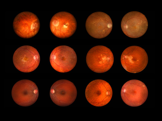
Fundus, 23/24
1 note
·
View note
Text
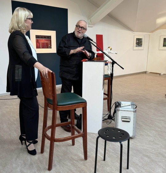

Obwohl Manuela Saß, die Bürgermeisterin der Stadt Werder (Havel), in einem anderen Termin gebunden war, kam sie kurz mit einer Ansprache in die Ausstellung.
Foto oben: Frank W. Weber stellt die Akteure des Abends vor. Olaf Möldner (sitzend) beantwortete Fragen des Kurators, die allen Besuchern mehr Informationen zur Fotoserie „China hautnah“ gaben. Am Abend wurden zwei Schenkungen an den Fundus der Stadtgalerie übergeben. Der russische und in Berlin lebende Kunsthandwerker Andrei Zaplatynski (links von Olaf Möldner) und der Leipziger Bildhauer Roland Wetzel (neben dem Kurator) übergaben je ein Werk dem Fundus.
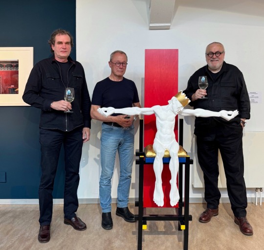
Roland Wetzel (Mitte) hatte 2022 eine Ausstellung in der Stadtgalerie und überließ nun seine Plastik des sitzenden „Nebukadnezar“ dem Fundus. Zwischenzeitlich wurden davon zwei Bronzen gegossen und das Original im Gipsaufbau ging jetzt an die Stadtgalerie. Der Thron des Königs kam aus der Hand von Andrei Zaplatynski. (links) Eine Adaption auf den bekannten Gerrit Rietveld Stuhl von 1916. Der Kunsthandwerker hatte den Stuhl der Stadtgalerie angeboten und geschenkt. Das goldene Sitzkissen des Königs Nebukadnezar passt wie in einem Teamwork abgesprochen exakt auf die Sitzfläche. Der Kurator dazu: „Nebukadnezar, als König umstritten, wird so mit einem modernen Thron in die Jetztzeit transportiert. Dieses neue Gesamtkunstwerk bietet somit neue Interpredationsmöglichkeiten aus der Geschichte auf das Heute. Nebukadnezar hat nun alle Zeit der Welt und darf, wie Pontius Pilatus in „Der Meister und Margarita“ von Michael Bulgakow, über seine Missetaten auf dem Thron sitzend nachdenken.“ Roland Wetzel dazu: „Nebukadnezar verkörpert das Prinzip des Scheiterns als Endpunkt einer Selbstüberschätzung.“ Andrei Zaplatynski redete auf Russisch, was von seiner Frau Elena übersetzt wurde.: „Es ist eine Ehre für meinen Stuhl, jetzt als Thron in der Kunstsammlung der Stadtgalerie zu dienen.“
Obwohl nicht zur Ausstellung „China hautnah“ gehörend wird diese Schenkung an den Fundus der Stadtgalerie während der gesamten Ausstellungszeit bis 23. Juni zu sehen sein.
Die Ausstellung von Olaf Mödner mit 45 ausbelichteten Fotografien und 350 Fotos in einer Bilderschau auf einem Monitor ist eröffnet. Die Bilderschau läuft als Endlosschleife mit 36 Minuten Dauer während der Ausstellung.
Wie immer Donnerstag, Samstag und Sonntag von 13-18 Uhr, bei freiem Eintritt zu besichtigen.
Am Himmelfahrtstag geöffnet, Pfingssamstag und -sonntag geöffnet. Nicht am Feiertag Pfingstmontag!
Die Fotografien sind als Abzug 40x65 cm für 150€/Stück zu erwerben. Olaf Möldner stiftet diese Einnahmen dem Kindergarten im Hohen Weg für Arbeitsmaterialien.
Fotos: ANDRIOTTA
#kunst geschoss#stadtgalerie#werder havel#fotografie#kunstgalerie#frank w weber#roland wetzel#andrei zaplatynski#olaf moeldner#nebukadnezar#plastik#china#china hautnah#fundus#kunstsammlung
0 notes
Text
Eye Fundus Camera: Revolutionizing Ophthalmic Imaging
Introduction to Eye Fundus Camera
Eye fundus cameras, also known as retinal cameras, are specialized devices used to capture images of the back of the eye, including the retina, optic disc, macula, and blood vessels. These images, referred to as fundus photographs, play a crucial role in diagnosing and monitoring various eye conditions, such as diabetic retinopathy, glaucoma, macular degeneration, and hypertensive retinopathy.
Importance of Eye Fundus Examination
A comprehensive eye examination typically involves the evaluation of the eye's posterior segment, which includes the retina and optic nerve head. Fundus examination allows ophthalmologists and optometrists to assess the health of these structures, detect abnormalities, and monitor disease progression over time.
Types of Eye Fundus Cameras
Traditional Fundus Cameras
Traditional fundus cameras are stationary devices typically found in ophthalmology clinics and hospitals. They offer high-resolution imaging capabilities and are suitable for detailed retinal examinations.
Handheld Fundus Cameras
Handheld fundus cameras provide flexibility and portability, allowing eye care professionals to capture fundus images outside of traditional clinical settings. These devices are particularly useful for screening programs and telemedicine applications.
Smartphone Fundus Cameras
Smartphone fundus cameras leverage the power of mobile technology to transform smartphones into cost-effective fundus imaging devices. With the use of specialized adapters or attachments, these cameras enable primary care providers and even patients themselves to capture fundus images conveniently.
How Eye Fundus Cameras Work
Fundus cameras utilize various imaging techniques, such as digital photography, scanning laser ophthalmoscopy, and optical coherence tomography, to capture detailed images of the eye's posterior segment. These images are then analyzed by eye care professionals to detect abnormalities and guide treatment decisions.
Benefits of Using Eye Fundus Cameras
Early detection of eye diseases
Monitoring disease progression
Guiding treatment decisions
Facilitating patient education
Enhancing collaboration among healthcare providers
Applications in Ophthalmology
Eye fundus cameras are indispensable tools in ophthalmic practice, with applications including:
Diabetic retinopathy screening
Glaucoma management
Age-related macular degeneration evaluation
Retinopathy of prematurity assessment
Optic nerve head analysis
Challenges and Limitations
Despite their numerous benefits, eye fundus cameras face certain challenges, such as:
Cost of equipment and maintenance
Training requirements for operators
Limited access in underserved areas
Image interpretation variability
Future Trends in Eye Fundus Imaging Technology
Advancements in imaging technology, such as artificial intelligence and telemedicine, are poised to revolutionize eye fundus imaging by improving accuracy, efficiency, and accessibility.
Comparison with Other Imaging Techniques
Compared to other imaging modalities like optical coherence tomography and fluorescein angiography, fundus photography offers a non-invasive and cost-effective approach to visualizing the retina and optic nerve.
Considerations for Purchasing an Eye Fundus Camera
When selecting an eye fundus camera, factors to consider include image quality, ease of use, compatibility with existing systems, technical support, and cost-effectiveness.
Tips for Efficient Use of Eye Fundus Cameras
Ensure proper patient preparation and positioning
Adjust camera settings for optimal image quality
Use appropriate imaging techniques for different eye conditions
Regularly calibrate and maintain equipment
Training and Certification for Eye Fundus Imaging
Eye care professionals should undergo specialized training and certification programs to acquire the necessary skills for operating and interpreting fundus images accurately.
Costs Associated with Eye Fundus Imaging
The initial investment in purchasing an eye fundus camera may vary depending on the type and features of the device. Additional costs include maintenance, accessories, and ongoing training.
Case Studies and Success Stories
Numerous studies have demonstrated the clinical utility and cost-effectiveness of eye fundus cameras in various healthcare settings, highlighting their role in improving patient outcomes and reducing healthcare costs.
Conclusion
Eye fundus cameras have transformed the landscape of ophthalmic imaging, enabling early detection, accurate diagnosis, and personalized management of eye diseases. As technology continues to evolve, these devices will play an increasingly vital role in preserving vision and enhancing patient care.
FAQs (Frequently Asked Questions)
Are eye fundus cameras safe for all patients?
Yes, fundus cameras are non-invasive and safe for most patients, including children and pregnant women.
Can fundus cameras detect all eye diseases?
Fundus cameras can detect a wide range of eye conditions, but they may not capture certain abnormalities that require specialized imaging techniques.
Is dilating the pupils necessary for fundus photography?
Dilating the pupils may be necessary to obtain clear fundus images, especially in cases where a detailed examination is required.
Are smartphone fundus cameras as accurate as traditional devices?
While smartphone fundus cameras offer convenience and accessibility, they may not provide the same level of image quality and diagnostic accuracy as traditional devices in all cases.
How often should fundus examinations be performed?
The frequency of fundus examinations depends on various factors, including age, medical history, and risk factors for eye diseases. It is best to consult with an eye care professional for personalized recommendations.
#eyecare#fundus examination#health#medical care#eye health#eyes#choroida#choroid#medical devices#medical equipment
0 notes
Text
Perfect rendition 😭😭❣️❣️❣️

findus my son i would do anything for him
226 notes
·
View notes
Text
Fluorescein Fundus Camera Market Size, Segments, Growth and Trends by Forecast to 2031
According to a new report published by The Insight Partners, titled, ” Fluorescein Fundus Camera Market Forecast | Share and Size – 2030″. The report provides a detailed analysis of the top investment pockets, top winning strategies, drivers & opportunities, Fluorescein Fundus Camera market size & estimations, competitive landscape, and changing market trends. The Fluorescein Fundus Camera…
View On WordPress
0 notes