#foliose coral
Explore tagged Tumblr posts
Text


my new pretty Siren Pirate OC <3
her bra and tail are based on Foliose Coral!
3 notes
·
View notes
Text
Not to be edgy or start anything, but what is the best word to get to say when you are describing the texture of lichens to your friends?
16 notes
·
View notes
Text


Parmotrema flavescens
Yesterday was the "first day of winter" in Iceland, which has me feeling some kind of way about the looming months of cold. I wish I could head south for the winter and hang out in the warm like our pal P. flavecens likes. This foliose lichen grows on rock in hot, humid areas of South America, southern North America, and Africa. It has round, loose lobes with crenulate, ciliate margins. It produces cylindrical and coral-like isidia on the lobe surface and margins. The upper surface is pale green-yellow, smooth or moderately cracked. The lower surface is dark brown toward the centers, and black and rhizinate toward the margins. There isn't a lot of info about this and other lichens native to the tropics and southern hemisphere, which means job security somewhere warm I guess? Maybe?
images: source
info: source
#lichen#lichens#lichenology#lichenologist#mycology#ecology#fungi#biology#nature#bryology#fungus#botany#symbiosis#symbiotic organisms#algae#phycology#Parmotrema flavescens#Parmotrema#I'm lichen it#lichen a day#daily lichen post
42 notes
·
View notes
Text
lichen informational post
because a lot of my mutuals seems to be unclear about lichen and there are so few accessible sources out there on these amazing beings :D

[ID: A grey stick with wolf lichen growing on it. Wolf lichen is very three-dimensional, with the rough appearance of many light green blood vessels or coral. End ID]
Image from Wikimedia Commons, taken by Katja Schultz.
What is lichen?
Put simply, lichen (pronounced LIE-ken or LITCH-en) is a bunch of different fungi and algae working together to make an organism that looks something like a mix between a plant and a fungus. It can have the appearance of moss sometimes, and often you will find species named “[adjective] moss”.
There are many types of lichen, which can broadly be categorized as fruticose (like the wolf lichen pictured above the cut), foliose (slightly less 3D and usually having a leafy appearence), crustose (clearly one solid form, but appearing plastered to the surface it’s found on), and leprose (a more powdery substance). There are a few more broad categories, including jello-like and stringy, but these are the most well-known.
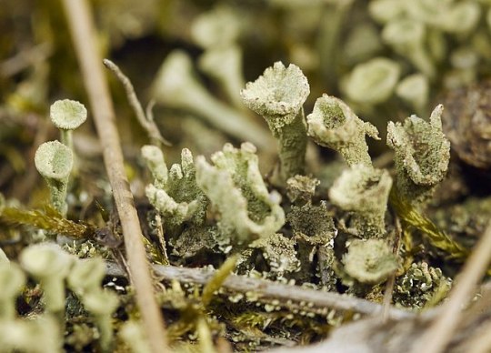
[ID: Trumpet Lichen growing among some small sticks. Trumpet lichen is light green, shaped rather like the mouth of a trumpet. It has a bumpy texture. End ID]
Image from Wikimedia Commons, taken by Roman.
Where can I find lichen?
Basically anywhere above sea level! Lichen can grow on almost any surface, and are mostly common on rocks and trees. They can also grow on toxic surfaces, though! Lichen do not have roots, but they do photosynthesize. They do not have any adverse affect on whatever they may find their home on. Lichen can also hang from surfaces.
Apart from what’s already been said, why is lichen so cool?
- We know of about 20,000 species of lichen so far!
- Lichen may be the longest-living organisms on earth, and are also some of the earliest organisms to grow after a natural disaster.
- Some species of lichen reproduce sexually, and some don’t, but they can all evolve.
- Lichen can be very susceptible to air pollution, especially when there are not many trees around to provide clean air. However, lichen can survive in space, if so moved.
- Lichen was not officially classified until 1867.
- Most lichen is made up of photosynthesizing organisms (cyanobacteria or algae) surrounded by the fungal organisms, but this is often flipped in gelatinous or stringy lichen!
Here is the link to the Wikipedia page, which is where I compiled this information from, if you would like to read more. I am also really really excited about this and can answer questions!!
https://en.wikipedia.org/wiki/Lichen
(tagging @cnnamonrolls because you said you’d never heard of lichen about a week back and I’m very excited to share this)
19 notes
·
View notes
Text
Coral Morphology & Colouration
Credit: Gaby Pilson
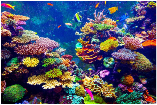
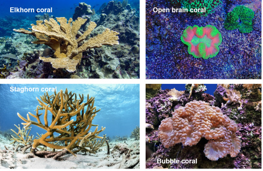
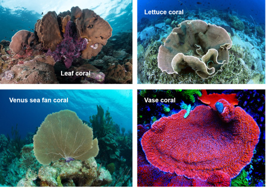
In this blog, we will explore various shapes of corals and the factors determining coral morphology. We will also know how colourful corals acquire diverse colouration. Before getting to the main points, let's take a look at various beautiful corals below.
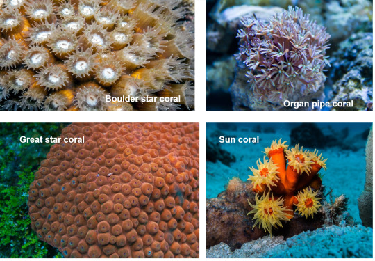
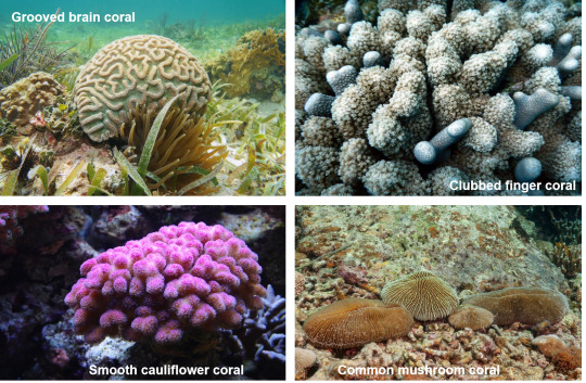
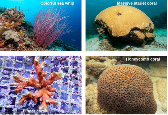

Credit: Gaby Pilson
Coral morphology
Within and among taxa, scleractinian corals vary in morphology, from the simple encrusting or hemispherical colonial shape to tree-like branching, bubble-like, finger-like or globular shapes (Zawada, Dornelas & Madin 2019).
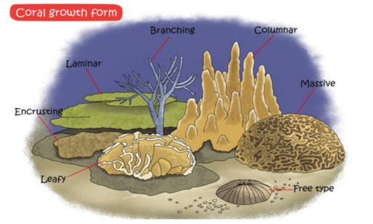
Credit: Liao, Xiao & Li
The variation and complexity in the morphology of corals can be affected by several factors, such as
environmental conditions (Zawada, Dornelas & Madin 2019)
biological process (Jackson 1979)
genetic constraints (Filatov et al. 2013)
partial mortality (Meesters, Wesseling & Bak 1996)
fragmentation of colony (Karlson 1986)
indeterminate growth (Sebens 1987)
The environmental conditions include free-flowing water for filter feeding and adequate sunlight for photosynthesis (Hoogenboom, Connolly & Anthony 2008; Kaandrop et al. 1996). Although most corals favour the mentioned environmental conditions, corals can also adapt to less optimal growing environments. Based on adaptability, various coral species' population density also differs in a particular environment.
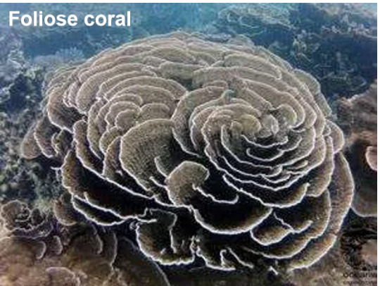
For instance, platy and foliose corals are the dominant species in deeper water or shallow, sheltered, high-turbid and low-light areas (Riegl, Heine & Branch 1996; Hallock 2005). Other coral species also grow in such regions, but their population densities are relatively lower (Zapalski et al. 2021). These two corals are more adaptable to low light because the platy and foliose morphologies maximize the surface area to harvest light for photosynthesis (Anthony, Hoogenboom & Connolly 2005; Kahng et al. 2012).
Credit: Khao Lak Explorer
Some corals, such as the staghorn and elkhorn corals, grow upwards to reduce and avoid competition for the benthic space (Jackson 1997). The upward growth allows these corals not to need to continuously occupy the benthic space to increase their standing biomass.

Credit: Gaby Pilson
Some corals, such as laminar and foliose corals, grow laterally on the benthos to diversify the risk, thereby reducing the colony's mortality (Jackson 1979).

Credit: Khao Lak Explorer (Foliose coral); Project Noah (Laminar coral)
Coral colouration
Corals are colourful. But how do corals acquire diverse colour patterns and intensities? Well, the coral colouration is determined by (Dove et al. 2006; Oswald et al. 2007):
endosymbionts
fluorescent proteins
The symbiotic dinoflagellates living in the coral cells give a brown colour to the surfaces of corals (Porter et al. 1984). Depending on the densities of the cells or photosynthetic pigments, the brown colour may get paler or darker (Porter et al. 1984).
The fluorescent proteins (FPs) in coral cells mainly refer to the green fluorescent protein-like proteins that can fluoresces under the visible or ultraviolet light to produce colouration in corals, such as the brilliant green, red and blue (Dove, Hoegh-Guldberg & Ranganathan 2001). FPs are suggested to be photo-protective (Salih et al. 2000) or photo-enhancing (Schlichter, Meier & Fricke 1994). In the photo-protective role, FPs can dissipate excess energy by scattering light and fluorescing to prevent photo-damage and photo-inhibition in coral symbionts in excessive sunlight (Salih et al. 2000). In the photo-enhancing role, FPs can absorb light with a shorter wavelength and re-emit it with a wavelength suitable for the endosymbiont of corals to carry out photosynthesis (Frade et al. 2008).
That's all for this blog. Hope you enjoy the read ^ ^
References
Anthony, KR, Hoogenboom, MO & Connolly, SR 2005, ‘Adaptive variation in coral geometry and the optimization of internal colony light climates’, Functional Ecology, vol. 19, pp.17-26.
Dove, SG, Hoegh-Guldberg, O & Ranganathan, S 2001, ‘Major colour patterns of reef-building corals are due to a family of GFP-like proteins’, Coral Reefs, vol. 19, pp.197-204.
Dove, S, Ortiz, JC, Enríquez, S, Fine, M, Fisher, P, Iglesias-Prieto, R, Thornhill, D & Hoegh-Guldberg, O 2006, ‘Response of holosymbiont pigments from the scleractinian coral Montipora monasteriata to short‐term heat stress’, Limnology and Oceanography, vol. 51, no. 2, pp.1149-1158.
Filatov, MV, Frade, PR, Bak, RP, Vermeij, MJ & Kaandorp, JA 2013, ‘Comparison between colony morphology and molecular phylogeny in the Caribbean scleractinian coral genus Madracis’, PLoS One, vol. 8, no. 8, p.e71287.
Frade, PR, Englebert, N, Faria, J, Visser, PM & Bak, RPM 2008, ‘Distribution and photobiology of Symbiodinium types in different light environments for three colour morphs of the coral Madracis pharensis: Is there more to it than total irradiance?’, Coral Reefs, vol. 27, pp.913-925.
Hallock, P 2005, ‘Global change and modern coral reefs: New opportunities to understand shallow-water carbonate depositional processes’, Sedimentary Geology, vol. 175, pp.19-33.
Hoogenboom, MO, Connolly, SR & Anthony, KR 2008, ‘Interactions between morphological and physiological plasticity optimize energy acquisition in corals’, Ecology, vol. 89, no. 4, pp.1144-1154.
Jackson, JBC 1979, ‘Morphological strategies of sessile animals’, in G Larwood & BR Rosen (eds), Biology and Systematic of Colonial Animals, Academic Press, Google ebooks, pp. 499-555.
Kaandorp, JA, Lowe, CP, Frenkel, D & Sloot, PM 1996, ‘Effect of nutrient diffusion and flow on coral morphology’, Physical Review Letters, vol. 77, no. 11, p.2328.
Kahng, SE, Hochberg, EJ, Apprill, A, Wagner, D, Luck, DG, Perez, D & Bidigare, RR 2012, ‘Efficient light harvesting in deep-water zooxanthellate corals’, Marine Ecology Progress Series, vol. 455, pp.65-77.
Karlson, RH 1986, ‘Disturbance, colonial fragmentation, and size-dependent life history variation in two coral reef cnidarians’, Marine Ecology Progress Series, vol. 28, pp.245-249.
Meesters, EH, Wesseling, I & Bak, RP 1996, ‘Partial mortality in three species of reef-building corals and the relation with colony morphology’, Bulletin of Marine Science, vol. 58, no. 3, pp.838-852.
Oswald, F, Schmitt, F, Leutenegger, A, Ivanchenko, S, D'Angelo, C, Salih, A, Maslakova, S, Bulina, M, Schirmbeck, R, Nienhaus, GU & Matz, MV 2007, ‘Contributions of host and symbiont pigments to the coloration of reef corals’, The FEBS Journal, vol. 274, no. 4, pp.1102-1122.
Porter, JW, Muscatine, L, Dubinsky, Z & Falkowski, PG 1984, ‘Primary production and photoadaptation in light-and shade-adapted colonies of the symbiotic coral, Stylophora pistillata’, Proceedings of the Royal Society of London. Series B. Biological Sciences, vol. 222, no. 1227, pp.161-180.
Riegl, B, Heine, C & Branch, GM 1996, ‘Function of funnel-shaped coral growth in a high-sedimentation environment’, Marine Ecology Progress Series, vol. 145, pp.87-93.
Salih, A, Larkum, A, Cox, G, Kühl, M & Hoegh-Guldberg, O 2000, ‘Fluorescent pigments in corals are photoprotective’, Nature, vol. 408, no. 6814, pp.850-853.
Schlichter, D, Meier, U & Fricke, HW, 1994 ‘Improvement of photosynthesis in zooxanthellate corals by autofluorescent chromatophores’, Oecologia, vol. 99, pp.124-131.
Sebens, KP 1987, ‘The ecology of indeterminate growth in animals’, Annual Review of Ecology and Systematics, vol. 18, pp.371-407.
Zapalski, MK, Baird, AH, Bridge, T, Jakubowicz, M & Daniell, J 2021, ‘Unusual shallow water Devonian coral community from Queensland and its recent analogues from the inshore Great Barrier Reef’, Coral Reefs, vol. 40, pp.417-431.
Zawada, KJ, Dornelas, M & Madin, JS 2019, ‘Quantifying coral morphology’, Coral Reefs, vol. 38, no. 6, pp.1281-1292.
Images or videos from external sources:
Gaby Pilson - https://outforia.com/types-of-coral/
Khao Lak Explorer - https://www.khaolakexplorer.com/coral-types/
Liao, Xiao & Li - https://link.springer.com/chapter/10.1007/978-94-024-1612-1_1
Project Noah - https://www.projectnoah.org/spottings/6319210
0 notes
Note
tells us more about lichenology please
i'm by no means a scientist or even educated in biology past the most rudimentary high school level so my understanding and study of lichens is that of an artist and layperson.
my focus has been on the use of lichens for textile dyeing specifically, but in order to ethically and correctly collect lichen for dyeing i have to be able to identify them at least by their genus for a few major reasons. primarily, as an artist, different species need to be processed in different ways to produce colours unique to them. some species don't produce any colour when processed one way and produce vivid colour in another; others produce wildly different colours in different contexts. secondarily, from an ecological perspective, its important to know how common/rare different lichen are so you don't unknowingly destroy a rare and old organism, and there are ways of harvesting parts of lichen without damaging the thallus or body of the lichen so it can continue to grow if you learn how to do so but it's best to collect lichen that have already fallen off their substrate or are on substrate soon to be destroyed (ex. fallen trees, lumber, etc.)

[image id: diagram showing the various common types of lichen growing on a bark or tree substrate: crustose or crusty lichen, foliose or leaf-like lichen, and frucitose or bushy lichen. the crustose lichen are very flat and bumpy, while the foliose lichen have flat little leaf-like parts branching across the bark. the bushy lichen sticks out from the bark and can resemble either a shrubby moss ball or a large branching piece of coral. /end id]
as organisms, lichen are weird because they're not one singular organism but rather a composite of algae or cyanobacteria and fungal spores that spontaneously develop a symbiotic relationship with each other and become something that is neither algae nor fungi but its own 'thing' altogether with unique characteristics. they are so complex that they can be considered mini ecosystems within themselves. they come in a variety of forms but the most common are crustose, foliose, and frucitose. many lichen species look very similar with minor differences that can only be ascertained through chemical testing (ie. placing a drop of specific chemicals on the thallus and seeing what colours appear), while others are more easily identifiable. some resemble moss and are often mistaken for moss, but they are fundamentally different organisms.
different species are native to different habitats and all lichen species use specific surfaces as their substrate (bark, rocks, soil, etc.) these are photos i took the other day in southern ontario, canada, and the lichens in both images use bark as their substrate. all of the lichen i've identified so far are foliose, or leaf-like, lichen.


[image id: the image on the left shows a bright yellow circular lichen that i've identified as xanthoria parietina (common sunburst lichen) while the image on the right shows pale grey branching lichens that i've identified as physcia stellaris (star rosette lichen). /end id]
there are 3 ways of processing lichen for dyeing: the boiling water method, ammonia method, and photo-oxidation method. boiling water method is exactly how it sounds and is the most common method used in natural dyeing; it typically produces yellows, oranges, and deep rusty browns/reds. the ammonia method requires filling a jar with a mixture of lichen, household ammonia (historically it was urine) and water and shaking the jar every day to oxygenate the mixture, turning the lichen acids into dye pigment; this is left to 'ferment' over the course of weeks or months, depending on the species, amount of oxygen, and amount of lichen. over time the mixture eventually produces dyes that are vivid neon pinks and purples. the final type is photo-oxidation, which only works with a few specific genera of lichen, as they contain an interesting UV-reactive acid that causes things dyed with them to go from pink to purple to bluish-grey when exposed to sunlight.

[image id: a flatlay showing seven bundles of various yarns dyed with lichen. the leftmost yarn is bare white and undyed, the next two are bright pink and purple, and the last four are various shades of greenish-browns and orange. in each bundle there is a thicker yarn that is highly pigmented and a few other thinner yarns that are more pale in colour. /end id]
there are different ways of figuring out the dyeing process required for each lichen species, the most involved of which is the same chemical test method mentioned above. i won't go into detail about the chemistry aspect because i don't have my book on it to reference right now, but essentially, specific chemical reactions tell you what dye pigments a lichen species will produce when processed. for example, the xanthoria (above left) is a species that contains the UV-reactive acid that means fibre dyed with it will photo-oxidize. the physcia is a boiling water method lichen that produces yellows dye.
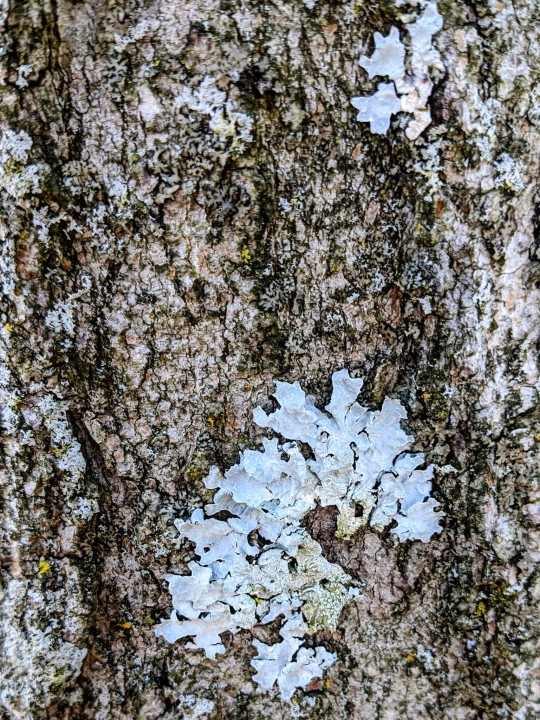
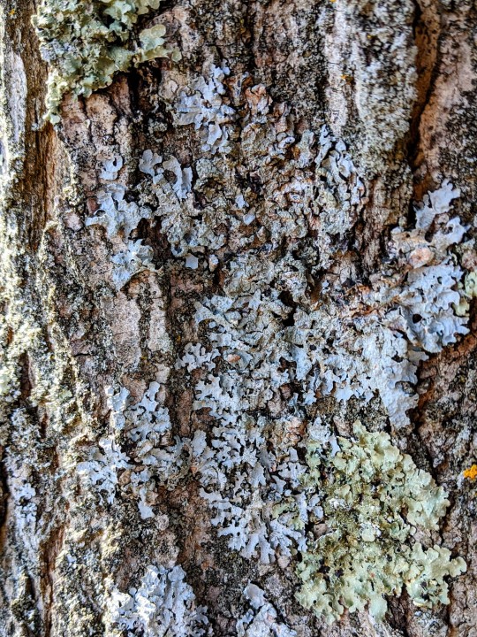
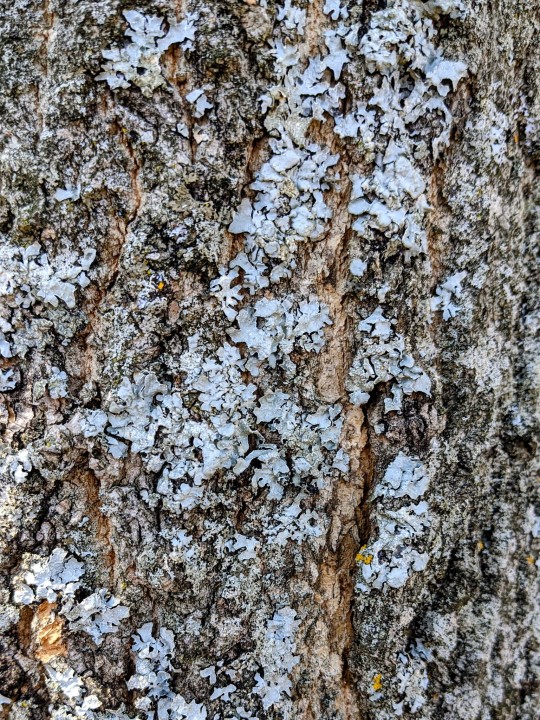
here are some other lichens i recently identified: the light grey flat branching lichen is called parmelia sulcata (common shield lichen) and it produces reddish-brown dye using the boiling water method. various indigenous peoples of north america use it as a medicine; the metis have used it to soothe the gums of teething babies and the wsanec people use it for a variety of ailments depending on the tree it was harvested from. the very similar looking pale green lichen is called flavoparmelia caperata (common greenshield lichen) and it makes a very pale yellowish-brown dye using the boiling water method.
i'm still learning and have yet to actually collect lichen for dyeing as i've been primarily practicing identification and learning more about the various dyeing processes and the chemistry of it in my free time so any of my identifications could be wrong but i feel fairly confident in them overall.
#long post#lichen#mycology#tex help#deepest apologies to anyone scrolling past this monstrosity of a post <3
360 notes
·
View notes
Text
Foliose coral garden
17 notes
·
View notes
Text
Carala: The Coastal Pokemon, water-type, first stage, a slug-like, bright green pokemon covered in rings of many long, neon purple tentacles, eyestalks are yellow-orange, “The regenerative abilities of Carala are lauded across the ?? region. Even if predators eat all the tentacles from it’s back, this pokemon will bounce back in a week or so.” “Carala live in large herds in an attempt to deter predators. The sight of these herds through the water, glowing masses of green and purple, have lead to myths of giant Carala living in the depths of the sea.”, evolves at lvl 24
Ogreef: The Coastal Pokemon, water/rock, final stage, a grey, ungulate-like creature made of stone, long-legged, tailless, sides and upper legs bare colonies of rust-red table coral, neck and back show Carala-like mane, head is dominated by almost flower-like growth of yellow-orange foliose coral, “Carala’s eyes have disappeared, growing and hardening into a singular sensory organ through which Ogreef experiences the world around it. It is unknown how this pokemon eats.” “Despite only having the one rock-like sensory organ on their head, Ogreef appear to have very heightened senses. Drops in water quality cause this usually sedentary pokemon to migrate, leaving hardier Carala in their wake.”
2 notes
·
View notes
Photo

Foliose Coral by Rosekampoong Foliose Coral
27 notes
·
View notes
Photo

A hard coral assemblage showcasing common growth forms: branching, massive, foliose, and solitary or mushroom. Year: 2016 Adobe Photoshop and Wacom Intuos
2 notes
·
View notes
Text
Expedition confirms Panaon Island’s rich marine biodiversity
#PHnews: Expedition confirms Panaon Island’s rich marine biodiversity
TACLOBAN CITY – Environmental group Oceana has reported rich marine biodiversity in Panaon Island in Southern Leyte as they embark on a 22-day expedition in the bid to help protect and sustainably manage the island’s underwater resource.
In a diary shared by Danny Ocampo, Oceana senior campaign manager, he said the corals in Panaon Island are “extensive and quite good” compared to other dive sites he has been to.
“Coral cover was very good in the shallows and as we went deeper between two coral outcrops, it was decorated with lots of crinoids, soft corals, and sea fans,” Ocampo said.
Among the marine species seen are octopuses, black tip sharks, batfishes, eel, sea turtles, sea snakes, large chevron barracudas, giant trevallies, and big red snappers, among others.
“In one of the marine protected areas, the first thing we saw upon entering the water was a huge shallow area of unbroken foliose coral extending up to the deeper part of the reef,” he added.
Ocampo is one of the experts deployed by Oceana for a 22-day expedition to assess the marine biodiversity of the island in Southern Leyte.
Oceana has started the assessment on Oct. 18, deploying a team composed of marine scientists, media production crew, and ship personnel who conducted the first of several dives off the waters of Panaon Island. The activity will conclude on Nov. 8.
They are on-board the expedition ship MV Discovery Palawan.
“Being situated at the Coral Triangle, the Philippines has several coral-rich sites that need attention. One of these is Panaon Island in Southern Leyte. While studies show a decline in coral cover in the Philippines, this particular site stands out and gives hope with its high diversity of coral species,” Oceana Philippines said in a statement posted on its website.
The team is currently assessing corals, seagrass, and fish using scientific survey methods.
Leading the expedition is Dr. Badi Samaniego, a seasoned marine scientist, he has done extensive research on reef, fish ecology, and marine protected areas in Galapagos Islands, Coral Triangle, French Polynesia, and the British Indian Ocean Territory, according to Oceana.
All crew and members of the expedition team have undergone coronavirus testing procedures while regular disinfection procedures and basic health protocols are strictly enforced on-board.
Panaon Island is a small island, lying south of Leyte, separated from Dinagat to the east, and Mindanao to the southeast by Surigao Strait. Panaon is about 30 kilometers long from north to south. The largest town is Liloan, which is connected by a bridge to the main Leyte Island. (PNA)
***
References:
* Philippine News Agency. "Expedition confirms Panaon Island’s rich marine biodiversity." Philippine News Agency. https://www.pna.gov.ph/articles/1120518 (accessed November 03, 2020 at 05:48PM UTC+14).
* Philippine News Agency. "Expedition confirms Panaon Island’s rich marine biodiversity." Archive Today. https://archive.ph/?run=1&url=https://www.pna.gov.ph/articles/1120518 (archived).
0 notes
Text


Parmeliella triptophylla
Black-bordered shingle lichen
I love a bizarre lichen. Just an absolute weirdo. Maybe because I suffer from the universal condition of feeling as if I am just an unknowable freak who can't possibly be like anyone else, but when I look at a lichen oddball like P. triptophylla here, I feel a kinship that supersedes our evolutionary distance. Why is this thing a weirdy? Well first off, look at those weird little baby lobes sitting somewhere between a squamule and lobule. Cause they are like, leaf-shaped with their upturned, indented margins, but are all spread out on top of that blue-black hypothallus (undifferentiated mycelium base connecting the thallus) so they look like a foliose lichen that's been pulled apart into little puzzle pieces. And then the lobes being a shiny gray-blue to brown with coral-like isidia along the margins to boot. It's just not like a lot of other lichens out there. It will occasionally produce apothecia with a red-brown to blackish disc and a pale margin. To top it all off, P. triptophylla is a cyanolichen (only about 10% of lichens are), and has a nostoc cyanobacteria as its photobiont partner. It grows on the bark of old trees and mossy rocks in humid, cool-temperate to boreal-montane deciduous forests across the northern hemisphere.
images: source | source | source
info: source | source | source
#lichen#lichens#lichenology#lichenologist#mycology#fungus#fungi#symbiosis#symbiotic organisms#cyanobacteria#ecology#biology#botany#bryology#systematics#taxonomy#life science#environmental science#natural science#nature#naturalist#beautiful nature#weird nature#I'm lichen it#Parmeliella triptophylla#Parmeliella#lichen a day#daily lichen post#trypo#trypophobia
22 notes
·
View notes
Text
Ecosystem Description

Coral reefs are unique ecosystems located underwater, specifically in salt water. They’re known for their many species of fish and reef-building corals that a re known as hermatypic because they have hard calcium carbonate skeletons. There are many species of corals, but they are all categorized by shape: Massive, Encrusting, Branching, and Foliose.
Source
Shimokawa, Shinya, et al. “Thermodynamics of Coral Diversity - Diversity Index of CoralDistributions in Amitori Bay, Iriomote Island, Japan and Intermediate Disturbance Hypothesis.” IntechOpen, IntechOpen, 21 Dec. 2015, https://www.intechopen.com/books/recent-advances-in-thermo-and-fluid-dynamics/thermodynamics-of-coral-diversity-diversity-index-of-coraldistributions-in-amitori-bay-iriomote-isla.
0 notes
Text
Revitalizing Biodiversity with 3D-Printed Coral Tiles
Following the 3D-printed footsteps of previous efforts to save coastal marine biodiversity utilizing technology and biomimicry, architects and marine scientists at the University of Hong Kong (HKU) have jointly developed a novel method for coral restoration making use of specially designed 3D-printed artificial ‘reef tiles’ for attachment by corals to enhance their chance of survival in the Hoi Ha Wan Marine Park in Hong Kong waters.
A project commissioned by the Agriculture, Fisheries and Conservation Department (AFCD) to combat bio-erosion of essential coral habitat home to more than 120 fish species, architects from HKU’s Robotic Fabrication Lab, under the Fabrication and Material Technologies Lab of the Faculty of Architecture, and marine scientists from the Swire Institute of Marine Science (SWIMS) of the Faculty of Science at HKU collaborated to develop reef tiles printed with intricate detailing optimized to invite live coral attachment.
The reef tiles were 3D-printed using reef-safe terracotta, using a robotic 3D clay printing method before firing each piece at 1,125 degrees Celsius to eventually be seeded with coral fragments. The finished pieces were then introduced within three selected sites across the Hoi Ha Wan Marine Park in Hong Kong waters.
Though beautiful in their own right, the intricate patterns of each tile aren’t merely aesthetic, but designed to represent different growth forms of branching patterns of three endemic coral species – staghorn, massive brain and foliose plating colony forms – and optimize restoration of the ailing coral communities.
The project printed out 128 pieces of reef tile, covering roughly 40 square meters in total. If proven successful, the team plans to expand their collaboration to new designs to promote seabed restoration in the region in other ways across the ocean floor landscape. Additional information about the project is over at HKU Architecture.
via http://design-milk.com/
from WordPress https://connorrenwickblog.wordpress.com/2020/09/11/revitalizing-biodiversity-with-3d-printed-coral-tiles/
0 notes
Text
Types of Coral
Whilst there are many different types of corals, they generally fall into one of eight different categories...

Massive corals: these are the mainstay of most reef and are boulder shaped. This category varies in size from the size of a normal egg, to and enormous two story house. They are very slow growing.

Foliose corals: these are again among some of the most recognisable corals and are characterised by broad plate-like flaps that stick out from the underlying coral substrate.

Table coral: again one of the most easily recognisable corals, characterised by a rising trunk and multiple fused branches that spread horizontally to create a table-like structure. These corals are one of the most commonly associated with coral bleaching.

Encrusting corals: These corals get their name from the fact that they form a crust on a substrate, and spread across the surface creating a thin colourful surface.

Elkhorn corals: as the name suggests, these types of corals resemble the branched horns of elk or deer. They tend to protrude vertically with many several off shooting side branches. The most common of these is the staghorn coral.

Digitate corals: this category of corals resembles fingers or large clumps of cigars, that have no other branches. They grow vertically and are generally not as common as most other types of corals.

Mushroom corals: as their name would suggest, these resemble the cup of a mushroom.

Branching corals: this type is most commonly associated with the most colourful small corals, and are made up of a tree-like structure with multiple branches and secondary branches.
0 notes
