#atomic microscope
Explore tagged Tumblr posts
Visit Tumblr Blog
Explore Tumblr blogs with no restrictions, modern design and the best experience.
Last Seen Tumblr Blogs


Fun Fact
Tumblr has 16.74 million mobile monthly users in the US.
Text
⚠️Vote for whomever YOU DO NOT KNOW⚠️‼️
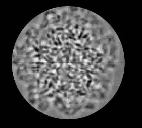

#ultimate obscure blorbo#polls#Round II#The Beast Homeworld#Homeworld: Cataclysm#can someone who knows the beast tell me what exactly im looking at?#is the beast only viable with atomic microscopes?#The Blok#Lego Nexo Knights
15 notes
·
View notes
Text
Earlier this week, I found out about a lit course that piqued my interest, but couldn't take unless I somehow raised 300 dollars in a week LMFAO so I just copied their syllabus and bibliography, now I'll recreate it. I'm gonna start with the book "Historia natural de los cuentos de miedo" written by Rafael Llopis, who seems to have been an influential Spainard critic and essayist who was also a PSYCHIATRIST LMAO like ok? I really need to read on the history of psy-chology-chiatry, but not yet lol
#you're welcomed to join me if u want#thoughts log#i actually did have to take a mandatory psychology 101 class in my last year of hs that included a small section on the history of it#but i was not going to school anymore lol I did read that unit to prepare to accredit it in exam but idr anything other than#the biggest difference between psychology/psychiatry and other sciences is that it lacked foundational paradigms#for example— you get the cell‚ microscopic pathogens/agents‚dna for biology#around which a lot of the existing body of knowledge purportedly got reorganized#there's the atom and subatomic particles for chemistry etc.#there's no such a thing for psych stuff and there's whole anti-psych books organized around this fact lol#I also need to read Kuhn's History of the scientific revolutions tbh lol sigh...
5 notes
·
View notes
Text
hey remember that sparkeater thing i mentioned
:)
fair warning, it's about sparkeaters and shit so like...expect descriptions of robot violence and gore lol
in addition, as i am a whore, there's gonna be smashin at some point too
i know it just says getaway/atomizer rn but...
...getaway's gonna end up getting passed around the crew LOL
#transformers#transformers idw#transformers mtmte#transformers fanfiction#valveplug#sparkeater au#transformers getaway#transformers atomizer#guest starring perceptor's wrecker trauma :D!#i love to torture my favorite microscope uwu#also featuring zef forcibly shoving organic/biological dynamics onto mechanical entities
8 notes
·
View notes
Text
ah piss, wheel out the sign, kronk
[Enter stage left: illuminated sign w obscenely large text and obnoxious squeaky trolley wheels that reads: "THIS USER IS SO STUPID THEY TOOK THIS LONG TO REALISE THAT THE BIT IN RENJU OKIURA'S BACKSTORY ABOUT ACTING AS LOOKOUT FOR THE YAKUZA AT THEIR STRING OF MURDERS UNDER COERCION IS A LESS THAN SUBTLE METAPHOR FOR HIS ROLE IN THE FAILED NUCLEAR FAMILY AND THE CYCLE OF ABUSE. IT'S LITERALLY ALL ABOUT COMPLICITY UNDER DURESS. BLOODY HELL. BOTTOM TEXT."]
#if you can get one of those huge microscopes that can see atoms maybe you can find my final brain cell#my brain is doing the velma 'my glasses my glasses! i cant see without my glasses!' gag#aitsf#aitsf spoilers#renju okiura#at least one day i might be remembered for being comically inept
13 notes
·
View notes
Text

me whenever i see ppl talk about their muses & pour so much love & thought into them
#the passion ppl have for their faves is so !!!#love to see it#picking them apart atom by atom#observing them under a microscope like the creatures they are#``ooc.#``mobile.
2 notes
·
View notes
Text

A Journey into the World of Microscopy: From Humble Beginnings to High-Tech Magnification
The science of looking into the hidden invisible Microscopy has transformed our understanding of the world around us. It can explore the universe beyond the reach of our naked eyes, with complex cellular structures, red blood cells, viruses and other viruses and microorganisms taking on amazing perspectives
The history of the microscope is a fascinating story of human curiosity, scientific genius, and relentless exploration. From the humble beginnings of simple magnifying glasses to the sophistication of modern electronic microscopes, the invention of microscopes has shaped our understanding of the microscopic world
In the 1600s, Dutch opticians such as Hans and Zachary Janssen are credited with inventing the first microscope. Known for this hybrid microscope, many lenses were used to magnify objects up to 30 times.At the end of the 17th century, Antony van Leeuwenhoek, Dutch draper some changed our perception of thumbnails. Armed with a well-made single-lens microscope, and explored the hidden reaches of nature. In 1674, Leeuwenhoek discovered microorganisms in lake water, which he aptly named “animalcules”. His discovery laid the foundations of biology and inspired generations of scientists. This incredible feat allowed him to uncover a hidden universe – the first sightings of bacteria, red blood cells, and other microorganisms.
Formation of the scientific environment (17th-19th centuries): Leeuwenhoek’s discoveries boosted scientific research. Robert Hooke, an English scientist, established these developments. In 1665, his book "Micrographia" recorded his observations with a compound microscope. Notably, the term "cell" was coined by Hooke when he examined cork tissue, laying the foundation for cell biology.Microscope systems flourished throughout the 18th and 19th centuries Joseph Lister and other scientists addressed the limitations of the early lenses, introducing improvements that reduced image distortion.
Beyond the Limits of Light: The Beginning of the New Age (19th-20th century): As the 19th century progressed, the limitations of optical microscopy became apparent and scientists yearned for a tool which can go deeper into cells. This research culminated in the development of the electron microscope in the 1930s. The 20th century was revolutionary with the invention of the electron microscope. Unlike light microscopes, which use visible light, electron microscopes use electron beams to achieve much higher magnification.Formation of the scientific environment (17th-19th centuries): Leeuwenhoek’s discoveries boosted scientific research. Robert Hooke, an English scientist, established these developments. In 1665, his book "Micrographia" recorded his observations with a compound microscope. Notably, the term "cell" was coined by Hooke when he examined cork tissue, laying the foundation for cell biology.Microscope systems flourished throughout the 18th and 19th centuries Joseph Lister and other scientists addressed the limitations of the early lenses, introducing improvements that reduced image distortion.
Beyond the Limits of Light: The Beginning of the New Age (19th-20th century): As the 19th century progressed, the limitations of optical microscopy became apparent and scientists yearned for a tool which can go deeper into cells. This research culminated in the development of the electron microscope in the 1930s. The 20th century was revolutionary with the invention of the electron microscope. Unlike light microscopes, which use visible light, electron microscopes use electron beams to achieve much higher magnification.
In the 1930s, German experts Max Knoll and Ernst Ruska made the first electron microscope. This tool let us see tiny things like cells and even atoms by using electron beams, not light, getting images many times bigger. This cool invention showed us the tiny parts inside cells, viruses, and stuff too small to see before. The 1900s brought even more cool microscopes. New kinds like phase-contrast and confocal microscopy let scientists look at live cells without using stuff that could hurt them. Now, the world of looking at tiny things is getting even better. Today, we have high-tech microscopes that use computers and lasers. These let us see and even change tiny things in ways we never could before.
Modern Microscopy's Diverse Arsenal - Today, the field of microscopy boasts a diverse range of specialized instruments, each tailored to address specific scientific needs. Here's a glimpse into some remarkable examples:
Scanning Electron Microscope (SEM): Imagine a high-tech camera that captures images using a beam of electrons instead of light. That's the essence of a SEM. By scanning the surface of a sample with a focused electron beam, SEMs generate detailed information about its topography and composition. This makes them ideal for studying the intricate structures of materials like insect wings, microchips, and even pollen grains.
Transmission Electron Microscope (TEM): While SEMs provide exceptional surface detail, TEMs take us a step further. They function by transmitting a beam of electrons through a very thin sample, allowing us to observe its internal structure. TEMs are the go-to instruments for visualizing the intricate world of viruses, organelles within cells, and macromolecules like proteins.
Confocal Microscopy: Ever wished to focus on a specific layer within a thick biological sample and blur out the rest? Confocal microscopy makes this possible. It utilizes a laser beam to precisely illuminate a chosen plane within the sample, effectively eliminating information from out-of-focus regions. This allows researchers to create sharp, three-dimensional images of cells, tissues, and even small organisms.
Atomic Force Microscopy (AFM): This technique takes a completely different approach, venturing into the realm of physical interaction. AFM employs a tiny cantilever, akin to a microscopic feeler, to physically scan the surface of a sample. By measuring the minute forces between the cantilever and the sample's surface, AFM can map its topography at an atomic level. This provides invaluable insights into the properties of materials at an unimaginable scale, making it crucial for research in fields like nanotechnology and surface science.
Fluorescence Microscopy: Imagine illuminating a sample with specific wavelengths of light and observing it glowing in response. That's the essence of fluorescence microscopy. This technique utilizes fluorescent molecules or tags that bind to specific structures within a cell or tissue. When excited by light, these tags emit their own light, highlighting the target structures with remarkable clarity. This allows researchers to visualize specific proteins, DNA, or even pathogens within biological samples.
Super-resolution Microscopy (SRM): Overcoming the limitations imposed by the wavelength of light, SRM techniques like STED (Stimulated Emission Depletion) and PALM (Photoactivated Localization Microscopy) achieve resolutions surpassing the diffraction limit. This allows researchers to visualize structures as small as 20 nanometers, enabling the observation of intricate cellular machinery and the dynamics of individual molecules within living cells.
Cryo-Electron Microscopy (Cryo-EM): This powerful technique takes a snapshot of biological samples in their near-life state. Samples are rapidly frozen at ultra-low temperatures, preserving their native structure and minimizing damage caused by traditional fixation methods. Cryo-EM has been instrumental in determining the three-dimensional structures of complex molecules like proteins and viruses, providing crucial insights into their function and potential drug targets.
Correlative Microscopy: Combining the strengths of multiple microscopy techniques, correlative microscopy offers a comprehensive view of biological samples. For instance, researchers can utilize fluorescence microscopy to identify specific structures within a cell and then switch to electron microscopy to examine those structures in high detail. This integrated approach provides a deeper understanding of cellular processes and their underlying mechanisms.
Light Sheet Microscopy (LSM): Imagine illuminating a thin slice of a sample within a living organism. LSM achieves this feat by focusing a laser beam into a thin sheet of light, minimizing photobleaching and phototoxicity – damaging effects caused by prolonged exposure to light. This allows researchers to observe dynamic processes within living organisms over extended periods, providing valuable insights into cellular behavior and development.
Expansion Microscopy (ExM): This innovative technique physically expands biological samples by several folds while preserving their structural integrity. This expansion allows for better resolution and visualization of intricate cellular structures that would otherwise be difficult to distinguish using traditional microscopy methods. ExM holds immense potential for studying the organization and function of organelles within cells.
Scanning Near-Field Optical Microscopy (SNOM): This innovative technique pushes the boundaries of resolution by utilizing a tiny probe that interacts with the sample at an extremely close range. SNOM can not only image the surface features of a sample with exceptional detail but also probe its optical properties at the nanoscale. This opens doors for research in areas like material science and photonics, allowing scientists to study the behavior of light at the interface between materials.
X-ray Microscopy: Stepping outside the realm of light and electrons, X-ray microscopy offers unique capabilities. By utilizing high-energy X-rays, this technique can penetrate deep into samples, making it ideal for studying the internal structure of dense materials like bones and minerals. Additionally, it allows for the visualization of elements within a sample, providing valuable information about their distribution and composition.
From revealing the building blocks of life to aiding in the development of new medicines, the microscope has played an undeniable role in shaping our scientific understanding. As technology continues to evolve, one can only imagine the future breakthroughs this remarkable invention holds in unveiling the secrets of our universe, both seen and unseen. These advancements hold the potential to revolutionize our understanding of biological processes, develop new materials with extraordinary properties, and ultimately pave the way for breakthroughs in medicine, nanotechnology, and countless other fields. As we continue to refine and develop novel microscopy techniques and the future holds immense promise for further groundbreaking discoveries that will undoubtedly revolutionize our perception of the world around us.
#science sculpt#life science#science#molecular biology#biology#biotechnology#artists on tumblr#microscopy#microscope#Scanning Electron Microscope#Transmission Electron Microscope#Confocal Microscopy#Atomic Force Microscopy#Fluorescence Microscopy#Expansion Microscopy#X-ray Microscopy#Super-resolution Microscopy#Light Sheet Microscopy#illustration#illustrator#illustrative art#education#educate yourself#techniques in biotechnology#scientific research#the glass scientists#scientific illustration#scientific advancements
7 notes
·
View notes
Text
started thinking about how D.U.F.U.S. works on a technological level and now i'm coming up with headcanons at speeds not yet known to man
#delete later#he runs on an atomic battery#quarkanite alloy = diamondium + graphene#nearly indestructible (but not 100%)#he's made of billions of microscopic nano-plates that can bend in any way or direction#basically a jewel of technology#but if he does break. oh boy#quarkanite is incredibly hard to make (not too mention of rare diamondium is)#his internals are... weird. the other bots have NO IDEA how to work on him#and finding a new battery once his current one runs out is gonna be a nightmare#quark was an absolute genius and a visionnary. it's a shame that accidentally fusing himself with a critter duck has been progressively an#continuously eating away at his physical and intellectual abilities
3 notes
·
View notes
Text
Agilent atomic force microscope for nanoscale imaging
Molecular Imaging presents the Agilent atomic force microscope—a top-tier solution for researchers seeking high-resolution nanoscale imaging. Designed with precision and versatility in mind, this tool empowers scientists to visualize and measure surfaces with atomic-level accuracy. Built for reliability and ease of use, it's a powerful asset in materials science, biology, and electronics research. Trust Molecular Imaging for industry-leading innovation and performance in atomic force microscopy.
0 notes
Text
Atomic Force Microscope (AFM) Market Size, Share, Trends, Demand, Future Growth, Challenges and Competitive Analysis
"Executive Summary Atomic Force Microscope (AFM) Market : Data Bridge Market Research analyses that the global atomic force microscope (AFM) market which was USD 504.60 million in 2022, is expected to reach USD 798.15 million by 2030, and is expected to undergo a CAGR of 5.9% during the forecast period of 2023 to 2030.
A credible Atomic Force Microscope (AFM) Market report provides with the relevant information about the niche and saves lot of time that may otherwise get wasted for decision making. A premium market research report acts as an innovative solution for the businesses in today’s changing market place. The report offers a thorough synopsis on the study, analysis and estimation of the market and how it is impacting the industry. This industry analysis report is built by keeping in mind businesses of all sizes. The world class Atomic Force Microscope (AFM) Market report is generated by thoroughly understanding business environment which best suits the requirements of the client.
The top notch Atomic Force Microscope (AFM) Market research report offers an array of insights about industry and business solutions that will support to stay ahead of the competition. A systematic investment analysis is also underlined in this widespread report which forecasts impending opportunities for the market players. The persuasive Atomic Force Microscope (AFM) Market report is an outcome of persistent and numerous efforts lead by knowledgeable forecasters, innovative analysts and brilliant researchers who carry out detailed and diligent research on different markets, trends and emerging opportunities in the consecutive direction for the business needs.
Discover the latest trends, growth opportunities, and strategic insights in our comprehensive Atomic Force Microscope (AFM) Market report. Download Full Report: https://www.databridgemarketresearch.com/reports/global-atomic-force-microscope-afm-market
Atomic Force Microscope (AFM) Market Overview
**Segments**
- Based on Offering: AFM Systems, AFM Probes, AFM Software - Based on Grade: Industrial Grade AFM, Research Grade AFM - Based on Type: Contact Mode AFM, Non-Contact Mode AFM, Tapping Mode AFM
The global Atomic Force Microscope (AFM) market is segmented based on offering, grade, and type. The offering segment includes AFM systems, AFM probes, and AFM software. AFM systems are the main components and most significant part of the market as they are used for imaging and analyzing samples at the nanoscale level. AFM probes are essential for the measurement process, and AFM software helps in controlling and analyzing the data obtained from AFM systems. In terms of grade, the market is divided into industrial grade AFM and research grade AFM. Industrial grade AFMs are designed for applications in industries such as semiconductor, materials science, and nanotechnology, whereas research grade AFMs are more commonly used in academic and research institutions. Lastly, based on type, the market is categorized into contact mode AFM, non-contact mode AFM, and tapping mode AFM, each offering specific functionalities and advantages for different applications.
**Market Players**
- Bruker Corporation - NT-MDT Spectrum Instruments - Park Systems Corp. - Hitachi High-Technologies Corporation - Oxford Instruments - WITec GmbH - Nanonics Imaging Ltd. - A.P.E. Research - Kosi Analytical - Nanosurf AG
The global Atomic Force Microscope (AFM) market consists of several key players that contribute significantly to market growth and innovation. Some of the prominent market players in the AFM industry include Bruker Corporation, NT-MDT Spectrum Instruments, Park Systems Corp., Hitachi High-Technologies Corporation, Oxford Instruments, WITec GmbH, Nanonics Imaging Ltd., A.P.E. Research, Kosi Analytical, and Nanosurf AG. These companies offer a wide range of AFM systems, probes, software, and services to cater to the diverse needs of customers across different industries and research domains. Continuous investments in research and development, strategic partnerships, and technological advancements are key strategies adopted by these market players to maintain their competitive edge in the global AFM market.
The global Atomic Force Microscope (AFM) market is experiencing significant growth due to the increasing demand for nanotechnology applications across various industries such as semiconductor, materials science, life sciences, and electronics. AFM systems are essential tools for studying and manipulating materials at the nanoscale level, enabling researchers and scientists to understand surface properties, mechanical characteristics, and chemical compositions of samples with high precision. The advancements in AFM technology, such as higher resolution imaging, faster scanning speeds, and enhanced automation features, are driving the adoption of AFM systems worldwide.
One of the key trends influencing the AFM market is the integration of artificial intelligence (AI) and machine learning algorithms into AFM software for improved data analysis, image processing, and automated measurement capabilities. This integration allows for faster data interpretation, reduction of user error, and optimization of experimental workflows. Additionally, the development of multimodal AFM systems that combine different imaging modes, such as tapping mode and contact mode, into a single platform is gaining traction in the market. These multimodal AFM systems offer users the flexibility to perform a wide range of imaging and characterization techniques on a single instrument, enhancing efficiency and versatility in research and industrial settings.
Moreover, the growing focus on sustainability and environmental impact is influencing the adoption of AFM systems for studying renewable energy materials, environmental samples, and green technologies. AFM techniques are being employed to analyze the surface properties of materials used in solar panels, batteries, and fuel cells, contributing to the development of clean energy solutions and eco-friendly products. The increasing investment in research and development activities related to AFM technology, including sensor miniaturization, automation, and data analytics, is expected to drive further innovation and market expansion in the coming years.
In terms of market competition, the key players in the AFM industry are continuously striving to enhance their product offerings, expand their global presence, and strengthen their customer relationships to gain a competitive advantage. Strategic collaborations, acquisitions, and product launches are common tactics employed by market players to stay ahead in the highly competitive AFM market. As the demand for nanoscale imaging and analysis solutions continues to rise across diverse end-user sectors, the AFM market is poised for sustained growth and technological evolution. The integration of advanced imaging techniques, such as correlative microscopy and spectroscopy, with AFM systems is expected to open up new opportunities for research, innovation, and commercial applications in the future.The global Atomic Force Microscope (AFM) market is a dynamic and competitive landscape driven by the increasing demand for nanotechnology applications across various industries. With the rise of advanced imaging and analysis solutions, such as AFM systems, the market players are continuously innovating to stay ahead in the competitive market. The integration of artificial intelligence (AI) and machine learning algorithms into AFM software is a key trend that is reshaping the market. This integration offers enhanced data analysis, image processing, and automated measurement capabilities, leading to faster data interpretation and optimized experimental workflows. Additionally, the development of multimodal AFM systems is gaining traction as it allows users the flexibility to perform various imaging and characterization techniques on a single platform, boosting efficiency in research and industrial settings.
Furthermore, the focus on sustainability and environmental impact is influencing the adoption of AFM systems for studying renewable energy materials and green technologies. AFM techniques are being utilized to analyze the surface properties of materials used in clean energy solutions, contributing to the development of eco-friendly products. This trend aligns with the increasing investment in research and development activities related to AFM technology, including sensor miniaturization, automation, and data analytics, driving further innovation in the market.
Market players in the AFM industry are actively engaged in enhancing their product offerings, expanding their global presence, and strengthening customer relationships to gain a competitive edge. Strategic collaborations, acquisitions, and product launches are common strategies deployed by market players to maintain their market position. As the demand for nanoscale imaging and analysis solutions continues to grow across diverse sectors, the AFM market is poised for sustained growth and technological evolution. The integration of advanced imaging techniques, such as correlative microscopy and spectroscopy, with AFM systems is expected to unlock new opportunities for research, innovation, and commercial applications in the future.
In conclusion, the global AFM market is characterized by innovation, technological advancements, and strategic partnerships driving growth and competition among market players. The increasing adoption of AI, machine learning, and multimodal AFM systems, along with the focus on sustainability and environmental considerations, are shaping the future trajectory of the market. With continuous developments in AFM technology and applications, the market is set to witness further expansion and evolution in the coming years, opening up exciting possibilities for research, industry, and scientific advancements.
The Atomic Force Microscope (AFM) Market is highly fragmented, featuring intense competition among both global and regional players striving for market share. To explore how global trends are shaping the future of the top 10 companies in the keyword market.
Learn More Now: https://www.databridgemarketresearch.com/reports/global-atomic-force-microscope-afm-market/companies
DBMR Nucleus: Powering Insights, Strategy & Growth
DBMR Nucleus is a dynamic, AI-powered business intelligence platform designed to revolutionize the way organizations access and interpret market data. Developed by Data Bridge Market Research, Nucleus integrates cutting-edge analytics with intuitive dashboards to deliver real-time insights across industries. From tracking market trends and competitive landscapes to uncovering growth opportunities, the platform enables strategic decision-making backed by data-driven evidence. Whether you're a startup or an enterprise, DBMR Nucleus equips you with the tools to stay ahead of the curve and fuel long-term success.
Key Coverage in the Atomic Force Microscope (AFM) Market Report:
Detailed analysis of Global Atomic Force Microscope (AFM) Marketby a thorough assessment of the technology, product type, application, and other key segments of the report
Qualitative and quantitative analysis of the market along with CAGR calculation for the forecast period
Investigative study of the market dynamics including drivers, opportunities, restraints, and limitations that can influence the market growth
Comprehensive analysis of the regions of the Atomic Force Microscope (AFM) Marketand their futuristic growth outlook
Competitive landscape benchmarking with key coverage of company profiles, product portfolio, and business expansion strategies
Browse More Reports:
Global Digital Dose Inhaler Market Global Automotive In-Cabin Air Quality Improvement Solutions Market Global Cognitive Computing Market Global Hearing Screening Diagnostic Devices Market Global Injector Pen Polymers Market North America Small Molecule Sterile Injectable Drugs Market Global Propionic Acid Market Global Solid-State Solar Cell Market Global Wastewater Secondary Treatment Equipment Market Global Ride Sharing Market Europe Mass Notification Systems Market Global Sesame Seed Oil Market Global q-PCR Reagents Market Global Fuse Market Global Silanes and Silicones Market Global Holter ECG Market Europe Biometrics in Government Market Global Digital Pet Care Products and Services Market Europe Microgrid Market Global Packaging Suction Cup Market Global Brown Rice Syrup Market Global Flooring Market Global Urgent Care Apps Market Global Specimen Collection Kit Market Global Security Fencing Market Global Enzyme Replacement Therapy Market Europe Lung Cancer Diagnostics Market Global Flexible Packaging Paper Market Global Yield Monitoring System Market Global Microalgae in Feed Market Global Secure Digital (SD) Cards Near Field Communications Market
About Data Bridge Market Research:
An absolute way to forecast what the future holds is to comprehend the trend today!
Data Bridge Market Research set forth itself as an unconventional and neoteric market research and consulting firm with an unparalleled level of resilience and integrated approaches. We are determined to unearth the best market opportunities and foster efficient information for your business to thrive in the market. Data Bridge endeavors to provide appropriate solutions to the complex business challenges and initiates an effortless decision-making process. Data Bridge is an aftermath of sheer wisdom and experience which was formulated and framed in the year 2015 in Pune.
Contact Us: Data Bridge Market Research US: +1 614 591 3140 UK: +44 845 154 9652 APAC : +653 1251 975 Email:- [email protected]
Tag
"
0 notes


Text
#laboratory equipment supplier in india#laboratory instruments#labindia instruments#semiconductor#biology#marketing#lab equipments#semiconductor devices#Plasma Etcher#Atomic Layer Deposition System#Spectroscopic Ellipsometer#TESCAN AMBER X#Labjal – Lab Net#Atomic Force Microscope
0 notes
Text
Atomic force microscope
An atomic force microscope (AFM) is a high-resolution imaging technique used to observe the surface of materials at the atomic scale. It operates by scanning a sharp probe, typically a few nanometers in diameter, over the surface of a sample.

0 notes
Text
Still my very favorite machine in physics and biotechnology research!
Feeling like a pro in Atomic Force Microscopy

...to be honest I probably should really tell myself that at this point I really am the professional in this method. After 4 years of working with 2 different atomic force microscopes, now I started with a 3rd one, again a new type from a different company.
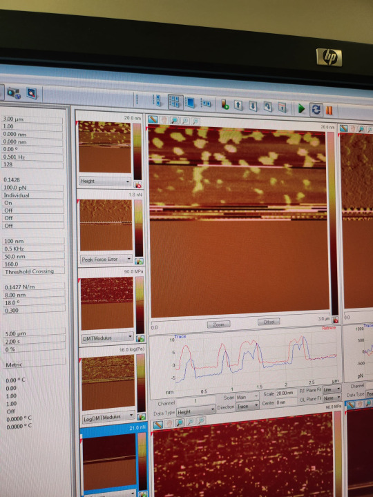
Only after 2 hours of training on the new machine, I could observe membranes of resistant bacteria all by myself. The membranes are the yellow pancakes sitting flat on the dark support. They are less than 8 nm high (0.000000008 m), as is visible in the blue and red profile lines. So it's super tricky to actually see them. Atomic force microscope touches the surface of my membranes and surrounding support with a tiny tip like with a finger and reconstructs the surface topology. On top of the small size, the cellular membranes are super soft so also the touching finger must be super soft to see them without damaging them.
#science#women in science#research#original content#postdoc#stem#biophysics#physics#academia#biology#technique#equipment#microscope#afm#atomic force microscope#cool science
29 notes
·
View notes
Text
Atomic Force Microscope

Atomic Force Microscope features a scan head and sample stage for robust anti-vibration performance, servomotor for precise scanning, and precision laser detection and probe alignment device. Shop online at Labtron.us
0 notes
Text
youtube
This is COOL
1 note
·
View note