#Skull fracture
Explore tagged Tumblr posts
Visit Tumblr Blog
Explore Tumblr blogs with no restrictions, modern design and the best experience.
Last Seen Tumblr Blogs


Fun Fact
There were a total of 171.5 billion posts on Tumblr in 2019.
Note
what happened to sans’ head?
Sans was investigating a viral rot infecting the forests near snowdin. When he got close it seemed mobile, and for a moment; sentient.

#fracturetale#original undertale au#undertale#UT#FT#sans#papyrus#snowdin#digital painting#digital illustration#digital art#digital comic#original comic#artwork#comic#sketch#messy sketch#kill me#sans undertale#sans fracturetale#bros cooked#concussion#skull fracture
3 notes
·
View notes
Text
KAFATASI KIRIKLARI

Kafatası kırığı, kafatası kırığı hakkında bilgiler: Kafatası üzerine düşme, kafaya sert bir cisimle vurma veya kafatasmı çarpma gibi kazalar kafatasındaki kemiklerin kırılmasına sebep olurlar. Bu cins kırıklar bugün trafik kazalarının çoğalmasından dolayı daha fazla görülmektedir. Kafatası kırıkları, beyin zarlarının ve beynin bu kazadan müteessir olup olmadığına göre çeşitli belirtiler gösterirler. Read the full article
3 notes
·
View notes
Text

Stay true to you ✊🏻
“Remember, being yourself is the only way to find out who truly cares and loves you for you.”
— TheGoodVibe
19K notes
·
View notes
Text
what are the surgical options for treating a skull fracture?
Skull fractures can vary in severity, and treatment options depend on factors such as the location and extent of the fracture. Here are some common surgical options for treating skull fractures: Craniotomy: This procedure involves creating a surgical opening in the skull to access the fractured area. Surgeons may use specialized tools to remove or repair damaged bone fragments, relieve pressure…
View On WordPress
0 notes
Text


max + jos verstappen x journal, day three by richard siken / manufacturing by alan r. shapiro / i am an atheist who says his prayers by karl shapiro / a thousand splendid suns by khaled hosseini / the merchant of venice by william shakespeare, and other assorted quotes
articles: x , x , x , x , x , x , x , x , x
this was a tough one, and i did my best to convey the right emotions in every slide. hopefully that comes across!
[ @carbonmono, @28ms28 , @nicaeno, @vesrtapen , @grise-and-rind , @mvlionheart, @dafunzies ]
#jos verstappen was charged as guilty with fracturing a guy's skull with a length of metal pipe at a karting track btw +#if the fact sophie had a restraining order against him (which he violated) wasn't enough#i think about that interview where max told journalists he needed the abuse and he doesn't think he'd be where he is without it 24/7#and that he doesn't actually consider it abuse. because that's all he knew as a kid.#the guy's a piece of shit but then again with who this sport's run by i can't say i'm surprised. so. father son webweave it is#f1#formula 1#formula one#max verstappen#mv1#mv33#f1 web weaving
117 notes
·
View notes
Text
youtube
#youtube#fight#man#road rage#dog#viral video#woman#artificial intelligence#artwork#sketch#fracture#skull
59 notes
·
View notes
Text
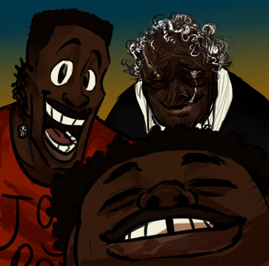
a little dp x dc art ;))

gotham in the ghost zone
#danny phantom#danny fenton#dc x dp#crossover#dp x dc#dp x dc crossover#gotham#ghost zone#just a little thing i made while i was on the painkillers#remember when i got hit with the chair?#well that got infected a little bit and when i went to get it checked out i had a fracture in my skull and neck#and i had just been trying to walk it off for two weeks#so they decided to confine me to bed rest hell with minimal blue light from screens#i absolutely did not listen to the second part#but i’m back now!#yay health
474 notes
·
View notes
Text

Via HeckoGecko
#lizard#reptiles#chameleon#art#comic#HeckoGecko#also you hold them wrong#and their ribcages fracture like a sugar skull
90 notes
·
View notes
Text
Yes I am normal. No I am not looking at types of skull fractures. No I am not searching up the severity and survivability of a depressed skull fracture and the associated injuries of one get out of my house
#for no reason in particular#tw mention of skull fracture#tw gore description#monoma neito#neito monoma#ignore these tags hahahahahhaahaahahahhahahahahahahahahahahahahahahah
12 notes
·
View notes
Text
i bet that even as humans they still refer to themselves as skeletons
#repostober#day 14 i think. i lost count in the catching up lol#undertale#sans#papyrus#gaster#dadster#blacktober#technically i drew this for blacktober but never posted it.. it just sat in my wips folder unsaved as an actual image until a few days ago#i meant to fully shade it and make a nice sky background and give sans lens blur but. alas. (ill probably finish it one day sfgkjf)#anayway i LOVE my designs for the human font family SM#sans has dwarfism and lets his hair naturally grow out and does nothing to take care of it (it is as soft as silk regardless)#papyrus is JACKED and has so many piercings and tattoos cus YOU KNOW HE WOULD IF HE HAD SKIN!!!! he always ALWAYS has a fresh cut and he-#gives me gender envy ANYWAYs-#gaster has monochromacy (monochromatic color blindness) and two sets of dentures#both his legs were amputated so he uses a wheelchair and he has scars that resemble burn scars and no nose#as well as two facial scars that connect to his eyes and mouth- his skull is also fractured under them#hes always wearing his favorite pair of glasses (that his sons bought him)#he loves tea and knitting so much................ he is a grandma#(visually hes lowkey based on my grandpa hgdfjd)#they are all such doofuses and they love each other so much..... papyrus can carry both of them at the same time and often does#when sans randomly falls asleep in public or when gaster has spent the last 17 hours in the librarby
90 notes
·
View notes
Text


Poldark 1.01
#ross poldark#getting his ass kicked#father carne does way more damage imo#ross can't be held responsible for stupidity going forward#since concussion and fractured skull#poldark whump
33 notes
·
View notes
Text


#undertale au#my ocs#sleepyverse#fracture skull point comic#tic toc#eraz#calcite#dream sans#underswap sans#ink sans#utmv au#utmv oc#au sans#muse's art
9 notes
·
View notes
Text
RED ROBIN FATAL BLOW


(Remade because the last one was absolute dog shit so here's a complete remake)
#this is just a triple skull fracture lol#mk#mortal kombat#memes#humor#art#fanart#mortal kombat 11#mk11#mortal kombat 1#mk1#mortal kombat oc#mk oc#mk oc red robin
24 notes
·
View notes
Text
That time Alex kept a YouTube channel & blog in 2010
Here's his accompanying blog and also an Observer article (along with some other fun stuff)

#alex horne#rosie the turtle is my favourite#I'm positive he's gonna live for a long time#He just needs to try a bit harder not to fracture his skull on taskmaster
13 notes
·
View notes
Text
what are the first aid measures for a suspected skull fracture?
When providing first aid for a suspected skull fracture, it is important to remember that immediate medical attention is crucial. However, there are a few steps you can take to assist the person until professional help arrives.What is a skull fracture? First and foremost, call emergency medical services right away. They are equipped to handle these types of emergencies and will provide the…
View On WordPress
0 notes
Text
Skull Fractures: Le Fort I-III
Le Fort fractures are a specific type of midfacial fracture which can be split into three types: Type I, Type II, and Type III. These three types vary in severity and presentation. They are most commonly caused by high-speed or high-impact forces.
Facial and Cranial Anatomy
Le Fort fractures of all types involve different bones which make up the facial structure. You can look up a picture of the skull if you don't know what I'm talking about, but you should have an idea of what's going on from. Anyways, the skull is 22 bones and that's too much to get into right now. Onto important stuff.
The mandible is not part of the skull and isn't part of what makes a Le Fort fracture, though facial trauma may also cause the fracture of this bone. It is also an important part of the facial anatomy and its misalignment with the other bones can help diagnose Le Fort fractures.
Le Fort Type I
Le Fort type I injuries are the least damaging type of Le Fort fracture, though they still cause significant damage to the facial structures. This type of fracture involves the maxilla and pterygoid plates (four structures that are up inside the skull/back of the mouth) primarily. Due to the involvement of the maxilla, it can also be referred to as a trans-maxillary fracture.
There are several mechanisms of injury for this type of fracture. Most commonly, it is caused by a downward blow to the face. This can be the result of a fall, assault (like stomping on someone's head), or motor vehicle accident, which causes the maxilla of the patient to impact a hard surface and detach from the rest of the facial structure.

The downward force on the face causes the fracture of the internal structures of the maxillary sinus as well as the pterygoid plates. The fracture extends to the maxillary antrum, or the antrum of Highmore, which is a large sinus cavity that sits superior to the upper teeth and posteriorly to the nose. All three walls of the sinus are fractured, and the maxillary structures break away in a palate-facial separation.
Type I fractures oftentimes present with swelling in the upper lip and ecchymosis (bruising) to the maxilla and cheeks. The most telling sign of maxillary dissociation is malocclusion of the teeth. This means that the upper teeth are not in alignment with the lower teeth, which can be seen in dissociations and fracture of the maxilla and mandible. The triangular area of the face that comes away from the cranium is made of the maxilla, palate, and pterygoid plates.
The soft tissue damage of this fracture is mostly within the oral cavity, sinuses, and nasal cavities. This can cause the respiratory ability of the patient to become compromised. Because of the damage to the nasal passages and the possibility of fracture of the inner bones of the skull, a nasopharyngeal airway (NPA) device is not recommended. NPAs are devices made of a pliable foam or rubber, which are inserted through the nares, and allow for the linkage of the nasal openings to the pharynx (throat). When heavy facial trauma, especially to the nasal area, is present, NPAs are not indicated. Instead, airway management should be done with an endotracheal tube, which goes into the trachea (windpipe). This bypasses the damage done to the oral or nasal cavities and allows for the establishment of respiratory function.
Other symptoms of the patient should be managed until they can reach a trauma center for further treatment, with the patient's airway, breathing, and cardiovascular function taking precedence.
Le Fort Type II
Type II fractures are more severe than Type I. They involve more facial structures and cause a greater portion of the face to separate from the skull. This type of fracture involves the nasion, which is commonly referred to as the bridge of the nose. It also involves the medial wall of the orbit and the inferior orbital rims. Importantly, this type of fracture does not involve the zygomatic bones (cheek bones).
This fracture is best traced beginning at the nasion. The fracture extends laterally to the medial wall of the orbit and downward into the inferior orbital rim. It then crosses downward into the maxilla. Inside of the structure, the fracture extends superiorly to the hard palate, and ends with the separation of the pterygoid buttress. The section of bone that breaks away from the cranium is pyramidal in structure.

The involvement of the orbit in this fracture increases the severity of this fracture because of the complexity of orbital bones as well as the involvement of the soft tissue of the eyes. The forces which are of a great enough magnitude to fracture this portion of the face can create severe damage to the delicate tissues inside the orbit, including blood vessels and the eyes themselves.
Type II fractures present distinctly from Type I fractures. In Le Fort Type II fractures, there will be mobility in the maxilla, but the nasal region will move with the maxilla away from the other facial structures. The increased severity also opens a possibility of cerebrospinal fluid (CSF) leakage from the nasal passages. CSF leakage can be confirmed using a piece of gauze to absorb some of the fluid. As the CSF dries, it will create a distinct two-ringed figure, which mucous does not. CSF is also very clear and watery compared to mucous, which is yellowish and sticky.
Due to the damage to the nasion and surrounding area, the intercanthal space (the distance between the corners of the eyes) has a high possibility of widening. This will lead to severe deformity of the normal facial structure. Bruising will be significant, with presentation of bruising on both orbital areas, or bilateral periorbital ecchymosis, sometimes called “racoon eyes.”
As with Type I fractures, an NPA is not indicated, particularly when CSF leakage is present. This indicates a fracture deep enough to compromise the integrity of the intracranial space. If an NPA is used in this case, even the soft tip of the device could worsen the intrusion into the cavity or cause severe damage to the sensitive nervous system structures. The airway should be established using an endotracheal tube instead. The patient should be transported with haste to the nearest trauma center.
Le Fort Type III
Type III fractures are the most severe type of Le Fort fracture. This type is also called a full facial-cranial separation. It involves the nasion, two walls of the orbit, the orbital rim, the zygomas, and the pterygoid plates.
The path of this fracture can be traced starting at the nasion. From there it extends laterally through the medial orbital rim and orbit. The fracture extends horizontally across the entire orbit and to the lateral rim. Finally, it extends downwards across the zygomatic arch and through the superior pterygoid plates. This fracture extends bilaterally through both sides of the facial structure5.

Due to the fracture extending horizontally through the orbit, there is a great risk of damage to the soft tissues of the eye. The fracture also extends parallel to the base of the cranium, and will cause a complete separation of the midfacial skeleton from the cranium. Because of this extreme dissociation, there is an increased risk of cerebrospinal fluid leakage. This leakage can result in infection or serious brain injury.
This type of fracture is most typically caused by large amounts of force to the nasion and superior maxilla. This force is of a great enough magnitude to cause a fracture to extend through the entirety of the facial structure in a posterior and downwards direction.
Many signs from Type I and II carry over, including bilateral periorbital ecchymosis, orbital edema, and buccal ecchymosis. However, Type III presents uniquely with a sign commonly called “dish-face deformity.” This condition results from the breakdown of the structure of the face, and results in a lengthened and shallowed face. There may also be a condition known as “orbital hooding” present, which is the drooping of the upper eyelid commonly seen when zygomatic structure is compromised. Battle’s sign may also be seen, which is the bruising of the mastoid region, or the region behind the ear. Enophthalmos, the sinking of the eyes posteriorly into the sockets, may also be seen. This is due to the severe damage to the orbital area. CSF may also leak out of the ears and nasal passages, called CSF otorrhea and CSF rhinorrhea, respectively. Blood may also be within the inner ear in a condition called hemotympanum.
The severe damage to the facial bones, along with intracranial hemorrhaging and CSF leakage will most likely cause a patient with a Type III fracture to be treated as critical. Airway integrity should be maintained with an endotracheal tube, but if the oral airway is compromised, stabilization can be difficult without a tracheostomy. A tracheostomy is a procedure where an opening is made in the neck into the trachea, where a breathing tube can be inserted. This bypasses any oral damage and allows for the respiratory function of the patient to be secured. The patient should be rushed to a trauma center, as the signs presenting give an impression of a condition that can be lethal.
Treatment
As with any trauma patient, those with Le Fort fractures should be stabilized to ensure the continuation of life before any effort is made to repair the damage to the face. This gives the patient’s airway, breathing, and circulatory systems precedence in their care. Once the patient reaches the emergency room of what is hopefully a trauma center, more specialized care can begin. This includes testing, scans, and stabilization.
Computerized tomography (CT) scans are very useful to determine the extent of the fracture and the type. These scans use several x-rays and an interpreting computer system to create several two-dimensional slices of the structures captured. This is important in severe facial injuries, as simple film x-rays do not typically provide enough data to accurately diagnose and see all of the damage done to the face. In Le Fort fractures, the most appropriate type of CT scan ordered is a non-contrast, fine cut scan with axial cuts. Contrast is a substance injected intravenously that allows for a better view of body structures on the scan, but it is not used in this case. Typically, contrast used in trauma cases is for abdominal injuries. The CT scan should be fine cut, which means that the slices of data gathered from the patient are 2 mm in width. This means more can be seen from the scan.
The two main areas of treatment in facial fractures are reduction and fixation. Reduction is the process of putting the structures back where they are meant to sit. Reduction can either be open or closed. Open reduction requires the “opening” of the face through surgical means. Typically, this means that the fracture was too complex to be reset through external manipulation. A closed reduction is done without exposing the bone. This is done externally through manipulation of the structures. Fixation is the process of keeping the bones in the correct place, typically through the use of metal plates, wires, and screws.
Intermaxillary fixation can be used to repair or stabilize the fracture. This should be done after all CT scans are completed. Intermaxillary fixation uses metal pieces screwed to the maxilla to demobilize it. Typically, metal plates are also screwed to the mandible and wire is connected between the mandible and the maxilla. This will reestablish proper occlusion of the teeth. Intermaxillary fixation can be used to stabilize the face while other surgeries are done, or as the treatment itself.
Facial reconstruction is most likely with Type III fractures. In a broad sense, facial reconstruction includes the reduction and fixation detailed previously. However, it also includes more fine and cosmetic reconstruction. After a more severe fracture and the resulting fixation, the skin and formation of the face can be altered from the patient’s original appearance. Due to this, there may be more extensive work required by a plastic surgeon after the initial treatment to repair the bone structure.
The goals of the fracture repair are to reestablish proper structure and restore the integrity of the face. The correct facial projection should be reclaimed, as to correct any deformity or dish-face condition. The sinus cavities should also be repaired so that they have proper function and location within the facial structure. The realignment of the orbital and nasal structures is also important to the overall soundness of the facial structure and function. Finally, malocclusion should be remedied, as proper occlusion of the teeth is necessary for not only facial structure, but the function of the teeth and mouth in chewing food.
Outcomes
The recovery of this type of fracture can be long and difficult, but the ultimate outcome is generally good. Mortality rates of patients with complex facial fractures, which includes Le Fort fractures, are estimated around 11.6%. Mortality of patients with simple facial fractures is estimated around 5.1%. For Le Fort fractures specifically, there are different mortality rates for each type. For Type I, there is a 0% mortality rate. For a Type II fracture, there is a 4.5% mortality rate. For Type III fractures, there is an 8.7% mortality rate.
Besides death, disability can also be the result of Le Fort fractures. The following conditions have been reported as a result of Le Fort fractures: difficulty breathing (31% of patients), difficulty chewing (40% of patients), vision issues (47% of patients), double vision (21% of patients), and excessive tearing and poor eye drainage (37% of patients). These disabilities can greatly affect the ability of the patient to return to normal activities. For Type I and II fractures, 70% of patients were able to return to work. For Type III fractures, only 58% reported being able to return to work. The facial deformities resulting from these fractures and their repair have an impact on the patient’s mental wellbeing. Of patients who had facial surgery, 89.1% reported satisfaction with the outcome of their appearance.
End Notes
Wow this was fucking long, wasn't it? I guess I just got going and couldn't stop. Now you know everything you need to know about Le Fort fractures, which was probably nothing. But anyways, I wanted to include the outcome of severe injuries like these because I feel like in fiction I don't see it enough. Like if you get that fucked up you're gonna need some help for a while. I also think I see too many NPAs in patients that don't need them or shouldn't have them, so I think that might be some valuable information. I'm gonna write on the structure of the nose eventually, but for the love of god don't stick anything up there, especially if someone has head trauma.
Thanks for reading :))
#medicine#med studyblr#medblr#head injury#whump writing#medical writing#fractures#skull fractures#whump#trauma#medical school#med student
5 notes
·
View notes