#RNA Special Feature
Explore tagged Tumblr posts
Note
You flatter me, dear Faust. Is intelligence the quality you find the most attractive in a romantic partner? If you find other features attractive, do let me know. I’m not above teasing you with a few more vague descriptions of myself.
As for my specialization in hematology, I originally did most of my work studying the immune system. Funnily enough, my first big research project was on the neuroimmune system and how RNA viruses affect it. My love for immunology first linked me to immunohematology, and then I also fell in love with looking at the little cells under the microscope.
Your suggestion to leave our schedules open sounds delightful. I'd love to hear if you have any recommendations for our first true experiment. Since you're so well-versed in Neurology, perhaps some somatosensory experimentation is in order.
- L
When a partner can challenge and teach you new things it creates a stimulating and rewarding environment. Knowledge, intelligence, skill are all great qualities, but they mean nothing without passion. People cannot be driven without passion, this makes them seem somewhat bland. So while intelligence is in the top qualities I find most attractive, it needs to be paired with passion. There are several qualities I find intriguing, but there's no need to tease me, I believe your qualities will shine on their own. What qualities do you find most attractive in a romantic partner?
It's clear that you truly enjoy your career. It seems like it was quite the adventure you went on to get to where you are today. It's cute that you're so fascinated by the wonders to be seen under the microscope. If you ever wish to discuss your research, please know that I am always available.
While I enjoy the praise, you give me too much credit. I was simply observing the development of epilepsy and cerebral palsy. Your suggestion of somatosensory is truly alluring. I believe you can achieve great results by using some sensory deprivation. Such as subjecting the person limited vision and hearing. This will enhance perception and reaction. Furthermore, since it will be the first round, let's start simple, vibrations, pressure, and pain. Now, which of us will be the subject for this round?
#ikemen rp#ikemen roleplay#ikevamp faust#ikemen vampire faust#cybird ikemen#ikemen games#ikemen series#ikemen vampire#cybird#faust ikemen#cybird series
2 notes
·
View notes
Text
How to Use an Oligo Analyzer: A Step-by-Step Guide
Regardless of specialization within the oligonucleotide manufacturing industry, getting the synthetic DNA or RNA right is imperative. This is where an Oligo Analyzer is a useful tool of choice. It’s imperative when working on DNA oligo synthesis, custom oligos or other processes such as oligonucleotide drug development that one makes use of an Oligo Analyzer to ensure one determines the properties of short nucleic acid sequences. This is important to ensure that your synthesized oligos have fulfilled the standard for research or therapeutic view.
Step 1: Input the Oligo Sequence
The first thing a user must do to use an Oligo Analyzer is enter the DNA or RNA sequence to be analyzed. Many of the analyzers available for use have an interface through which the sequences may be keyed in by the user or uploaded from the synthesizing software. Make sure that the sequence is correct because mistakes can influence analysis.

Next, your sequence will be analysed by the Oligo Analyzer, which can predict its physical and chemical properties. This includes aspects such as melting temperature (Tm), GC content and molecular weight from the two organisms. Such properties are important when selecting oligos for experiments or oligonucleotide drug candidates.
Step 2: Investigate Melting Temperature (Tm)
A key value when using an Oligo Analyzer is melting temperature. It suggests the temperature, which affects fifty percent of the DNA strands' separation. The Tm plays a significant role in simplifying oligonucleotide manufacturing and the development of drugs by assisting in understanding your oligos' behavior under different experimental conditions.
If the Tm of your Tma is too low then the oligos will not be able to bind to their target sequence efficiently. If it is set high, they might bond, which would complicate matters in the experiment being conducted. Depending on the Tm results you may swap or add some of the nucleotides to design the effective oligos for your study.
Step 3: Evaluate GC Content
There is much more to learn about Oligo Analyzer, and the following steps involve looking at the GC content. This fraction measures the dosages of guanine (G) and cytosine (C) bases in your oligonucleotide. Interestingly, the stability in the oligo increases with the GC percent, thus enhancing binding to any complementary DNA or RNA strand.
Many oligo synthesis companies employ this analysis to ensure the high quality of their products. In any case, extremes in GC content should be avoided, as they can reduce the oligo's efficiency in research or drug substance manufacturing.
Step 4: Search for More Levels
The Oligo Analyzer generates information regarding the secondary structures of compounds in addition to its other features. Unwanted interactions occur within the oligo itself, leading to the folding or looping of the structure. These secondary structures can have detrimental effects that influence the way that the oligo is able to perform, for secondary structures are known to be particularly problematic in sensitive areas of research, such as that of oligonucleotide drugs.

The analyzer can help shift the sequence to decrease these structures, so the oligos should be arranged properly according to their function.
Conclusion
An oligo analyzer must be used to ensure accurate and functional oligos for use in research or therapeutic applications. From the input of the sequence to determining its melting temperature and GC content, all stages contribute to the control of the final product quality. These tools are godsends when engaged in oligo synthesis and design within a research lab or when participating in a CDMO drug development project. Veliter Bio offers a wide range of oligo synthesis and analysis services to guarantee your oligo synthesis process will be accurate from the ground up.
0 notes
Text
Exploring the Power of MAGNA™ Technology

The field of biomedical studies is constantly evolving, driven by the need to recognize the complicated interactions at the molecular level that affect fitness and disorder. One of the most transformative gear in recent years is MAGNA™ Technology, an innovative platform developed by Depixus. This progressive generation is converting how scientists have a look at biomolecular interactions, specifically in terms of RNA-targeting, imparting unparalleled insights that pave the way for next-technology treatments. In this weblog, we’ll dive into the colossal capability of MAGNA™ Technology and explore the way it’s reshaping RNA-focused drug discovery and biomolecular research.
What is MAGNA™ Technology?
At its centre, MAGNA™ Technology allows the special exploration of Magna Biomolecular Interactions-the interactions between proteins, RNA, and other key molecules inside cells. These interactions are vital for cell features and, while disrupted, regularly lead to disorder. Understanding these complex relationships is vital for growing new therapeutics, specifically those targeting unique RNA molecules, which are emerging as key gamers in contemporary drug discovery.
Depixus Magna™ gives the clinical network an effective tool to seize, analyse, and visualise these biomolecular interactions in real time. By offering an excessive-decision, multi-dimensional view of ways biomolecules behave within their local environments, MAGNA™ units the degree for breakthroughs in RNA-concentrated on and other molecular interventions.
The Role of RNA Targeting Technology
The focus on RNA in drug discovery has been growing dramatically in recent years, and for true purpose. RNA performs an important function in gene expression, carrying the genetic instructions from DNA to synthesise proteins. Misregulation or mutation in RNA can result in numerous illnesses, inclusive of cancer and genetic problems.
RNA Targeting Technology is a promising technique that allows researchers to without delay target RNA molecules, in place of the proteins they encode. By intervening on the RNA stage, scientists can prevent the manufacturing of dangerous proteins, thereby addressing the root purpose of certain illnesses. This targeted technique opens up new avenues for treating conditions that have been formerly considered "undruggable."
Here’s where Depixus Magna™ becomes a sport changer. The advanced technology provided by using MAGNA™ lets in for the perfect mapping and look at RNA interactions, assisting researchers perceive how specific RNA molecules interact with proteins or other goals. This insight is important for the development of medication which can modulate or disrupt those interactions, leading to novel therapies for sicknesses starting from neurodegenerative disorders to most cancers.
Magna Biomolecular Interactions Unlocking the Mysteries of Cellular Communication
Magna Biomolecular Interactions are the interactions between diverse biomolecules, which include RNA, DNA, and proteins, that govern the fundamental tactics of lifestyles. These interactions are not best critical for preserving ordinary cell functions but also play key roles inside the onset and progression of illnesses.
Using MAGNA™ Technology, researchers can investigate these interactions in approaches that were previously unimaginable. The ability to have a look at molecular interactions with such depth permits scientists to identify new drug objectives and apprehend how existing drugs have interaction with their objectives at a molecular stage.
For example, in RNA Targeted Drug Discovery, know-how how small molecules or therapeutic dealers have interaction with RNA is important for growing pills that could correctly inhibit or beautify RNA functions. MAGNA™ makes it possible to visualise those interactions, offering insights which could accelerate the discovery of RNA-centred treatment options.
The Impact of MAGNA™ on RNA Targeted Drug Discovery
RNA-centred treatment options have gained excellent momentum, leading to current advances in technology and the developing recognition of RNA’s role in various illnesses. MAGNA™ Technology is poised to accelerate this progress by providing the tools important for deeper exploration of RNA interactions.
In the context of RNA Targeted Drug Discovery, MAGNA™ enables researchers to:
Identify new RNA goals: With its high-resolution evaluation talents, MAGNA™ allows scientists to discover previously unknown RNA molecules or interactions that would serve as drug goals.
Characterise drug-RNA interactions: Understanding how capacity pills bind to or have an effect on RNA is critical for optimising drug applicants. MAGNA™ provides the specified interaction profiles wished for this step.
Optimise RNA-concentrated on pills: By reading how RNA-targeted capsules have an effect on their supposed targets, researchers can refine those treatment options to improve efficacy and reduce side consequences.
The future of drug discovery lies in our potential to target RNA correctly, and Depixus Magna™ Technology is mainly the manner in making this imaginative and prescient a fact.
A Bright Future for Biomolecular Research and Therapeutics
The integration of MAGNA™ Technology into biomolecular research and RNA-centred drug discovery is using a wave of innovation. By allowing scientists to examine Magna Biomolecular Interactions with unparalleled elements, MAGNA™ isn't always best expanding our know-how of how cells speak but also offering new opportunities for developing life-saving treatments.
As the medical network continues to include RNA-targeted cures, the insights provided with the aid of Depixus Magna™ Technology may be instrumental in shaping the future of medicine. Whether it’s through figuring out new drug objectives or optimising present treatments, MAGNA™ is pushing the limits of what’s viable in molecular biology and drug discovery.
Conclusion
MAGNA™ Technology from Depixus is revolutionising the manner we approach RNA-focused drug discovery and biomolecular interactions. With its present day platform for reading RNA interactions and developing focused treatment options, Depixus Magna™ is poised to lead the following generation of innovation in biomedical studies.
To analyse extra approximately how MAGNA™ Technology can rework your studies or drug discovery efforts, Contact us today.
Reposted Blog Post URL: https://petrickzag.weebly.com/blog/exploring-the-power-of-magnatm-technology
#MAGNA™ Technology#Depixus Magna#RNA Targeting Technology#Magna Biomolecular Interactions#RNA Targeted Drug Discovery
0 notes
Text
Key Factors to Consider While Picking a Lab Centrifuge
Choosing the appropriate centrifuge for your laboratory can make all the difference when it comes to yielding good efficiency, dependability, and suitability for various applications. In this guide, we outline the key consideration factors that would help you make informed choices as you pick the right centrifuge for your needs.
Understanding Centrifuges and Their Role in the Lab
A lab centrifuge becomes indispensable in those laboratories where the separation of fluids, gasses, or liquids, based on density, becomes necessary. Various models of centrifuges are used in the laboratory, starting with blood, DNA, RNA, or other cellular components separations. The correct model will depend on your specific application, throughput, and long-term requirements.
Neuation offers a range of models with the aim of solving diversified laboratory needs, namely,
iFuge CS2P NXT
iFuge FX2P
iFuge L1000
iFuge L400 NXT
iFuge L400P
Below are the key factors to consider when picking the perfect centrifuge for your lab:
1. Type of Samples
Different centrifuges are optimized for specific sample types. Blood, DNA, RNA, and other biological materials may require varying speeds, rotor configurations, and temperature controls. Identifying your sample types will guide you in choosing the right model. For example, high-speed centrifuges are typically needed for molecular biology applications, whereas clinical labs may prefer low-speed models for blood sample separation.
2. Volume and Throughput
The main factor for consideration should be the volume and the number of samples you will be processing at any one time. Some centrifuges are designed for smaller volumes but at high throughput, whereas others handle larger volumes with a limited number of samples to be processed at one time. The iFuge L1000, for example, offers a large capacity of 4 x 250 mL for laboratories dealing in bulk sample processing.
3. Rotor Types
Centrifuge rotors are highly variable concerning the type of sample that one intends to process and the required application. The main types of rotors include:
Fixed-Angle Rotators: Can be operated at high speeds for faster separation of samples, thus generally used in units such as iFuge FX2P.
Buckets on swing-out rotors: Buckets on swing-out rotors work satisfactorily for applications that involve sedimentation and gradient separation. Models accommodating such include iFuge CS2P NXT and iFuge L1000.
Specialized Rotors: These are designed for specific tasks, such as cell culture or microplate centrifugation.
4. Maximum Speed and RCF
The centrifuge speed, conventionally measured in RPM, and the RCF are critical in such separations. The applications will fall into one category or another based on the speed and the RCF of a centrifuge, enabling its functionality in sample separations. As an example, the iFuge FX2P is for very demanding applications because of its unusually high maximum speed of 20,000 RPM at an RCF of 28,000g.
5. Refrigerated vs. Non-Refrigerated
For sensitive, temperature-dependent samples, a refrigerated centrifuge is required. Models include the iFuge CS2P NXT and the iFuge L1000, which boast of refrigeration that ensures sensitive samples are protected even on extended centrifugation. Variants, without refrigeration, include types like the iFuge L400P, best used when the separation of samples is rather basic, where temperature control is not of major concern.

6. Imbalance Detection and Lid Lock Features
Safety is the most important feature of any laboratory equipment. The presence of features like imbalance detection and secure lid locking prevents an accident in centrifugation. This, along with all other models in the Neuation series including iFuge CS2P NXT and iFuge FX2P has imbalance detection and lid locks that ensure safe operation of these.
7. Space Considerations
For space-saving without giving up performance, compact models include the iFuge L400 NXT and the iFuge CS2P NXT. These could be a good option for space-constrained laboratories, while major equipment such as the iFuge L1000 would be better suited to bigger laboratories.
8. User Interface and Ease of Maintenance
Centrifuges with user-friendly interfaces also minimize the learning curve for laboratory technicians and ensure smooth operation. In that regard, the iFuge FX2P and iFuge CS2P NXT models allow for intuitive digital setting of parameters with ease. Besides, ease of maintenance will be a factor to consider in ensuring long-term performance and cost efficiency.
9. Cost-Efficiency and After-Sales Support
Balancing performance with cost efficiency is relevantly important at a purchase decision. The iFuge L400 NXT and the iFuge L400P strike a reasonable balance between cost and performance; hence, these are excellent choices for labs with constrained budgets. Besides, a centrifuge from a reputable brand like Neuation assures one of reliable after-sales support in regard to maintenance, repairs, and calibration.
Sample type, throughput, rotor type, speed, refrigeration, and general safety features are considerations that will go into a purchasing decision when it comes to a lab centrifuge. For each of these requirements, Neuation, a brand of Accumax, has an extensive range of centrifuges. Be it high-volume processing, sensitive biological samples, or budget-friendly options, Accumax provides your laboratory with specific needs-oriented centrifuges with precision and reliability.
Read More:- Key Factors to Consider While Picking a Lab Centrifuge.
0 notes
Text
What are the Principles of ETO Sterilizer Machine?
The Key Principles of ETO Sterilizer Machines
1. Chemical Action
The fundamental principle of ETO sterilization is its chemical action. Ethylene oxide gas is a potent antimicrobial agent that works by penetrating the item being sterilized and disrupting the DNA and RNA of microorganisms. This disruption prevents the microorganisms from replicating and effectively kills them. The gas diffuses through porous materials and is capable of reaching all surfaces of the items being sterilized, ensuring comprehensive disinfection.

2. Penetration
One of the unique advantages of ETO sterilization is its ability to penetrate various materials, including complex and porous items. The gas can reach intricate areas of equipment, ensuring that even the most challenging parts of an instrument are sterilized. This makes EtO sterilization particularly useful for items with intricate designs or those that are heat-sensitive.
3. Temperature and Pressure
EtO sterilization operates at relatively low temperatures, typically between 30°C and 60°C (86°F and 140°F). This is crucial for sterilizing heat-sensitive materials that cannot withstand the high temperatures used in steam sterilization. The process usually occurs at atmospheric pressure or slightly higher pressures, which is ideal for items that could be damaged by extreme pressure conditions.
4. Time
The duration of the EtO sterilization cycle is another critical principle. The process generally takes several hours, as the gas needs sufficient time to penetrate and sterilize the items effectively. The typical cycle includes phases such as preconditioning, exposure, and aeration. Each phase is crucial to ensure that the items are properly sterilized and that any residual EtO gas is safely removed before the items are used.
5. Aeration
After the sterilization phase, the items must go through an aeration process. This step is vital for removing any residual ethylene oxide gas that may be harmful if inhaled or in contact with the skin. The aeration process can take several hours and is conducted in a well-ventilated area or specialized aeration chamber. Proper aeration ensures that the items are safe to handle and use after sterilization.
6. Safety Considerations
Ethylene oxide is a flammable and toxic gas, so safety is a paramount concern in EtO sterilization. EtO sterilizer machines are equipped with safety features to handle the gas safely, including leak detection systems, proper ventilation, and automated controls to ensure safe operation. Operators are trained to follow strict protocols to manage the gas and protect their health and safety.
What is Ethylene Oxide Sterilization?
Ethylene Oxide (EtO) sterilization is a chemical process used to sterilize heat-sensitive medical equipment, surgical instruments, and other items that cannot withstand high temperatures. Unlike steam sterilization, which uses heat and pressure, EtO sterilization relies on the gas form of ethylene oxide to achieve sterility.
Conclusion
Ethylene Oxide sterilizer machines play a critical role in ensuring that medical and laboratory equipment remains free from harmful microorganisms, especially when dealing with heat-sensitive items. Understanding the principles of EtO sterilization — chemical action, penetration, temperature and pressure, time, aeration, and safety considerations — helps in appreciating the complexity and effectiveness of this method.
For more details, please contact us!
Website :- www.cssdtechnologies.com
Email :- [email protected], [email protected]
Call Now :- +91–9873069138, +91–8896456000
Address :- Plot №173 /3, Hall №5, Rajendra Nagar Industrial Area, Ghaziabad-201007, Uttar Pradesh, India
#ETO Sterilizer#best eto sterilizer supplier in delhi ncr#best eto sterilizer manufacturer in india#eto sterilizer supplier in delhi ncr#eto sterilizer supplier in india#eto sterilizer manufacturer in delhi ncr#ETO Sterilizer Manufacturer#cssd technologies#instech systems#eto sterilizer manufacturer in india
1 note
·
View note
Text
Solar system
Solar system
Where more than billions of small and big bodies are continuously revolving around the sun as the center
Despite having more than billions of small and big bodies, nothing like life has been found anywhere in the solar system without the earth
It is said that the entire solar system is something like a reaction, even the sun
Is the entire solar system something like a reaction
Solar system
Wikipedia
https://en.wikipedia.org › wiki › Solar_System
The sun is a special star that maintains a balanced balance by fusion of hydrogen into helium at its center,
Is our solar system like an atom?
The Bohr or solar system model of the atom states that the atom has a nucleus around which there are many electrons in orbits, which is similar to the solar system.8 February 2022
So this means that all these organismic powers on earth may also be something like a reaction
Is life made up of a reaction
You can even consider cellular reproduction itself to be a complex set of autocatalytic reactions – a key feature of life. “Our research shows that this autocatalytic reaction network known as the formose reaction, which produces a range of sugars, can be coerced into making other compounds needed for RNA synthesis.15 Nov 2023
Ingredients for Life | Why Europa
NASA's Europa Clipper (.gov)
https://europa.nasa.gov › why-europa › ingredients-for-...
Life is abundant on Earth, but we haven’t yet found it anywhere else in the universe. How do we search for life beyond our home planet? Scientists say we should look for three key ingredients that make life possible: liquid water, chemistry, and energy. Also, life takes time to develop. We should look for life on worlds where sufficient time has passed for life to get started.
Jupiter’s icy moon Europa may have these essential ingredients and is as old as Earth. NASA is sending the Europa Clipper spacecraft to conduct a detailed exploration of Europa and investigate whether the icy moon, with its subsurface ocean, has the capability to support life. Understanding Europa’s habitability will help scientists better understand the potential for finding life beyond our planet and guide us in our search.
What is astrobiology?
Astrobiology is the study of the origin, evolution, and distribution of life in the universe. This multidisciplinary field investigates the extremes of life on Earth to inform its search for life in the universe. It encompasses characterizing habitable environments in preparation to search for life. Learn more at NASA Astrobiology ›
Water
Liquid water tops the list of ingredients for life, and Europa has lots of it. Scientists think Europa has a salty ocean beneath its icy crust with about twice as much water than all of Earth's oceans combined. Water dissolves nutrients for organisms to eat, transports important chemicals within living cells, supports metabolism, and allows those cells to get rid of waste. Scientists are confident there's a rocky seafloor at the bottom of Europa’s ocean. Hydrothermal activity could possibly supply chemical nutrients that could support living organisms.
The best evidence that there's an ocean at Europa was gathered by NASA's Galileo spacecraft, which orbited Jupiter from 1995 to 2003. While Europa has no magnetic field of its own, when the Galileo spacecraft made 12 close flybys of Europa, its magnetometer detected a magnetic field within Europa as Jupiter's powerful magnetic field swept past the moon. Scientists think the most likely cause of this magnetic signature is a global ocean of salty water.
Europa's bright, icy surface is unlike anything seen on Earth. It’s the smoothest body in the solar system, with few towering mountains or deep basins. Ridges and grooves crisscross the surface, breaking up the landscape. Many of these features coincide with long, curving streaks that are dark and reddish in color – some stretching across the surface in great arcs over 600 miles (1,000 kilometers) long. Elsewhere, domes, pits, and jumbles of icy blocks hint that warm ice may be rising from deep below.
Europa's surface has signs there may be an ocean beneath it. Images of Europa’s surface show patterns of cracks and ridges that suggest a global ocean allowing for large tides that deform the surface. The largest two impact structures on Europa show concentric patterns, which suggests impacts that may have penetrated through Europa’s ice shell into liquid water. In addition, Europa’s surface geology suggests that warm ice has risen upward through the ice shell, likely from near an ice-ocean interface.
Models also suggest that Europa's icy shell gets stretched and released by the tug of Jupiter's gravity as Europa orbits the giant planet. This squeezing in and out is called tidal flexing, and it creates heat inside Europa. In fact, the tidal flexing is likely creating enough heat inside Europa to maintain the liquid ocean beneath the surface.
Chemistry
Along with water, life as we know it also needs certain chemical elements – the building blocks of life – including carbon, hydrogen, nitrogen, oxygen, phosphorus, and sulfur. These elements are common in the universe and make up 98% of living matter on Earth by combining to form organic molecules essential to life. Scientists think these elements were likely incorporated into Europa as the moon formed. Later, asteroids and comets collided with the moon and may have left more organic materials.
All life on Earth is built from organic molecules, but simply finding organic molecules does not mean these molecules are linked to life. The molecules also can be made in lots of ways that do not involve living organisms. However, finding these types of molecules on Europa would help scientists determine if the ingredients for life have ever existed on the icy moon.
Some of these essential chemical elements may be located within Europa’s icy shell today. Other essential chemical elements may originate from Europa’s core and the weathering of the moon’s rocky interior. Tidal flexing is a heating system that can cycle water and nutrients among the moon's rocky interior, ice shell, and ocean. This could create a watery environment rich with chemistry conducive to life.
Energy
The third ingredient for life is energy. All lifeforms need energy to survive. On Earth, most of that energy comes from the Sun. For example, plants grow and thrive through photosynthesis, a process that converts sunlight into energy. The energy is transferred to humans, animals, and other organisms when the plants are eaten.
But the type of life that might inhabit Europa likely would be powered purely by chemical reactions instead of by photosynthesis, because any life at Europa would exist beneath the ice, where there is no sunlight.
Europa's surface is blasted by radiation from Jupiter. That's a bad thing for life on the surface – it couldn't survive. But the radiation may create fuel for life in an ocean below the surface.
The radiation splits apart water molecules (H2O, made of oxygen and hydrogen) in Europa's extremely tenuous atmosphere. The hydrogen floats away and much of the oxygen stays behind and may bind to other elements. Oxygen is a very reactive element, which means it could potentially be used in chemical reactions that release energy. If the oxygen somehow makes its way to the ocean, it could react with other chemicals to possibly provide chemical energy for microbial life.
Europa’s ocean is also probably in direct contact with warm rock at the seafloor. As Europa revolves around the gas giant, the icy moon’s interior flexes. The flexing forces energy into the moon’s interior, which then seeps out as heat (think of how repeatedly bending a paperclip generates heat). The more the moon’s interior flexes, the more heat is generated.
The interaction with warm rock could supply hydrogen and other chemicals to the ocean. While the energy input for life on Earth comes primarily from the Sun, Europa’s energy input might come from surface chemistry and water-rock interactions on the seafloor.
Also, if Europa’s rocky ocean floor is heated by tidal flexing, that process could potentially be supplying energy in the form of available chemical nutrients in hydrothermal vents. We know this type of process is possible because we see something similar in hydrothermal vents here on Earth. Vents on Earth were first discovered in 1977 on the Galapagos Rift, an area deep in the Pacific Ocean, far from the coast of South America. Since then, many such vent systems have been found on Earth’s ocean floors. The discovery revolutionized our understanding of life on Earth and is considered one of the most important discoveries ever made in ocean science.
Translate Hindi
सौरमंडल
जहां अरबों से अधिक छोटे बड़े पिण्ड जो सूरज को केंद्र बनाकर लगातार घूमे जा रहे है
इतने अरबों से अधिक छोटे बड़े पिण्ड होते हुए भी धरती बिना सौरमंडल की कहीं भी जीवन जैसा कुछ नहीं मिला
कहा जाता है संपूर्ण सौरमंडल ही रिएक्शन जैसा कुछ ही है यहां तक की सूरज भी
क्या संपूर्ण सौरमंडल रिएक्शन जैसा कुछ ही है
सौर मंडल
विकिपीडिया
https://en.wikipedia.org › wiki › Solar_System
सूर्य एक विशिष्ट तारा है जो अपने केंद्र में हाइड्रोजन के हीलियम में संलयन द्वारा संतुलित संतुलन बनाए रखता है,
क्या हमारा सौर मंडल एक परमाणु जैसा है?
परमाणु के बोहर या सौर मंडल मॉडल में कहा गया है कि परमाणु में एक नाभिक होता है जिसके चारों ओर कक्षाओं में कई इलेक्ट्रॉन होते हैं, जो सौर मंडल के समान है।8 फरवरी 2022
तो इसका मतलब है धरती में यह सारे ऑर्गेनिज्म वाली शक्तियां भी शयद रिएक्शन जैसा कुछ ही होगा
क्या जीवन किसी रिएक्शन से ही बने है
आप सेलुलर प्रजनन को भी ऑटोकैटेलिटिक प्रतिक्रियाओं का एक जटिल समूह मान सकते हैं - जीवन की एक प्रमुख विशेषता। "हमारे शोध से पता चलता है कि फॉर्मोज़ प्रतिक्रिया के रूप में जाना जाने वाला यह ऑटोकैटेलिटिक प्रतिक्रिया नेटवर्क, जो कई प्रकार की शर्करा का उत्पादन करता है, को आरएनए संश्लेषण के लिए आवश्यक अन्य यौगिक बनाने के लिए मजबूर किया जा सकता है। 15 नवंबर 2023
जीवन के लिए सामग्री | क्यों यूरोपा
NASA का यूरोपा क्लिपर (.gov)
https://europa.nasa.gov › why-europa › ingredients-for-...
पृथ्वी पर जीवन प्रचुर मात्रा में है, लेकिन हमने इसे अभी तक ब्रह्मांड में कहीं और नहीं पाया है। हम अपने गृह ग्रह से परे जीवन की खोज कैसे करें? वैज्ञानिकों का कहना है कि हमें तीन प्रमुख अवयवों की तलाश करनी चाहिए जो जीवन को संभव बनाते हैं: तरल पानी, रसायन विज्ञान और ऊर्जा। साथ ही, जीवन को विकसित होने में समय लगता है। हमें उन दुनियाओं पर जीवन की तलाश करनी चाहिए जहाँ जीवन शुरू होने के लिए पर्याप्त समय बीत चुका है।
बृहस्पति के बर्फीले चंद्रमा यूरोपा में ये आवश्यक तत्व हो सकते हैं और यह पृथ्वी जितना ही पुराना है। नासा यूरोपा क्लिपर अंतरिक्ष यान को यूरोपा का विस्तृत अन्वेषण करने और यह जांच करने के लिए भेज रहा है कि क्या बर्फीले चंद्रमा, अपने भूमिगत महासागर के साथ, जीवन का समर्थन करने की क्षमता रखता है। यूरोपा की रहने की क्षमता को समझने से वैज्ञानिकों को हमारे ग्रह से परे जीवन खोजने की क्षमता को बेहतर ढंग से समझने और हमारी खोज में मार्गदर्शन करने में मदद मिलेगी।
एस्ट्रोबायोलॉजी क्या है?
एस्ट्रोबायोलॉजी ब्रह्मांड में जीवन की उत्पत्ति, विकास और वितरण का अध्ययन है। यह बहु-विषयक क्षेत्र ब्रह्मांड में जीवन की खोज को सूचित करने के लिए पृथ्वी पर जीवन की चरम सीमाओं की जांच करता है। इसमें जीवन की खोज की तैयारी में रहने योग्य वातावरण की विशेषताएँ शामिल है��। NASA एस्ट्रोबायोलॉजी पर अधिक जानें ›
पानी
तरल पानी जीवन के लिए सामग्री की सूची में सबसे ऊपर है, और यूरोपा में इसकी बहुत अधिक मात्रा है। वैज्ञानिकों का मानना है कि यूरोपा की बर्फीली परत के नीचे एक नमकीन महासागर है जिसमें पृथ्वी के सभी महासागरों की तुलना में लगभग दोगुना पानी है। पानी जीवों के खाने के लिए पोषक तत्वों को घोलता है, जीवित कोशिकाओं के भीतर महत्वपूर्ण रसायनों का परिवहन करता है, चयापचय का समर्थन करता है, और उन कोशिकाओं को अपशिष्ट से छुटकारा पाने की अनुमति देता है। वैज्ञानिकों को विश्वास है कि यूरोपा के महासागर के तल पर एक चट्टानी समुद्र तल है। हाइड्रोथर्मल गतिविधि संभवतः रासायनिक पोषक तत्वों की आपूर्ति कर सकती है जो जीवित जीवों का समर्थन कर सकती है। यूरोपा में एक महासागर होने का सबसे अच्छा सबूत नासा के गैलीलियो अंतरिक्ष यान द्वारा एकत्र किया गया था, जिसने 1995 से 2003 तक बृहस्पति की परिक्रमा की थी। हालाँकि यूरोपा का अपना कोई चुंबकीय क्षेत्र नहीं है, जब गैलीलियो अंतरिक्ष यान ने यूरोपा के 12 नज़दीकी फ़्लाईबाई किए, तो इसके मैग्नेटोमीटर ने यूरोपा के भीतर एक चुंबकीय क्षेत्र का पता लगाया क्योंकि बृहस्पति का शक्तिशाली चुंबकीय क्षेत्र चंद्रमा से गुज़रा। वैज्ञानिकों को लगता है कि इस चुंबकीय हस्ताक्षर का सबसे संभावित कारण नमकीन पानी का एक वैश्विक महासागर है। यूरोपा की चमकदार, बर्फीली सतह पृथ्वी पर देखी गई किसी भी चीज़ से अलग है। यह सौर मंडल का सबसे चिकना पिंड है, जिसमें कुछ ही ऊँचे पहाड़ या गहरे बेसिन हैं। रिज और खांचे सतह को काटते हैं, जिससे परिदृश्य टूट जाता है। इनमें से कई विशेषताएँ लंबी, घुमावदार धारियों से मेल खाती हैं जो गहरे और लाल रंग की होती हैं - कुछ सतह पर 600 मील (1,000 किलोमीटर) से ज़्यादा लंबी बड़ी चापों में फैली हुई हैं। दूसरी जगहों पर, गुंबद, गड्ढे और बर्फीले ब्लॉकों के ढेर संकेत देते हैं कि गर्म बर्फ शायद नीचे से ऊपर उठ रही है।
यूरोपा की सतह पर इस बात के संकेत हैं कि इसके नीचे एक महासागर हो सकता है। यूरोपा की सतह की छवियों में दरारें और लकीरें दिखाई देती हैं जो वैश्विक महासागर का सुझाव देती हैं जो सतह को विकृत करने वाले बड़े ज्वार को अनुमति देती हैं। यूरोपा पर सबसे बड़ी दो प्रभाव संरचनाएं संकेंद्रित पैटर्न दिखाती हैं, जो उन प्रभावों का सुझाव देती हैं जो यूरोपा के बर्फ के खोल से होकर तरल पानी में घुस गए होंगे। इसके अलावा, यूरोपा की सतह के भूविज्ञान से पता चलता है कि गर्म बर्फ बर्फ के खोल से ऊपर की ओर उठी है, संभवतः बर्फ-महासागर इंटरफेस के पास से। मॉडल यह भी सुझाव देते हैं कि यूरोपा का बर्फीला खोल बृहस्पति के गुरुत्वाकर्षण के खिंचाव से फैलता और निकलता है क्योंकि यूरोपा विशाल ग्रह की परिक्रमा करता है। इस निचोड़ को अंदर और बाहर ज्वारीय लचीलापन कहा जाता है, और यह यूरोपा के अंदर गर्मी पैदा करता है। वास्तव में, ज्वारीय लचीलापन संभवतः यूरोपा के अंदर इतनी गर्मी पैदा कर रहा है कि सतह के नीचे तरल महासागर बना रहे। रसायन विज्ञान पानी के साथ-साथ, जैसा कि हम जानते हैं कि जीवन को कुछ रासायनिक तत्वों की भी आवश्यकता होती है - जीवन के निर्माण खंड - जिसमें कार्बन, हाइड्रोजन, नाइट्रोजन, ऑक्सीजन, फॉस्फोरस और सल्फर शामिल हैं। ये तत्व ब्रह्मांड में आम हैं और जीवन के लिए आवश्यक कार्बनिक अणुओं को बनाने के लिए मिलकर पृथ्वी पर 98% जीवित पदार्थ बनाते हैं। वैज्ञानिकों का मानना है कि ये तत्व संभवतः चंद्रमा के बनने के साथ ही यूरोपा में शामिल हो गए होंगे। बाद में, क्षुद्रग्रह और धूमकेतु चंद्रमा से टकराए और संभवतः अधिक कार्बनिक पदार्थ छोड़ गए होंगे। पृथ्वी पर सभी जीवन कार्बनिक अणुओं से बने हैं, लेकिन केवल कार्बनिक अणुओं को खोजने का मतलब यह नहीं है कि ये अणु जीवन से जुड़े हैं। अणुओं को कई तरीकों से भी बनाया जा सकता है जिसमें जीवित जीव शामिल नहीं होते हैं। हालांकि, यूरोपा पर इस प्रकार के अणुओं को खोजने से वैज्ञानिकों को यह निर्धारित करने में मदद मिलेगी कि क्या जीवन के लिए तत्व कभी बर्फीले चंद्रमा पर मौजूद थे। इनमें से कुछ आवश्यक रासायनिक तत्व आज यूरोपा के बर्फीले खोल के भीतर स्थित हो सकते हैं। अन्य आवश्यक रासायनिक तत्व यूरोपा के कोर और चंद्रमा के चट्टानी आंतरिक भाग के अपक्षय से उत्पन्न हो सकते हैं। ज्वारीय लचीलापन एक हीटिंग सिस्टम है जो चंद्रमा के चट्टानी आंतरिक भाग, बर्फ के खोल और महासागर के बीच पानी और पोषक तत्वों को चक्रित कर सकता है। यह जीवन के लिए अनुकूल रसायन से भरपूर जलीय वातावरण बना सकता है।
ऊर्जा
जीवन के लिए तीसरा घटक ऊर्जा है। सभी जीवों को जीवित रहने के लिए ऊर्जा की आवश्यकता होती है। पृथ्वी पर, अधिकांश ऊर्जा सूर्य से आती है। उदाहरण के लिए, पौधे प्रकाश संश्लेषण के माध्यम से बढ़ते और पनपते हैं, एक प्रक्रिया जो सूर्य के प्रकाश को ऊर्जा में परिवर्तित करती है। जब पौधे खाए जाते हैं तो ऊर्जा मनुष्यों, जानवरों और अन्य जीवों में स्थानांतरित हो जाती है।
लेकिन यूरोपा में रहने वाले जीवन का प्रकार संभवतः प्रकाश संश्लेषण के बजाय पूरी तरह से रासायनिक प्रतिक्रियाओं द्वारा संचालित होगा, क्योंकि यूरोपा में कोई भी जीवन बर्फ के नीचे मौजूद होगा, जहाँ सूर्य का प्रकाश नहीं है।
यूरोपा की सतह बृहस्पति से विकिरण द्वारा नष्ट हो जाती है। यह सतह पर जीवन के लिए एक बुरी बात है - यह जीवित नहीं रह सकता। लेकिन विकिरण सतह के नीचे एक महासागर में जीवन के लिए ईंधन बना सकता है।
विकिरण यूरोपा के अत्यंत कमजोर वातावरण में पानी के अणुओं (H2O, ऑक्सीजन और हाइड्रोजन से बना) को अलग करता है। हाइड्रोजन बह जाता है और अधिकांश ऑक्सीजन पीछे ��ह जाती है और अन्य तत्वों से बंध सकती है। ऑक्सीजन एक बहुत ही प्रतिक्रियाशील तत्व है, जिसका अर्थ है कि इसका उपयोग संभावित रूप से रासायनिक प्रतिक्रियाओं में किया जा सकता है जो ऊर्जा जारी करते हैं। अगर ऑक्सीजन किसी तरह समुद्र में पहुँच जाती है, तो यह अन्य रसायनों के साथ प्रतिक्रिया करके संभवतः सूक्ष्मजीवी जीवन के लिए रासायनिक ऊर्जा प्रदान कर सकती है। यूरोपा का महासागर संभवतः समुद्र तल पर गर्म चट्टान के सीधे संपर्क में भी है। जैसे-जैसे यूरोपा गैस के विशालकाय ग्रह के चारों ओर घूमता है, बर्फीले चंद्रमा का अंदरूनी भाग लचीला होता है। लचीलापन ऊर्जा को चंद्रमा के अंदरूनी भाग में धकेलता है, जो फिर गर्मी के रूप में बाहर निकलता है (सोचें कि कैसे बार-बार पेपरक्लिप को मोड़ने से गर्मी उत्पन्न होती है)। चंद्रमा का अंदरूनी भाग जितना अधिक लचीला होगा, उतनी ही अधिक गर्मी उत्पन्न होगी। गर्म चट्टान के साथ संपर्क महासागर को हाइड्रोजन और अन्य रसायनों की आपूर्ति कर सकता है। जबकि पृथ्वी पर जीवन के लिए ऊर्जा इनपुट मुख्य रूप से सूर्य से आता है, यूरोपा का ऊर्जा इनपुट सतह के रसायन विज्ञान और समुद्र तल पर पानी-चट्टान की बातचीत से आ सकता है। इसके अलावा, अगर यूरोपा के चट्टानी महासागर तल को ज्वारीय लचीलेपन से गर्म किया जाता है, तो यह प्रक्रिया संभावित रूप से हाइड्रोथर्मल वेंट में उपलब्ध रासायनिक पोषक तत्वों के रूप में ऊर्जा की आपूर्ति कर सकती है। हम जानते हैं कि इस प्रकार की प्रक्रिया संभव है क्योंकि हम पृथ्वी पर हाइड्रोथर्मल वेंट में कुछ ऐसा ही देखते हैं। पृथ्वी पर वेंट की खोज सबसे पहले 1977 में गैलापागोस रिफ्ट पर की गई थी, जो दक्षिण अमेरिका के तट से दूर प्रशांत महासागर में एक क्षेत्र है। तब से, पृथ्वी के महासागर तल पर ऐसे कई वेंट सिस्टम पाए गए हैं। इस खोज ने पृथ्वी पर जीवन के बारे में हमारी समझ में क्रांति ला दी और इसे महासागर विज्ञान में अब तक की सबसे महत्वपूर्ण खोजों में से एक माना जाता है।
0 notes
Text
How to Select the Right Pipette Tip: Step-by-Step Guide?
When working inside a laboratory setting, accuracy and precision of liquid handling are considerably of great importance for reliable results. One of the key components that play a significant role in this process is the pipette tip. Thus, choosing the right pipette tip can influence the outcome of your experiments by ensuring that the volumes are transferred accurately and contamination is minimized. This step-by-step guide will help you navigate the selection process to find the perfect pipette tip for your needs.
Step 1: Understand Your Pipetting Needs
Before selecting a pipette tip, it's essential to understand the specific requirements of your experiments. Consider the following questions:
Volume range are you working with: Different pipette tips are designed for different volume ranges. Ensure the tip you select matches the volume capacity of your pipette.
Type of liquid you are handling: Viscous, volatile, or corrosive liquids may require specialized tips.
Working with sensitive samples: For applications like PCR, RNA/DNA work, or cell culture, contamination prevention is critical.
Step 2: Choose the Correct Tip Size
Pipette tips come in various sizes, typically indicated by their volume capacity. Common sizes include:
10 µL tips for very small volumes.
200 µL tips for general laboratory use.
1000 µL tips for larger volumes.
Make sure to select a tip that fits within the range of your pipette. Using an inappropriate tip size can lead to inaccurate measurements and potential damage to the pipette.
Step 3: Consider Tip Compatibility
Not all pipette tips are universal. Ensure that the tips you select are compatible with your pipette brand and model. Many manufacturers provide compatibility charts or guidelines to help you match the correct tips with your pipettes. Using incompatible tips can lead to poor fitting, leakage, and inaccurate results.
Step 4: Evaluate Tip Quality
The quality of the pipette tip can significantly impact your pipetting accuracy and precision. Look for the following features:
Smooth Surface: High-quality tips have a smooth surface that ensures accurate volume delivery and prevents liquid displacement.
Low Retention: Some tips are specifically designed to minimize sample retention, which is crucial when working with valuable or limited samples.
Certification: For sensitive applications, choose tips that are certified DNase/RNase-free and pyrogen-free.
Step 5: Decide on Filtered vs. Non-Filtered Tips
Filtered tips have a barrier that prevents aerosols and liquid handling from contaminating the pipette. This is particularly important for:
Preventing Contamination: When working with biological samples or hazardous materials.
Protecting the Pipette: Filtered tips can protect the pipette from corrosive or volatile substances.
Sensitive Applications: Such as molecular biology or microbiology, where contamination can severely affect results.
Non-filtered tips are suitable as general laboratory equipment for they are used where contamination is less of a concern and can be more cost-effective.
Step 6: Assess Ergonomics and Ease of Use
Pipetting can be a repetitive task, and ergonomic design is crucial to reduce strain and improve user comfort. Consider:
Ease of Tip Attachment and Ejection: Tips should fit securely and eject easily without requiring excessive force.
Grip Design: Some tips come with a beveled or extended design that can improve grip and accuracy.
Step 7: Consider Bulk vs. Pre-Sterilized Packaging
Pipette tips are available in various packaging options, including bulk packs, racks, and pre-sterilized packs. Choose based on your workflow:
Bulk Packs: Cost-effective and suitable for high-throughput laboratories.
Racks: Convenient for everyday use, ensuring tips remain sterile and easy to access.
Pre-Sterilized Packs: Ideal for applications requiring strict sterility, such as cell culture or clinical diagnostics.
Step 8: Evaluate Cost and Budget
While it’s important to ensure quality and compatibility, budget considerations cannot be ignored. Assess the cost per tip and consider purchasing in bulk if it aligns with your usage and storage capabilities. However, avoid compromising on quality to save costs, as this can lead to inaccurate results and increased long-term expenses.
Step 9: Seek User Feedback and Reviews
Consult with colleagues or look for reviews and feedback from other users. Their experiences can provide valuable insights into the performance and reliability of different pipette tips.
Step 10: Conduct a Trial Run
If possible, obtain samples of different pipette tips and conduct a trial run in your laboratory. This allows you to evaluate firsthand how well the tips fit, their accuracy, and overall usability.
Selecting the right pipette tip is a crucial step in ensuring accurate and reliable results in the laboratory. By following this comprehensive guide, you can make informed decisions that align with your specific requirements, enhance your pipetting accuracy, and maintain the integrity of your experiments. Remember, the right pipette tip not only supports precision but also contributes to a smoother and more efficient laboratory workflow.
0 notes
Text
Pathways of Understanding: Unraveling the Mysteries of Pathology
Introduction
In the intricate tapestry of healthcare, pathology serves as a cornerstone, unraveling the mysteries of disease and guiding clinicians toward accurate diagnoses and effective treatments. From examining tissue samples under the microscope to unraveling the genetic underpinnings of disease, pathologists play a pivotal role in advancing medical science and improving patient outcomes. In this blog, we embark on a journey to explore the pathways of understanding in pathology, delving into its significance, methodologies, and transformative impact on healthcare.
The Significance of Pathology
Pathology, derived from the Greek words "pathos" (disease) and "logos" (study), is the branch of medicine concerned with the study of disease processes and their effects on the body. Pathologists are medical doctors who specialize in diagnosing diseases by examining tissues, cells, and bodily fluids under the microscope. Their findings provide critical insights into the nature of diseases, guiding clinicians in determining appropriate treatments and prognoses.
Publish your papers in our journals. Kindly submit your research work to the Global Journal Of Pathology & Laboratory Medicine, which provides a great way to expose your research work to the world. Kindly submit your research work here: https://ucjournals.com/global-journal-of-pathology-laboratory-medicine/ WhatsApp Us: +447723493307
Pathology encompasses a wide range of disciplines, including surgical pathology, cytopathology, hematopathology, microbiology, and molecular pathology. Each discipline focuses on different aspects of disease diagnosis and management, from identifying cancerous tumors to detecting infectious agents and genetic mutations.
Methodologies in Pathology
Pathologists employ a variety of methodologies to analyze tissue samples and make accurate diagnoses. These methodologies include:
Histopathology: Histopathology involves the microscopic examination of tissue specimens to identify abnormal cellular changes indicative of disease. Using specialized stains and techniques, pathologists can distinguish between different cell types, assess tissue architecture, and detect pathological features such as inflammation, necrosis, and malignancy.
Cytopathology: Cytopathology involves the examination of cells obtained from various body sites, such as the cervix, lungs, and thyroid gland, to screen for cancer and other abnormalities. Techniques such as Pap smears and fine needle aspiration (FNA) biopsies are commonly used in cytopathology to detect cancerous or precancerous changes in cellular morphology.
Publish your papers in our journals. Kindly submit your research work to the Global Journal Of Pathology & Laboratory Medicine, which provides a great way to expose your research work to the world. Kindly submit your research work here: https://ucjournals.com/global-journal-of-pathology-laboratory-medicine/ WhatsApp Us: +447723493307
Molecular Pathology: Molecular pathology focuses on the genetic and molecular alterations underlying disease processes. By analyzing DNA, RNA, and protein biomarkers, molecular pathologists can diagnose genetic disorders, predict treatment responses, and guide targeted therapy decisions in conditions such as cancer and infectious diseases.
Microbiology: Microbiology involves the identification and characterization of microorganisms, including bacteria, viruses, fungi, and parasites, that cause infectious diseases. Pathologists use culture, staining, and molecular techniques to isolate and identify pathogens from clinical specimens, aiding in the diagnosis and treatment of infections.
Transformative Impact on Healthcare
The contributions of pathology to healthcare are far-reaching and profound. By providing accurate diagnoses, prognostic information, and predictive biomarkers, pathologists empower clinicians to deliver personalized and evidence-based care to patients. Pathology also plays a crucial role in disease surveillance, outbreak investigation, and public health monitoring, helping to identify emerging threats and implement timely interventions.
Moreover, advances in pathology have led to the development of targeted therapies and precision medicine approaches that revolutionize the treatment of diseases such as cancer. By identifying specific molecular alterations driving disease progression, pathologists can guide clinicians in selecting the most effective therapies for individual patients, leading to improved outcomes and reduced side effects.
Challenges and Opportunities
Despite its critical importance, pathology faces challenges that must be addressed to optimize its impact on healthcare. These challenges include workforce shortages, limited access to pathology services in underserved areas, and the need for ongoing education and training to keep pace with rapidly evolving technologies and methodologies.
However, with these challenges come opportunities for innovation and collaboration. The integration of artificial intelligence, digital pathology, and telepathology holds promise for enhancing diagnostic accuracy, efficiency, and accessibility. By leveraging these technologies and fostering interdisciplinary partnerships, pathologists can continue to drive advancements in healthcare and improve patient outcomes.
Publish your papers in our journals. Kindly submit your research work to the Global Journal Of Pathology & Laboratory Medicine, which provides a great way to expose your research work to the world. Kindly submit your research work here: https://ucjournals.com/global-journal-of-pathology-laboratory-medicine/ WhatsApp Us: +447723493307
Conclusion
Pathology serves as a guiding light in the labyrinth of healthcare, illuminating the pathways of understanding and leading clinicians toward effective diagnosis, treatment, and prevention of diseases. From the microscope to the molecular level, pathologists unravel the mysteries of disease, providing insights that shape medical practice and transform patient care. As we continue to explore the frontiers of pathology, let us embrace curiosity, innovation, and collaboration. By working together to overcome challenges, harness emerging technologies, and advance scientific knowledge, we can unlock new insights into disease processes, improve diagnostic accuracy, and ultimately, enhance the health and well-being of individuals and communities worldwide.
0 notes
Text
An integrated single-nucleus and spatial transcriptomics atlas reveals the molecular landscape of the human hippocampus
The hippocampus contains many unique cell types, which serve the structure's specialized functions, including learning, memory and cognition. These cells have distinct spatial topography, morphology, physiology, and connectivity, highlighting the need for transcriptome-wide profiling strategies that retain cytoarchitectural organization. Here, we generated spatially-resolved transcriptomics (SRT) and single-nucleus RNA-sequencing (snRNA-seq) data from adjacent tissue sections of the anterior human hippocampus across ten adult neurotypical donors. We defined molecular profiles for hippocampal cell types and spatial domains. Using non-negative matrix factorization and transfer learning, we integrated these data to define gene expression patterns within the snRNA-seq data and infer the expression of these patterns in the SRT data. Using this approach, we leveraged existing rodent datasets that feature information on circuit connectivity and neural activity induction to make predictions about axonal projection targets and likelihood of ensemble recruitment in spatially-defined cellular populations of the human hippocampus. Finally, we integrated genome-wide association studies with transcriptomic data to identify enrichment of genetic components for neurodevelopmental, neuropsychiatric, and neurodegenerative disorders across cell types, spatial domains, and gene expression patterns of the human hippocampus. To make this comprehensive molecular atlas accessible to the scientific community, both raw and processed data are freely available, including through interactive web applications. http://dlvr.it/T671M2
0 notes
Text
What is Lesch-Nyhan disease and how cord blood banking can help?

I am ready to enroll in cord blood banking NOW and get my special discount!

By clicking on either buttons, you are agreeing to our TOS and disclaimers and will be redirected to an affiliate cord blood banking provider. We might get financial compensation if you sign up with them through our affiliate links. Unlock your special discounts by adding your promo code.CORD300 in the coupon field to get $300 OFF cord blood and tissue banking. OR cord200 to get $200 OFF if you are getting cord blood banking only. I want more information on cord blood banking

Lesch-Nyhan disease is a rare but serious genetic disorder that affects approximately 1 in 380,000 individuals worldwide. This disorder, also known as LND, is caused by a mutation in the HPRT1 gene, which results in a deficiency of the enzyme hypoxanthine-guanine phosphoribosyltransferase (HPRT). This enzyme is essential for the production of purines, which are vital for the proper functioning of cells. Without enough purines, the body is unable to break down uric acid, leading to its accumulation in the body and causing a variety of physical and neurological symptoms. These symptoms include severe intellectual disability, self-injurious behavior, and movement disorders. Unfortunately, there is currently no cure for Lesch-Nyhan disease, and treatment is mainly focused on managing the symptoms. However, recent research has shown promising results in using cord blood banking as a potential treatment option for LND. In this article, we will explore the causes and symptoms of Lesch-Nyhan disease, as well as how cord blood banking can potentially provide a solution for affected individuals and their families.
Understanding Lesch-Nyhan disease: a genetic disorder.
Lesch-Nyhan disease is a rare genetic disorder characterized by a deficiency of the enzyme hypoxanthine-guanine phosphoribosyltransferase (HPRT). This enzyme plays a crucial role in the recycling of purines, which are essential building blocks of DNA and RNA. Without adequate levels of HPRT, there is an accumulation of uric acid in the body, leading to the development of severe symptoms. Individuals with Lesch-Nyhan disease commonly experience neurological and behavioral abnormalities, such as involuntary muscle movements, intellectual disability, self-injurious behaviors, and gout-like symptoms. The disorder primarily affects males, as it is linked to a mutation on the X chromosome. Despite the challenges posed by Lesch-Nyhan disease, ongoing research and advancements in medical science, including cord blood banking, offer hope for potential future therapies and improved management of the condition.
Symptoms and diagnosis of Lesch-Nyhan.
Symptoms of Lesch-Nyhan disease usually manifest within the first year of life. One of the hallmark features of the condition is the presence of abnormal involuntary movements, also known as choreoathetosis. These movements often affect the face, limbs, and trunk, and can vary in severity from mild to severe. Additionally, individuals with Lesch-Nyhan disease may also exhibit dystonia, which is characterized by sustained muscle contractions leading to abnormal postures and repetitive movements. Behavioral abnormalities are another prominent feature, with self-injurious behaviors being particularly common. These behaviors can range from biting fingers and lips to head banging and self-hitting. Intellectual disability is also a characteristic of Lesch-Nyhan disease, varying in severity among affected individuals.Diagnosing Lesch-Nyhan disease typically involves a combination of clinical assessment and laboratory testing. The presence of characteristic symptoms, such as choreoathetosis, self-injurious behaviors, and intellectual disability, can provide initial indications for further investigation. Laboratory tests, including measurement of hypoxanthine-guanine phosphoribosyltransferase (HPRT) enzyme activity in blood, urine analysis to assess uric acid levels, and genetic testing to identify mutations in the HPRT gene, can help confirm the diagnosis. Additionally, prenatal testing is available for families with a known history of Lesch-Nyhan disease, allowing for early detection and informed decision-making.It is important to consult with healthcare professionals specialized in genetic disorders for an accurate diagnosis and to develop a comprehensive management plan for individuals with Lesch-Nyhan disease. Early intervention and multidisciplinary care involving neurologists, geneticists, psychologists, and other healthcare providers can help optimize the quality of life for individuals affected by this rare genetic disorder.
Importance of early detection and treatment.
Early detection and treatment play a crucial role in managing Lesch-Nyhan disease and improving the long-term outcomes for affected individuals. Identifying the disease at an early stage allows for prompt intervention and implementation of appropriate therapies. Early detection enables healthcare professionals to closely monitor the progression of symptoms and provide targeted interventions to alleviate the associated challenges, such as the self-injurious behaviors and motor abnormalities. Additionally, early diagnosis enables physicians to provide genetic counseling and support for affected families, empowering them with knowledge and resources to better manage the condition. By recognizing the signs and symptoms early on, healthcare providers can offer comprehensive care and interventions that can significantly enhance the quality of life for individuals with Lesch-Nyhan disease. Hence, underscore the importance of early detection and prompt treatment for optimal disease management.
Exploring the role of cord blood banking.
Cord blood banking has emerged as a promising avenue in medical research and treatment options for various genetic disorders. While Lesch-Nyhan disease is a rare genetic condition with no cure, cord blood banking holds potential in offering viable solutions for affected individuals. Cord blood, rich in hematopoietic stem cells, possesses the unique ability to differentiate into various types of cells and tissues. This characteristic makes it a valuable resource for potential stem cell transplantation, which has shown promising results in the treatment of certain genetic and acquired disorders. Exploring the role of cord blood banking in the context of Lesch-Nyhan disease involves considering the potential benefits of stem cell transplantation in mitigating the symptoms and improving the quality of life for affected individuals. Research and ongoing studies in this field aim to unravel the therapeutic potential of cord blood stem cells, providing hope for future advancements in the management and treatment of Lesch-Nyhan disease.
Preservation of stem cells for future use.
Preservation of stem cells for future use is a crucial aspect of advancing medical research and treatment options. Stem cells are unique cells with the potential to develop into different types of cells in the body. By preserving these cells, scientists and medical professionals can harness their regenerative properties and explore their potential in various therapeutic applications. Stem cell preservation can be achieved through methods such as cord blood banking, which involves collecting and storing stem cells from umbilical cord blood at the time of childbirth. This allows for the long-term availability of a valuable source of stem cells that can be used in potential treatments for a range of diseases and conditions. By preserving stem cells, we can unlock a world of possibilities for future advancements in personalized medicine and regenerative therapies.
Potential use of stem cells in Lesch-Nyhan treatment.
Stem cell research has shown promising potential in the treatment of Lesch-Nyhan disease, a rare genetic disorder characterized by neurological and behavioral abnormalities. The underlying cause of this condition is a deficiency of the enzyme hypoxanthine-guanine phosphoribosyltransferase (HPRT), which leads to the accumulation of uric acid and subsequent neurological damage. Stem cells, with their ability to differentiate into different cell types, offer a potential solution for Lesch-Nyhan treatment. Researchers are exploring the possibility of using stem cells to regenerate or repair damaged neurons in the brain, potentially improving the symptoms and quality of life for individuals affected by this debilitating disease. While further research is needed, the potential use of stem cells in Lesch-Nyhan treatment represents an exciting avenue for future therapeutic interventions.
Advantages of using cord blood.
Cord blood, which is obtained from the umbilical cord after childbirth, is a rich source of hematopoietic stem cells (HSCs) that have the ability to regenerate and differentiate into various blood cell types. Utilizing cord blood for therapeutic purposes offers several advantages. Firstly, cord blood collection is a non-invasive and painless procedure that poses no risk to the mother or baby. Secondly, cord blood is readily available at the time of birth, eliminating the need for invasive procedures or waiting for a compatible donor. Furthermore, cord blood contains a higher concentration of stem cells compared to other sources, ensuring an ample supply for potential transplantation. Additionally, cord blood is less likely to be rejected by the recipient's immune system, reducing the risk of graft-versus-host disease. These advantages make cord blood banking a valuable resource for potential treatment options, including Lesch-Nyhan disease, where the regenerative properties of cord blood stem cells can potentially aid in repairing damaged neurons and improving patient outcomes.
Cord blood banking process explained.
The process of cord blood banking involves several steps to ensure the safe collection, processing, and storage of valuable stem cells. Firstly, after obtaining informed consent from the parents, a trained healthcare professional will collect the cord blood immediately after the baby's delivery. This collection process is quick and painless, as it occurs after the umbilical cord has been clamped and cut.Once collected, the cord blood is transported to a specialized laboratory where it undergoes rigorous testing and screening for infectious diseases and quality assurance. The stem cells are then isolated and separated from the other components of the cord blood, such as red blood cells and plasma, through a process called centrifugation.After isolation, the stem cells are cryopreserved using specialized freezing techniques that allow them to be stored at ultra-low temperatures. These cryopreservation methods help maintain the viability and potency of the stem cells over long periods of time, ensuring their potential use in future therapies.To facilitate easy retrieval and tracking, the cord blood units are carefully labeled and stored in secure and monitored storage facilities. These facilities are equipped with state-of-the-art technology to maintain optimal storage conditions, including temperature and humidity control, to preserve the integrity of the cord blood units.When needed for transplantation or therapy, the stored cord blood unit is thawed and prepared for use by a qualified medical professional. The stem cells can then be infused into the patient's bloodstream, where they have the potential to replace damaged or diseased cells and promote tissue regeneration.In summary, the cord blood banking process offers a valuable opportunity to collect and store precious stem cells for potential therapeutic use. Through careful collection, processing, and storage, cord blood banking provides a reliable resource for future medical treatments, including the potential treatment of conditions such as Lesch-Nyhan disease.
How cord blood can provide hope.
Cord blood banking offers a promising source of hope for individuals and families affected by various medical conditions. The stem cells present in cord blood have the potential to treat a wide range of diseases and disorders, including those that are genetic or acquired. These stem cells can be used in transplantation procedures to replace damaged or dysfunctional cells in the body, promoting healing and regeneration. This opens up possibilities for patients with life-threatening conditions, such as certain cancers, immune disorders, and blood disorders, to receive potentially life-saving treatments. The availability of stored cord blood enables medical professionals to access these valuable stem cells when needed, providing a ray of hope for those facing challenging medical circumstances.
Resources for families with Lesch-Nyhan.
For families dealing with Lesch-Nyhan disease, finding resources and support can be crucial in navigating the challenges associated with this condition. One valuable resource is the Lesch-Nyhan Syndrome Children's Research Foundation, which provides information, educational materials, and support networks for affected families. They offer resources on managing symptoms, accessing appropriate medical care, and connecting with other families who are going through similar experiences. Additionally, organizations like the National Organization for Rare Disorders (NORD) and the Genetic and Rare Diseases Information Center (GARD) can provide further information and assistance in understanding Lesch-Nyhan disease, including potential treatment options and available clinical trials. Seeking out these resources and support networks can help families affected by Lesch-Nyhan disease feel empowered and better equipped to navigate the unique challenges they may face.In conclusion, Lesch-Nyhan disease is a rare genetic disorder that can severely impact an individual's quality of life. However, with advancements in medical technology, cord blood banking has emerged as a promising treatment option for this condition. By preserving a newborn's cord blood, we can potentially provide a cure or alleviate symptoms for those affected by Lesch-Nyhan disease. In addition, cord blood banking also offers hope for future treatments and research for other genetic disorders. It is crucial to spread awareness about the importance of cord blood banking and its potential to improve lives and change the course of rare diseases like Lesch-Nyhan. Let us continue to support and invest in this life-saving technology for a healthier and brighter future.
FAQ
What is Lesch-Nyhan disease and how does it affect individuals who have it?Lesch-Nyhan disease is a rare genetic disorder characterized by a deficiency of the enzyme hypoxanthine-guanine phosphoribosyltransferase (HGPRT), leading to the build-up of uric acid in the body and resulting in severe neurological symptoms like involuntary muscle movements, self-injury behavior, intellectual disability, and kidney stones. Individuals with Lesch-Nyhan disease often exhibit compulsive self-mutilating behaviors such as lip and finger biting. Treatment primarily focuses on symptom management and supportive care, as there is currently no cure for the condition.How can cord blood banking help individuals with Lesch-Nyhan disease?Cord blood banking can potentially help individuals with Lesch-Nyhan disease by providing access to stem cells that could be used in future treatments such as stem cell therapy. These stem cells have the potential to replace damaged cells in the body, potentially offering a way to alleviate symptoms or even cure the disease. By storing cord blood at birth, individuals with Lesch-Nyhan disease can have a valuable resource available for potential future treatments or clinical trials that may arise.What are the potential benefits of using cord blood stem cells in treating Lesch-Nyhan disease?Cord blood stem cells have the potential to treat Lesch-Nyhan disease by replenishing deficient enzymes and repairing damaged tissues, potentially alleviating symptoms and improving the quality of life for affected individuals. These stem cells can differentiate into various cell types and replace dysfunctional cells, offering a promising therapeutic approach for this genetic disorder. Additionally, cord blood stem cells have low rejection rates and are readily available, making them a viable option for treatment in Lesch-Nyhan patients.Are there any limitations or risks associated with using cord blood for treating Lesch-Nyhan disease?While cord blood stem cell therapy shows promise for treating Lesch-Nyhan disease, there are limitations and risks to consider. One limitation is the need for further research to establish its long-term efficacy and safety. Risks may include graft failure, infection, or the development of graft-versus-host disease. Additionally, the availability of suitable cord blood units may be limited, and the treatment cost could be high. Close monitoring and careful consideration of these factors are essential when considering cord blood therapy for Lesch-Nyhan disease.How Read the full article
0 notes
Link
0 notes
Text
Fluorescent Dye, Global Market Size Forecast, Top 11 Players Rank and Market Share
Fluorescent Dye Market Summary
According to the new market research report “Global Fluorescent Dye Market Report 2023-2029”, published by QYResearch, the global Fluorescent Dye market size is projected to grow from USD 949.1 million in 2023 to USD 1311 million by 2029, at a CAGR of 5.5% during the forecast period.
Fluorescent dyes are special chemicals that have the ability to absorb visible or ultraviolet light and re-emit longer wavelength fluorescent light. This ability makes fluorescent dyes very useful in a variety of applications, including fields such as biomedical research, cell labeling, drug screening, materials science, and labeling technology.
Key features of fluorescent dyes include:
Absorption and Emission Wavelengths: Fluorescent dyes absorb light from shorter wavelengths (usually ultraviolet or visible) and then re-emit fluorescent light at longer wavelengths. The wavelength of this emitted light usually depends on the molecular structure of the dye.
Brightness of Emitted Light: Fluorescent dyes typically emit fluorescence with high brightness, making them powerful tools for detecting and observing tiny structures and biomolecules.
Excitation light source: Fluorescent dyes require an external light source to excite them to emit light, usually using UV lamps, lasers, or visible light sources.
Fluorescent color: Different fluorescent dyes can emit different colors of fluorescence, from purple to red, and even near-infrared light.
Stability: A good fluorochrome is usually stable enough to last in an experiment or application.
Fluorescent dyes are widely used in life science research to label and track biomolecules such as proteins, DNA, and RNA in order to visualize and analyze their location and activity. Fluorescent dyes are classified into protein-based fluorescent dyes, organic fluorescent dyes, and organic polymers. In addition, fluorescent dyes are also widely used in the fields of material science, environmental monitoring, food industry and safety marking. Different fluorescent dyes have different properties and are suitable for a variety of different application needs.

Figure. Global Fluorescent Dye Market Size (US$ Million), 2018-2029

Based on or includes research from QYResearch: Global Fluorescent Dye Market Report 2023-2029.
Market Drivers:
Increased Demand in Life Science Research: Fluorescent dyes are widely used in the fields of biomedical research, cell biology, and molecular biology to label and visualize biomolecules, and developments in these areas are driving the growth of the fluorescent dyes market.
Medical Diagnosis and Therapy: Fluorescent dyes have potential applications in medical diagnosis, tumor labeling, surgical navigation, and drug delivery, and the demand for new fluorescent dyes is growing for these applications.
Materials Science and Nanotechnology: Fluorescent dyes are used in materials science to research and develop new materials, especially in the fields of optoelectronics and nanotechnology.
Environmental monitoring: Fluorescent dyes can be used to monitor water quality, air quality and environmental pollution, and these applications are of great value in the field of environmental protection.
Food and Beverage Industry: Fluorescent dyes are used to detect contaminants in food and for quality control to ensure product quality and safety.
Restraint:
Cost and Availability: High-performance fluorochromes are often expensive, which may limit adoption for some applications. Also, some specific types of dyes may be in short supply.
Stability and Toxicity Issues: Some fluorochromes may become destabilized or toxic with prolonged use or at high concentrations. This may limit their use in certain applications.
Market competition: The fluorescent dye market is highly competitive, and continuous innovation and development of new dyes are required to meet market demand.
Opportunity:
Research and development of novel dyes: Development of more stable, brighter, and more sustainable fluorescent dyes will provide market opportunities. These dyes could be used in a wider range of applications, including life sciences, energy storage and optoelectronics.
Custom dyes: Services that provide custom fluorescent dyes and labeling systems to meet the needs of specific applications will be a growth opportunity. This personalized approach can meet the requirements of different customers.
Development of biological imaging technology: With the continuous advancement of biological imaging technology, the demand for more sensitive, multi-spectral fluorescent dyes will increase. This will drive the development of new dyes and imaging techniques.
Increased demand for environmental monitoring: As environmental pollution issues intensify, the demand for fluorescent dyes capable of efficient monitoring and detection will continue to grow.
Figure. Fluorescent Dye, Global Market Size, The Top Five Players Hold 50% of Overall Market

Based on or includes research from QYResearch: Global Fluorescent Dye Market Report 2023-2029.
This report profiles key players of Fluorescent Dye such as Thermo Fisher (Life Technologies)、BD Biosciences、Merck Millipore、Bio-Rad Laboratories、PerkinElmer (BioLegend, Inc)
In 2022, the global top five Fluorescent Dye players account for 55.5% of market share in terms of revenue. Above figure shows the key players ranked by revenue in Fluorescent Dye.
Figure. Fluorescent Dye, Global Market Size, Split by Product Segment


Based on or includes research from QYResearch: Global Fluorescent Dye Market Report 2023-2029.
In terms of product type, type one is the largest segment, hold a share of 50.5%.
Figure. Fluorescent Dye, Global Market Size, Split by Application Segment


Based on or includes research from QYResearch: Global Fluorescent Dye Market Report 2023-2029.
In terms of product application, application one is the largest application, hold a share of 44.16%.
Figure. Fluorescent Dye, Global Market Size, Split by Region (Production)


Based on or includes research from QYResearch: Global Fluorescent Dye Market Report 2023-2029.
Figure. Fluorescent Dye, Global Market Size, Split by Region


Based on or includes research from QYResearch: Global Fluorescent Dye Market Report 2023-2029.
About The Authors
Ziyi Fan
Lead Author
Consumer Goods,
Equipment & Parts, Packaging, etc.
About QYResearch
QYResearch founded in California, USA in 2007.It is a leading global market research and consulting company. With over 16 years’ experience and professional research team in various cities over the world QY Research focuses on management consulting, database and seminar services, IPO consulting, industry chain research and customized research to help our clients in providing non-linear revenue model and make them successful. We are globally recognized for our expansive portfolio of services, good corporate citizenship, and our strong commitment to sustainability. Up to now, we have cooperated with more than 60,000 clients across five continents. Let’s work closely with you and build a bold and better future.
QYResearch is a world-renowned large-scale consulting company. The industry covers various high-tech industry chain market segments, spanning the semiconductor industry chain (semiconductor equipment and parts, semiconductor materials, ICs, Foundry, packaging and testing, discrete devices, sensors, optoelectronic devices), photovoltaic industry chain (equipment, cells, modules, auxiliary material brackets, inverters, power station terminals), new energy automobile industry chain (batteries and materials, auto parts, batteries, motors, electronic control, automotive semiconductors, etc.), communication industry chain (communication system equipment, terminal equipment, electronic components, RF front-end, optical modules, 4G/5G/6G, broadband, IoT, digital economy, AI), advanced materials industry Chain (metal materials, polymer materials, ceramic materials, nano materials, etc.), machinery manufacturing industry chain (CNC machine tools, construction machinery, electrical machinery, 3C automation, industrial robots, lasers, industrial control, drones), food, beverages and pharmaceuticals, medical equipment, agriculture, etc.
0 notes
Quote
We describe convergent evidence from transcriptomics, morphology and physiology for a specialized GABAergic neuron subtype in human cortex. Using unbiased single nucleus RNA sequencing, we identify ten GABAergic interneuron subtypes with combinatorial gene signatures in human cortical layer 1 and characterize a novel group of human interneurons with anatomical features never described in rodents having large, “rosehip”-like axonal boutons and compact arborization. These rosehip cells show an immunohistochemical profile (GAD1/CCK-positive, CNR1/SST/CALB2/PVALB-negative) matching a single transcriptomically-defined cell type whose specific molecular marker signature is not seen in mouse cortex. Rosehip cells in layer 1 make homotypic gap junctions, predominantly target apical dendritic shafts of layer 3 pyramidal neurons and inhibit backpropagating pyramidal action potentials in microdomains of the dendritic tuft. These cells are therefore positioned for potent local control of distal dendritic computation in cortical pyramidal neurons.
Transcriptomic and morphophysiological evidence for a specialized human cortical GABAergic cell type - PMC
0 notes
Text
Decoding Magna™ Biomolecular Interactions

In the world of scientific research and drug discovery, knowledge biomolecular interactions is critical for unravelling the complexities of biological structures. These interactions play a pivotal function in diverse cell tactics, from sign transduction to gene regulation. One of the most advanced equipment for decoding those interactions is MAGNA™ Biomolecular Interactions era advanced by using Depixus. In this blog, we’ll discover how this contemporary era is revolutionising the field and what it approach for the destiny of biomedical studies.
The Importance of Biomolecular Interactions
Biomolecular interactions seek advice from the numerous methods wherein molecules inside a mobile interact with every other. These interactions can dictate cell features, impact disorder mechanisms, and effect healing consequences. Understanding how proteins, nucleic acids, and other biomolecules interact is important for identifying new drug targets and designing greater effective treatments.
For years, researchers have sought methods to better examine these interactions to benefit insights into how they affect organic systems. Traditional methods, even as treasured, frequently fall quick in phrases of resolution and complexity. This is wherein MAGNA™ Biomolecular Interactions technology comes into play.
Unveiling Magna™ Technology
MAGNA™ Technology represents a sizable development within the have a look at of biomolecular interactions. Developed by using Depixus, this brand new technology presents an unparalleled level of detail and accuracy in studying how biomolecules engage inside their native environments.
Magna Interaction is on the coronary heart of this generation, offering a comprehensive platform to explore and visualise the dynamic interactions among proteins, nucleic acids, and different biomolecules. By leveraging superior techniques and proprietary algorithms, MAGNA™ allows researchers to seize high-decision interplay information, which is critical for information complex biological techniques.
Here’s how Depixus Magna™ Technology sticks out in the field:
1. Enhanced Resolution and Sensitivity
MAGNA™ Technology gives an extraordinary level of decision when reading biomolecular interactions. By utilising current detection methods and excessive-throughput abilities, researchers can acquire particular interplay profiles that screen previously unseen components of biomolecular conduct.
2. Comprehensive Interaction Mapping
One of the key features of MAGNA™ is its capacity to create special maps of biomolecular interactions. These maps help scientists apprehend how different molecules within a cell engage with one another, presenting insights into cell pathways, signalling networks, and gene law mechanisms.
3. Integration with MAGNA Genome
MAGNA™ is designed to seamlessly combine with the MAGNA Genome, imparting a holistic view of biomolecular interactions inside the context of genomic statistics. This integration lets in researchers to correlate interplay information with genetic statistics, leading to a deeper knowledge of ways genetic versions influence biomolecular interactions and sickness states.
4. Versatility and Application
The versatility of Depixus Technology makes it applicable to a extensive variety of studies areas. Whether reading protein-protein interactions, protein-DNA interactions, or RNA interactions, MAGNA™ can be adapted to various experimental setups and biological structures. This flexibility ensures that researchers can tailor their research to address unique scientific questions and demanding situations.
5. Impact on Drug Discovery and Development
The capability to decode biomolecular interactions with excessive precision has widespread implications for drug discovery and improvement. Understanding the special interactions between drug targets and their partners permits scientists to layout extra centred and powerful treatments.
For example, by utilising MAGNA™ Biomolecular Interactions generation, researchers can pick out capability drug goals with extra accuracy and check how capability pills engage with those targets. This knowledge can lead to the development of medication that are extra powerful and have fewer aspect results.
Moreover, the mixing of interaction data with genomic records can find new therapeutic opportunities by means of revealing how genetic versions influence drug reaction and ailment susceptibility.
6. Future Directions and Innovations
As Depixus Magna™ Technology continues to adapt, future advancements are probable to enhance its skills even similarly. Innovations in detection methods, records analysis algorithms, and integration with different omics technology will retain to push the boundaries of what’s feasible in biomolecular interaction research.
Researchers can assume even extra insights into complex organic structures, leading to new discoveries and improvements in customised medication. The continued development of MAGNA™ Technology promises to transform the landscape of biomedical studies and drug discovery.
Conclusion
MAGNA™ Biomolecular Interactions technology by way of Depixus represents a breakthrough inside the take a look at of biomolecular interactions. By providing more advantageous decision, complete interplay mapping, and integration with genomic records, MAGNA™ is poised to drive sizeable improvements in medical studies and drug improvement.
As we continue to unlock the mysteries of biomolecular interactions, Depixus stays at the forefront, offering present day tools and technology to guide the clinical community.
To study extra about how Depixus Magna™ Technology can decorate your studies, contact us today.
Reposted Blog Post URL: https://hubpages.com/health/magnatm-biomolecular-interactions
#Magna Technology#MAGNA™ Biomolecular Interactions#Magna Interaction#Depixus Technology#Magna Genome#Depixus Magna™ Technology
0 notes
Text
BIO STUDYING CRAMING LOL
A gene pool refers to the combination of all the genes (including alleles) present in a reproducing population or species.
GEOGRAPHICAL CONTEXT: where populations are as speciation proceeds applies to all organisms
Sympatric speciation: Speciation without geographic isolation
Allopatric (different places) speciation = Speciation after geographic evolution (Live in different places and there's no gene flow between them)
Isolation populations adapt to local conditions in different regions
Reproductive isolation can evolve as accidental by product
They can't/won't interbreed if they meet again

Where does biodiversity come from?
Diversity within species -> microevolution
Where does it go?
Extinction (loss of species)
Family contains 1 to many genera
Genus contains 1 to many species
Speciation + extinction: Biodiversity increases and decreases
Mass extinction: Rapid loss of large % of biodiversity
There have been 5 so far
Biodiversity takes over 10s of million years to recover
Biggest example: Permian-Triassic extinction
750% of families lost
Best supported reason why: run away climate change
Anthropocene Extinction
New geological age, beginning @ industrial evolution
Ex: Insects (group with great biodiversity)
Extinction will have reverberating effect on biodiversity of other groups

Ways complex traits can evolve:
1) Simple transformation series: (what it does) stays the same
1A) Series of (gradual) stages from ancestral to present day forms
Trait function stays the same
Ex: Horse feet: evolution towards use of middle toe for walking
1B) Plausibility argument
Do prospectively intermediate stages exist in different species
Ex: Eye evolution in molluscs (squids in this case)
Flat retina: Cells with opsin on skin
Cup shared retina: Detects light direction
Pinhole pupil, lens, iris: improves acuity (at diff light levels)
2. Functional shift: Function changes as trait evolves
2A) A trait with 2 functions specialize to do only one of those functions
2B) 2 traits sharing the same function specialize to do diff functions, or one is lost
3. Simplify a more complex trait
Ex: Insects have 2 pairs of wings
All flies have 1 pair of wings, reduced hindwings (also a functional shift)
Irreducibly complex? Eliminate all three!

ORIGIN OF LIFE
SOMETHING TO UNLEARN: ORGINATED IN 'PRIMORDIAL SOUP' OF CHEMICAL BUIDLING BLOCKS AND LIGHTNING STRIKES. THIS DID NOT HAPPEN
Geological context:
1) Age of Earth: ~ 4.6 billion years old
2) Oldest bit of crust: Zircon crystals 4.37 billion years old
3) Oldest rocks: ~4.2 million tears old
(Biological) Life originated:
4) First candidate for biochemical signs of life: 3.8 billion years ago
5) Oldest fossil so far: 3.465 billion years old (looks like fused cells)
Sun gradually becomes dominant energy source for life
02 is a waste product
Cyanobacteria: 2.7 billion years ago. May have had primitive photosynthesis.
Atmosphere reaches O2 concentration 2.4 billion years ago
6) O2 rich atmosphere: ~2.4 billion years ago
Earliest modern photosynthesis ~2.7 billion years ago
Present day earth: Highly environmental O2 concentration
Pre-biotic Earth: Earliest rocks with fossils, lowest O2 environmental concentration
Energy sources before sunlight:
Key feature of life: Highly organized chemical reactions
Requires chemistry-based energy sources
Creating/breaking chemical bonds to release energy
Ex: Photosynthesis uses light to create chemical bonds
Energy-rich chemical environments deep sea vents:
Warm (not hot) temperatures -> faster reactions
Sources of "building blocks" for nucleotides, lipids, amino acids
Many inorganic catalyst to build them and facilitate other pre-biotic reactions
EX: Lost City, hydrothermal vent (low seepage rates)

ORIGIN OF LIFE
"Tree of Life": Phylogeny of all organisms
Last Universal Common Ancestor
Must of had:
DNA
RNA
With genetic code
Proteins
With 20 amino acids
Plasma (cell)
Membrane
Cell cycle (mitosis, cell division, etc)
Biochemistry of complex metabolism: glycolysis, Krebs cycle, etc
Evolution is a chicken and egg problem, DNA, RNA, proteins, membranes, and metabolism had to all arise and work together at the same time.
RNA WORLD HYPOTHESIS

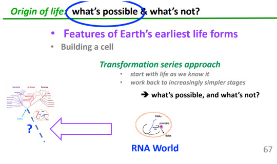

Natural selection began as soon as RNA began to self-catalyze
RNA molecules are different
Some RNA molecules copy themselves better than others because of their nucleotide sequences
Nucleotide sequence differences are inherited
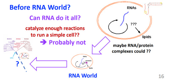
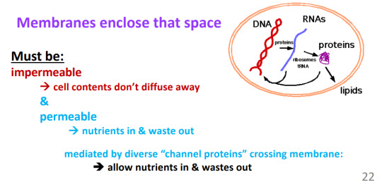

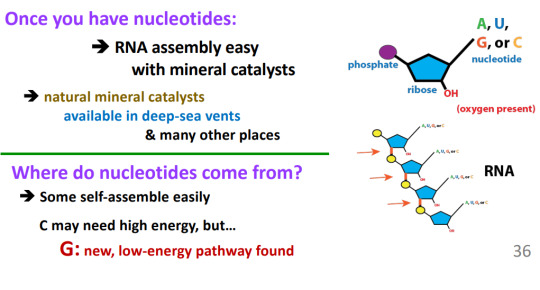
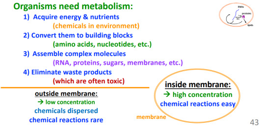

I GIVE UPPPPPPPPPPPPPPP

Evolutionary tree of the homins
Bipedalism -> walked up right
1) Sahelanthropus: 6.5 - 7 mya
Evidence: Femur structure, hip joint, foreman located under head's center of gravity
2) Ardipithecus: 5 mya
Opposite toes=climbing and walking
Monkey like
Key insights to what great ape ancestors might look like
FACT: Complex trait can revert functionally, but rarely to original morphology
Did humans evolve from chimpanzees? No.
Austrapolic afarenis
"Lucy"
Fat, human like foot
Australopithecus garnhi
2.6 mya
Earliest manufactured stone tools
Homo-habilus
Large brain size evolving
Large brain possible when high meat diet
As many as 9 species of human coexisted at the modern humans evolved (7 shown here, 2 more known from DNA)
Homonaledi
South Africa
350,000 - 200,000 years ago
Deep in South African caves
May have used fire (buried and sacrificed their dead)
and 2 hobbits
Died out around when modern humans arrived
On islands till humans got to them
They...
Independently evolved from Australopithecus
Small brains
Flat feet, so no arch

DNA-based phylogeny
Last 800,000 years
DNA extracted from bones
Neanderthals: Eastern and Western
Lived from 400,000 to 35,000 years ago
Lived in Europe and North-Central Asia
Had pale skin and red hair
Denisovan: North-Central Asia
Lived from 400,000 to 70,00 years ago
DNA reveals: alleles for darker skin
Known for a few teeth and small bones
Ancient human species interbreed.
At least 6 cases of interbreeding in early humans
One girl from Denisova cave had a Deni father and Neanderthal mother
Look at bone structure and chromosome
Modern human chromosomes carry small pieces of other species DNA
Sub-Saharan African: from mystery hominin #2
East Asian and Europeans: From Neanderthals and Denisovans
INTROGRESSION INTO MODERN HUMANS -> NOBODY IS PURE HUMAN!
East and Central Asians, and Europeans: From Neanderthals
1.5 - 3% of Neanderthals DNA in Europeans
Many deleterious: risk increase for heart disease, skin cancer, and depression
Benefits: pathogen resistance, and more expected
What can mitochondria and Y chromosomes tell us?
Mitochondria:
Have their own DNA = mDNA
Inherited from mother via egg
Y chromosomes:
Inherited via father only
MODERN HUMAN MDNA
High diversity of human mDNA variants:
Color: Major groups of genotype, each with their own minor variants
Where did first ancestors of all modern humans live?
Africa
"Mitochondrial Eve"
Woman who carried common ancestor of all humans' mtDNA
2 daughters -> one mutation
208,000 and 60,000 years ago (2014 estimate)
Y-Chromosome Adam
Man who carried common ancestor of all men's y chromosomes
-230,00 and 60,000 years ago
Difference not statistically different (2015 estimate)
Difference not statistically different

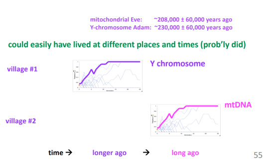

0 notes
Text
Features of yeast RNA polymerase I with special consideration of the lobe binding subunits
Pubmed: http://dlvr.it/SxPK5R
0 notes