#EPI (ElectroPhotonic Imaging)
Explore tagged Tumblr posts
Text
Electrography


(Kirlian photograph of a fingertip, 1989 & a Coleus leaf)
"Kirlian photography is a collection of photographic techniques used to capture the phenomenon of electrical coronal discharges. It is named after Semyon Kirlian, who, in 1939, accidentally discovered that if an object on a photographic plate is connected to a high-voltage source, an image is produced on the photographic plate. The technique has been variously known as "electrography", "electrophotography", "corona discharge photography" (CDP), "bioelectrography", "gas discharge visualization (GDV)", "electrophotonic imaging (EPI)", and, in Russian literature, "Kirlianography".
Kirlian photography has been the subject of scientific research, parapsychology research, and art. Paranormal claims have been made about Kirlian photography, but these claims are rejected by the scientific community. To a large extent, it has been used in alternative medicine research."
wikipedia
#kirlian photography#electrography#energy#aura#electricity#electromagnetism#bioelectrography#coronadischarge photography#technology#photography#frequency#soul#life#electric universe#spirit#medicine
6 notes
·
View notes
Text
The History of Bioelectrography

Kirlian photograph of the electromagnetic discharge between two fingertips.
The Bioelectrography History
Since 500BC HISTORY OF GDV/EPI BIOELECTROGRAPHY KIRLIANGRAPHY, ELECTROPHOTONICS see more about Different names of Bioelectrography 500BC – Testing of genuine amber by static process in China 1541-1603 – Electroscope, William Gilbert

William Gilbert William Gilbert, also known as ‘Gilberd’, was a famous researcher in magnetism. He was famous during the time of Queen Elizabeth I and is best known for his publication, ‘De Magnete’. Credited as one of the originators of the term of electricity, William Gilbert is also known as the father of electricity, magnetism and electrical engineering. He travelled extensively and wrote many publications such as ‘Magnetisque Corporibus’ and ‘ET de Magno Magnete Tellure’ during his lifetime. Apart from being a scientist, Gilbert led a parallel career as an astronomer. He studied the moon’s surface without a telescope and concluded that the craters were in fact land, and the white patches on the moon’s surface were water bodies. One of his other significant contributions was when he pointed out that the motion of the skies occurred due to the rotation of the earth. One of the first people to try to map the markings of the moon’s surface, Gilbert was a celebrated astronomer and scientist. His theories on magnetism and electricity had also been the subject of controversy for many of his successors.

electroscope An electroscope is an early scientific instrument that is used to detect the presence and magnitude of electric charge on a body. It was the first electrical measuring instrument. The first electroscope, a pivoted needle called the versorium, was invented by British physician William Gilbert around 1600. The pith-ball electroscope and the gold-leaf electroscope are two classical types of electroscope that are still used in physics education to demonstrate the principles of electrostatics. A type of electroscope is also used in the quartz fiber radiation dosimeter. Electroscopes were used by the Austrian scientist Victor Hess in the discovery of cosmic rays. Electroscopes detect electric charge by the motion of a test object due to the Coulomb electrostatic force. Since the electric potential or voltage of an object with respect to ground equals its charge divided by its capacitance to ground, an electroscope can be regarded as a crude voltmeter. However, the accumulation of enough charge to detect with an electroscope requires hundreds or thousands of volts, so electroscopes are only used with high-voltage sources such as static electricity and electrostatic machines. Electroscopes generally give only a rough, qualitative indication of the magnitude of the charge. An instrument that measures charge quantitatively is called an electrometer. 1602-1686 – Electrostatic machine, Otto von Guerricke & Hauksbee
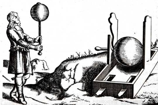
Otto Von Guerickes Sulphur Ball Otto von Guericke's electrostatic machine evolved into increasingly improved instruments in the hands of later scientists. Otto von Querricke (1602-1686), the Burgomeister of Magdeburg best known for his demonstration of the effect of atmospheric pressure on evacuated bodies (the Magdeburg Hemispheres), also showed the electrical effects were obtained by rubbing glass. Originally, he used a globe of sulphur mounted on a shaft, with the hand rubbing the rotating sphere. The sulphur was cast in a spherical shell of glass that was subsequently broken away; soon it was discovered that glass, and not sulphur, was the key ingredient of the demonstration. About 1700 Francis Hauksbee the Elder suggested that a glass cylinder be used in place of the sphere. In the early 1700s, Francis Hauksbee designed his own electrostatic generator, a feat stemming from his studies of mercury. 1661-1713 - Resulted in the reproduction of electrical phenomena in the laboratory 1622 – Magnetic declination varies with time, Edmund Gunter 1671 – Electric organ of torpedo fish studied, Francesco Redi 1672 – Living tissues react to environment, Francis Glisson 1702 – Air at low pressure glows during an electrical discharge, Hauksbee 1704 – Electrons, particles, wave forms. Newton 1729 – Photometry, Pierre Bouger 1729 – Electric current, Stephen Gray 1731 – Anything can be charged with static electricity if isolated by non conductive materials, Stephen Gray 1733 – Two types of static electric charge, like charges repel whilst unlike charges attract, (later opposed by Benjamin Franklin), Charles Francois de Cistemay 1746 – Leyden Jar (Leiden) for storing static electricity Pieter Van Musschenbrooek & E G Pieter got a shock when using it suggesting a connection between lightning, VonKleist 1747 – A pointed conductor draws off an electric charge from a charged body, Benjamin Franklin 1747 – First electrometers, Abbe Jean-Antoine Nollet (Paris) 1756 – Electricity and the origin of light and the wave theory, Mikhail Valilievich Lomonosov 1766 – That all nerves follow a path through the spinal column to the brain and that the nerves stimulate the muscles, A Von Haller 1766 – Improved electrometer, Horace Benedict de Saussure (Suisse) 1766 – Chart of magnetic inclination, Johna Wicke (German) 1766 - Joseph Priestley, English explorer, registered the colored circles which are obtained by electrical discharge on a metal surface («Priestly's rings»). 1771 – Tissue conduction of electricity, Luigi Galvani 1775 – Early electrical condensers, Alessandro Volta 1777 – Electrographic images, G C Lichtenberg 1777 – GERMANY GEORGE CRHISTOPHER LICHTEMBERG
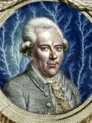
GEORGE CRHISTOPHER LICHTEMBERG Georg Christoph Lichtenberg was a German physicist, satirist, and Anglophile. As a scientist, he was the first to hold a professorship explicitly dedicated to experimental physics in Germany. He obtained, with dust particles, under Statics Electricity, anything that we can consider as being primitive “Bioelectrographic Images” and those primitive images, in electrified dust were named, by him, as Electro-graphics”. In 1777 a German physicist George Lichtenberg touched a metal electrode covered with glass and connected to voltage with his finger while experimenting with the electrical machine. And suddenly a burst of sparkles flew all around. This was magically beautiful, although a little bit frightening. Lichtenberg jerked back the finger and then repeated the experiment. The finger placed on the electrode was shining with bright blue light and treelike sparkles dispersed from it. Lichtenberg, being a real academic scientist, investigated the behavior of this fluorescence in detail, although he substituted a grounded wire for a finger. The effect was the same, which later suggested an idea that some special energy exists in the body, and first electrical then torsion properties were attributed to it. Articles by Lichtenberg, masterfully done in German, are still cited in books on gas discharge. Further research demonstrated that electrical fluorescence was not so rarely met in nature.
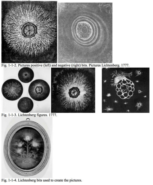
Lichtenberg figures 1777-Lichtenberg, Georg Christoph. De Nova Methodo Naturam Ac Motum Fluidi Electrici Investigandi (Concerning the New Method Of Investigating the Nature and Movement of Electric Fluid). Göttinger Novi Commentarii, Göttingen, 1777. 11 1778-Lichtenberg, G.C. "Super nova methodo motum ac naturum fluidi electrici investigandi," Soc. Reg. Sc. Gottingensis, 1778, T.8, p.168-180. 1779-Lichtenberg, G.C. Commentatio posterior. Commentationes Soc. Reg. Sc. Gott. Glassis mathematicae T.1. p.65-79. 1779. 1779-G.C. Lichtenberg. Zweite Abhandlung uber eine neue Methode, die Natur und die Bewegung der elektrischen Materie zu erforschen. Ebd., Class. Math. tom. I, ad annum 1778, S.65 (1779) (Pup 56). The effect of discharge in the high voltage field observed in the experiments Tesla, Rengo and D'Arsonval, at voltages above 30 kV (especially good discharge visible after a 100kV). Two major invention that allowed to implement a method of photographing images of various objects in the high-frequency field: 1839 - invention of photography Daguerre, 1851 - creation Ruhmkorff coil. 1839-Jacques Daguerre, a French researcher, has published a method for producing an image on a copper plate covered with silver. 1842-G. Karsten, Berlin. Germany. He put a coin on a glass plate and gave it a few sparks from the electric machine. If you then breathe on the glass, it was seen the image of the coin. He called these figures «electrical breath figures». 1851-Heinrich Daniel Ruhmkorff, German inventor. In 1851, he patented the first version of its induction coil. This further coil is widely used for electrophotographic images.
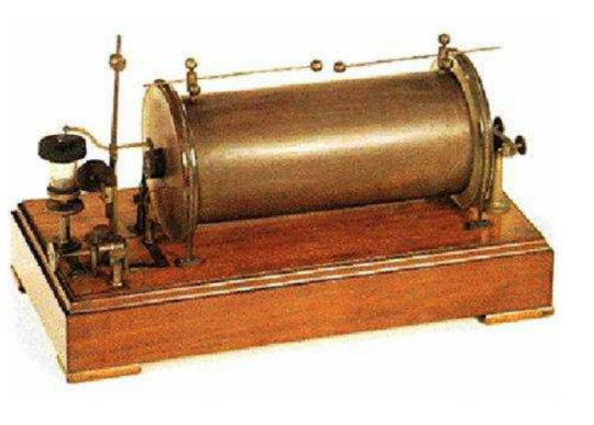
The History of Bioelectrography - Coil Ruhmkorff
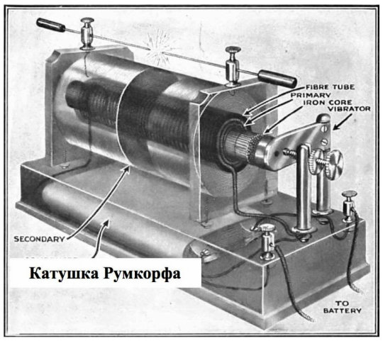
Coil Ruhmkorff - The History of Bioelectrography The device Ruhmkorff coil. The primary winding of the coil, consists of several tens of turns of thick wire wound around the core, and is energized through the electrochemical cell (chemical current source). An important element of the chopper coil is in the form of a hammer, which is attracted by the core to create the primary winding of the magnetic field due to flow through it from the DC power source. Thus, the hammer breaks the circuit and the magnetic field disappears, the hammer returns to its original state, closing the circuit again. The change in the magnetic field reacts secondary winding consisting of thousands of turns of a thin wire, is wound over the primary winding. This leads to a second winding of high instantaneous currents of different directions (closing / opening). Due to a member of the condenser coil, the coil stores energy in a magnetic field, which further increases the currents in both windings, and allows the air gap between the punch pin of the secondary winding. 1876 - I. Goldstein Gittorfor (1850-1931), German physicist, received a specially designed discharge tube coin image using it as a cathode. These experiments were carried out under reduced pressure of the gaseous medium. Relief cathode (coins) was seen in the light of the cathode-ray fluorescence on the opposite wall of the cathode of the discharge tube. 1871 - Cromwell Fleetwood Varley (1828-1883), English engineer-electrician. The interrelation of the phenomena of electricity and spiritualism, studied electrical discharge in gases. 1877 - Lachinov Dmitry Alexandrovich (1842-1902), Russian physicist and electrical engineer, SPGU, Professor of Forest Institute, St. Petersburg. Since 1877 Lachinov worked on the gas discharge visualization. Building on its cycle of meteorological research, continuing to work on the study of the electric arc and pictures in the late 1870s and early 1880s Lachinov published in the "Russian Invalid" a number of articles dealing with different aspects of the research programs, their complex application. In the summer and autumn of 1887 in the physics laboratory of the Forest Institute Lachinov simulated form of atmospheric electricity, differentiation electrodischarges in a gaseous environment. With the assistance of the photographer V.Monyushko photographed or recorded on the plate bromzhelatinovoy direct impact sparks. During the first experiments filmed bright discharge (spark induction coil connected to a capacitor) or dim when entered in a long chain of the resistance gave a discharge discharge. The second and third series of experiments was carried out without a camera, the category of sliding along the surface of the dry bromzhelatinovoy plate and left her a trail that the manifestation is made visible, nothing else, as one of the first examples of the so-called gas discharge visualization. On the progress and results of the experiments reported in V.Monyushko V (photographic) department Russian Technical Society (St. Petersburg Engineering Society) October 9, 1887. He spoke about the possibility of photographing using a variety of metal objects spark. October 27, 1887 Lachinov posts made in Russian Physical and Chemical Society (RFHO). Lachinov DA He invented a device for detecting defects of electrical insulation. 1878 DA Lachinov A new way of photographing. Russian invalid. 1878. №14. 1879 DA Lachinov Electrophotography. Russian invalid. 1879. №98. 1880 DA Lachinov Phosphorescence and its application to the photos. Russian invalid. 1880. №331. 1880 – UNITED STATES OF AMERICA NIKOLA TESLA

Nikola Tesla He showed, in a public presentation, a luminous halo around human body and another objects while exposed to a strong electromagnetic field with high A.C. voltage and high frequency. But he considered this subject only as a scientific curiosity that received the general name of “Corona Effect”. In the Nineteenth century enigmas of electricity were opening to people. One of the great inventors was Nicola Tesla, from whom we now have lamps and television sets. He invented the generator of alternating current. However, if it had not been him, somebody else would have done it. Inventions come to life when a social need for them appears. Then different people simultaneously and independently start arriving at the same ideas. This is connected with the fact that the ideas have their logic of development, and the developers shall only intuitively feel this logic. After raising good money with his patents, Nikola Tesla began the mysterious experiments on energy transfer without wires. He did not finish his developments and died in destitution, but up to now enthusiasts have been trying to investigate his ideas. We get used to our technical progress and reap its fruits with pleasure, but is it the only possible way of development? At the peak of his career Tesla liked to give public lectures and impress the audience with the following experience. The light was turned off in the room, Tesla turned on the generator of his own design, stood on the platform-electrode, and his body got wrapped in the glow. The hair stood on end, glowing rays of light radiated in the space. The experiment was very effective, though not all those who wished managed to repeat it: as a matter of fact, their glow was much less and for some people even missing. Not in vain was it said that Nikola Tesla had special energy state. Further research did not go much beyond investigations of the glow of fingers, sometimes ears, nose and other prominent parts of the body. Is it possible to reproduce Tesla’s experiments and make all the body glow? Yes, it is. But is it necessary? Powerful equipment, which is not safe if not handled properly, is required for such an experiment. Moreover, the stronger electrical glow, the more ozone is generated in the air. A high concentration of ozone is far from being healthy. 1887 - DA Lachinov "Russian Invalid". 1887, №220, №225. November 26th 1887 - DA Lachinov On studies of electrical discharges through photography. ZhRFKhO. 1887, issue 8. s.438. 1888 DA Lachinov On studies of electrical discharges through the photos. Journal of Russian PhysicoChemical Society. 1888 of the Financials. vol.3. s.44-49. 1888 DA Lachinov "Electricity". 1888, №1-2. s.1-7. 1888 DA Lachinov "Yearbook of the St. Petersburg Forestry Institute." 1888 vol.3. s.169-179. 1888 ZRTO. 1888, Issue 1. s.42-48. 1902 DA Lachinov (Obituary). Herald of experimental physics and elementary mathematics. 1889, Czech B. Navratil coined the word "electrography" 1896, a French experimenter, H. Baraduc, created electrographs of hands and leaves Hippolyte Ferdinand Baraduc (1850 - 1909) was a French physician and magnetist parapsychologist, and highly recognized for photographing thought and feelings with iconographies. 1892 – RUSSIA Jakob. J. NARKIEVITCH-JODKO

J. J. NARKIEVITCH-JODKO On an electricized plate, he obtained a type of photo that he simply named Electric Photography, in black and white colors, and started to investigate, through this technique, the human potencialities, but he did not continue his work. Later on, already during the middle of XXth Century, his work was taken back by M. Pogorelski, in Russia, and by B. J. Navratil, in Czechoslovakia, but with no other significative consequence. A significant contribution to the study of these photographs was made by a talented Byelorussian scientist Jacob Narkevich-Yodko in the end of the Nineteenth century. He was an independent landowner and spent most of his time on his estate above the river Neman. There he actively experimented with electricity, applying it in agriculture and medicine. A straight parallel with modern medicine can be drawn from the description of experiments on the stimulation of plants with electrical current, on electrotherapy, and magnetism by J. Narkevich-Yodko. But the scientific achievements of our time are not just “the new as a well-lost old”. This is a new convolution of perception. 1893 - Russia “Electrophotosphenes and energography as a proof of existence of the physiological polar energy”. This was the name of a small book by a doctor from St. Petersburg, Messira Pogorelsky, where he described his experiments in bioelectrography. Book, published in 1893. Many photographs of the glow of fingers and toes, ears and nose show how the pattern of fluorescence varies when the psychic state of a person changes. However, this work was far from being the first one. 1865 - Narkevitch-Yodko finished Minsk provincial classical gymnasium and went abroad. 1869 - entered the medical faculty of the University of Paris. He studied in Vienna, Paris, Florence. 1872 - he returned home, he conducts scientific experiments. 1888 - Metoostantsiya transferred to the estate of over-Neman (80km south-west of Minsk). 1902. Issue 335. s.259. http: /www.vofem.ru/ru/cat/113/ (Russian) 1904 – BRAZIL Father Roberto Landell de Morua (A Brazilian Jesuit Catholic Priest)
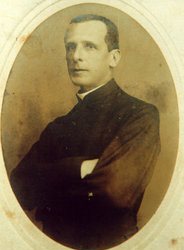
Father Roberto Landell de Morua Besides to be a priest, he was a Physicist too and, in Porto Alegre (RS), he invented an engine that he named “Bioelectrographic Machine”. He did take some hundreds of photos and named the halo around the human body as “Perianto”. He researched on this subject during 8 (eight) years, until 1912, when he was obliged by Catholic Church to stop those researches. In Brazil, very similar experiments were performed by a Catholic monk, padre Landell de Morua. A monk’s life left a lot of free time, after reading prayers and performing rituals. Padre de Morua invented the technique of photoregistration of electrical glow and started giving lectures and writing to social leaders in order to attract attention to his offspring. Invention of padre de Morua produced much rapt attention, congratulations, banquets, but was not widespread. Then the little priest invented the radio (practically simultaneously with Popov and Markoni), but again he was unable to draw in large crowds. Even the military. FIRST SCIENTIFIC RESEARCHES AT INTERNATIONAL LEVEL In the beginning of the Twentieth century nobody even recalled the mysterious glow. There were many other problems: wars, revolutions, breakthroughs in physics, discovery of antibiotics and roentgen rays – everybody was sure that it was very close to the outright victory. Only by 1930’s the life more or less came right. And here appeared the mysterious glow again. And, as if by chance, it was discovered anew, but there is a rule behind every chance. 1904/12 – BRAZIL We can consider that Fr. Landell was the pioneer of the first scientific and systematic researches on the Bioelectrography field, during 8 years, at worldwide level. However, Catholic Church, in that period, did not allow he continued his researches because of purely doctrinary reasons and confiscated almost all of his notes, but many of those reports escaped and are in a safe place, the current Fr. Roberto Landell de Moura Museum, in Porto Alegre (RS) – Brazil. 1939 - Around the World In 1939, two Czechs S. Pratt and J. Schlemmer. published photographs showing a curious glow or aura around leaves. The same year, the Russian electrical engineer Semyon Kirlian and his wife Valentina developed their own technique after observing a patient who was receiving medical treatment from a high-frequency electrical generator. Electrotherapy was popular at the time and they had noticed that when the electrodes were brought near the patient’s skin, there was a glow similar to that seen in an electrified tube filled with neon. Kirlian photography consisted of placing photographic film on top of a conducting plate, and attaching another conductor to a hand, leaf, or other part of a plant. When the conductors were energized by a high frequency high voltage power source, the resulting image showed a silhouette of the object surrounded by an aura of light. 1939 - Experiments in psychics Practical studies in direct writing (automatic) supernormal photography and other phenomena by F. W. Warrick Golinger, Skotographs, Nengraphy, Nensha, Thoughtography, Spirit Photography, Themography, ectoplasm

Semyon & Valentina Kirlian Semyon & Valentina Kirlian 1939 – RUSSIA Semyon and Valentina Kirlian In Krasnodar, former URSS, he re-invented the Bioelectrographic Camera, now renamed as Kirlian Camera, and started systematic and scientific researches, helped by several Soviet Scientists. Those researches were revealed to the rest of the world only in 1960, during a Congress on Parapsychology. Semyon Kirlian spent most part of his life with his wife Valentina in a poor two-room apartment at the corner of Gorky and Kirov streets in Krasnodar. The wooden two-story house where they had started their family life was swept away by progress – a building program turned the small provincial town on the banks of the Kuban river into an industrial center. Kirlians were deeply carried away with the experiments with auras of live subjects, and since 1939 they had worked hard. The only rest they could afford was walking hand in hand under the trees and along blossoming fields so typical of the South Russian cities. The Kirlians published the results of their experiments for the first time in 1958, and in 1961 reported that the characteristics of fingertip auras not only varied in different people, but was also affected by their emotional status. If someone felt very anxious or was in an opposite state of deep relaxation during meditation, there was a corresponding change in the size and intensity of the glow. Their work was virtually unknown in the West until 1970, when two Americans, Lynn Schroeder and Sheila Ostrander published their book, “Psychic Discoveries Behind the Iron Curtain”. Russian books about Bioelectrography 1967 – BRAZIL Prof. Newton Milhomens

Prof. Newton Milhomens At the end of 1967, in Brasilia (DF), he built his first Kirlian Camera, based on a Soviet electronic scheme, starting his scientific researches in Psychological Clinics, in 1968, and, later on, in Hospitals. Moving to Rio de Janeiro (RJ), in 1981, in that city he continued his researches. Currently, he is living in Curitiba (PR), ever since 1983. Professor Newton Milhomens books 1969 – GERMANY

Dr. Peter Mandel Dr. Peter Mandel YEARS 1970s It was published the book “Psychic Discoveries Behind the Iron Curtain” that was writen by the American press-women Sheyla Strander and Lynn Schroeder, that popularized worldwide the Semyon Kirlian researches. So, hundreds of persons, in all countries, started to build Kirlian Cameras and began to research with this new device, some of them in an amateur manner but another persons used a systematic and scientific manner. Unfortunately, this subject was divulged by International Press with sensationalism considering the Kirlian Photo as a mystic and supernatural subject. 1970 – BRAZIL Dr. Paulo de Castro Teixeira, a Homeopathic Pharmaceut from do Rio Preto (SP), built his Kirlian Camera and take hundreds of Kirlian Photos of several persons, before and at each 15 minutes, after those persons had ingested any homeopathic remedy, and he found several types of modifications, in the Kirlian Photos. He published several books, in Portuguese, all about his researches in this field and, till today, he is living in S?o Jos? do Rio Preto (SP), where he is the owner of a Pharmaceutic Industry that produces Homeopathic remedies. 1972 – BRAZIL Dr. Hernani G. Andrade He always was considered the greatest divulger of Spiritism in Brazil. He built his Kirlian Camera, in San Paulo (SP), and took many hundreds of Kirlian Photos of plants and human beings intending to prove the existence of Spirit with them, not only in human beings, but even in bacteria too. He only divulged his researches in Spiritist Magazines and Congresses mainly at the State of S?o Paulo (SP) where is considered the greatest one. 1974 - IKRA IKRA – The International Kirlian Research Association 1974 scientists from different countries came together in an international association for the study of Kirlian effect (The International Kirlian Research Association, IKRA) (Drexel University). The organization was formed in December 1974 at a seminar in the Community Hospital in Brooklyn, NY for the purpose of standardization and assistance at all stages of research into the Kirlian phenomenon. Dr. Benjamin Shafiroff, New York College of Medicine, USA President of the Association IKRA.
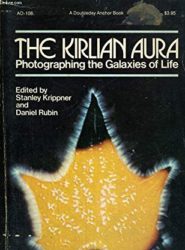
1974 – UNITED STATES OF AMERICA Dr. Stanley Krippner, PhD Psychologist and Parapsychologist, published the book “The Kirlian Aura” in which he described all that was known in that period, about Kirlian Photos, under a scientific approach.

The Probability Of The Impossible: Scientific Discoveries and Explorations of the Psychic World 1974 - The Probability Of The Impossible: Scientific Discoveries and Explorations of the Psychic World by Thelma Moss. In The Probability of the Impossible, Dr. Thelma Moss presents a rare picture of a parapsychologist at work in her laboratory and in the field. She explores the seemingly incredible assumptions of parapsychologists and carefully shows how the scientific psychical researcher attempts to recreate, capture, and analyze the elusive phenomena of the paranormal. Step by step, she leads the reader along a continuum from everyday experiences through events that happen only rarely and under "special" circumstances on to the extraordinary, indeed the seemingly impossible, experiences of both well-known and little-heralded psychics. She convincingly presents arguments for a psychic world as "real" as the material one.
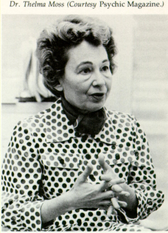
Dr. Thelma Moss 1975 – UNITED STATES OF AMERICA Dr. Thelma Moss, PhD Psychologist and Professor of University of California, she started her researches on Kirlian Photos and published the book “The Probability of the Impossible”, where she reported her initial researches. Later on, she published another book “The Electric Body” in which she described her last researches in that University and showed to all, the reasons which provoked her removal, all of them were the result of pure preconception.
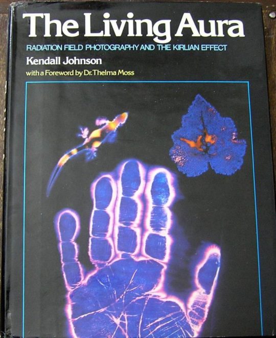
The living aura - Radiation field photography and the Kirlian effect 1975 - The living aura: Radiation field photography and the Kirlian effect by Kendall L Johnson 1978 - IUMAB IUMAB – International Union of Medical and Applied Bioelectrography, the maximum and the greatest Organization, at worldwide level, on Bioelectrography. Today, IUMAB is recognized by UNO/WHO and is considered by this UNO’s Organization as the maximum Bioelectrographic Organization on world whose rules, recommendations, directives and other acts are valid and must be observed in Bioelectrographic researches, courses, standards, etc. The International Union of Medical and Applied Bio-Electrography was formed in 1978 to help standardize equipment, research methods, and data acquisition. Researchers such as German naturopath and acupuncturist Peter Mandel and Newton Milhomens in Brazil developed their own way of interpretation of Kirlian photography of human fingers and toes. Peter Mandel was one of the first, who energized certain acupuncture points by using different colored lights to achieve a desired response. Mandel's Energy Analysis Emission diagnostic system utilized Kirlian photography and his Esogetic Colorpuncture therapy is believed to restore yin and yang equilibrium. All of these modalities, as well as non-invasive laser acupoint stimulation, have been used with varying degrees of success in thousands of patients over the years.

1979 - USA The Body Electric: A Personal Journey into the Mysteries of Parapsychological Research, Bioenergy and Kirlian Photography by Dr. Thelma Moss The Body Electric tells the fascinating story of our bioelectric selves. Robert O. Becker, a pioneer in the filed of regeneration and its relationship to electrical currents in living things, challenges the established mechanistic understanding of the body. He found clues to the healing process in the long-discarded theory that electricity is vital to life. But as exciting as Becker’s discoveries are, pointing to the day when human limbs, spinal cords, and organs may be regenerated after they have been damaged, equally fascinating is the story of Becker’s struggle to do such original work. The Body Electric explores new pathways in our understanding of evolution, acupuncture, psychic phenomena, and healing. 1983 – BRAZIL It was published, in Brazil, the book “Kirlian Photos – How to Interpret”, in Portuguese, whose author was Prof. Newton Milhomens, the first, on world, to teach how to diagnose almost all about the main problems of organic and psychic health using Kirlian Photos, where he described in details the way and the results of his researches, since 1968. 1983 – BRAZIL Newton Milhomens founded at Curitiba (PR) , his Industry and started to produce his first models of Kirlian Cameras which were initially sold to Brazilian doctors, psychologists and therapeuts.
Electrographic Imaging in Medicine & Biology by Ion Dumitrescu 1983 - Romania Electrographic Imaging in Medicine & Biology by Ion Dumitrescu 1985 – RUSSIA Dr. Konstantin Korotkov, PhD Physicist, Professor of the University of Saint Petersburg, after innumerable researches with a team of scientists of that University, he discovered that Kirlian Effect is the result of the ionization of gases and vapors exhaled by our skin, through the pores. He named this model GDV (Gas Discharge Visualization) or, simply, GDV Technique, Electrophotonic Imaging, EPI. 1986 – BRAZIL At Curitiba (PR), the Ist Brazilian Congress on Kirliangraphy was realized with more than 250 Brazilian researchers on Kirliangraphy that came from all Brazil. During this Congress, it was approved by unanimous decision the “Newton Milhomens Standard” as the Brazilian Official Standard of Kirliangraphy. 1987 – BRAZIL It was published (in Portuguese) in Revista do Hospital das Foras Armadas (Magazine of the Brazilian Armed Forces Hospital), a scientific magazine on Military Medical Area, a Scientific Article entitled “Kirliangraphic Diagnosis on Oncology” that was written by two military doctors from Curitiba (PR): – Drs. J?lio Grott and H?lio Grott Filho. They discovered a sign that they named “fracture”, which is the sign able to diagnose cancer in human beings. 1987 – ARGENTINA Prof. Newton Milhomens was invited to dictate some lectures at Buenos Aires to hundreds of persons. By this way, Kirliangraphy with a scientific approach was introduced in that South American country. AROUND THE WORLD – YEARS 1987/95 During this period of 8 years, hundreds of articles, theses of post-graduation, and several books using Kirlian Photos, were published at several countries on world, including Brazil. Some of those books were written in an amateuristic manner but other ones, however, were written seriously with many scientific and systematic foundation and accurate statistic treatment. Thus, the scientific phase of Kirliangraphy was starting. 1989 – PORTUGAL Prof. Newton Milhomens was invited to dictate a lecture on Kirliangraphy at Lisbon, Portugal, during a Congress on Parapsychology, sponsored by Escola Superior de Biologia e Sa? de (Superior School of Biology and Health) and so, he introduced Kirliangraphy with a scientific approach in Portugal. During this period, he taught Kirliangraphy as an official matter subject of the course of Naturopatic Medicine in that Portuguese University. 1990 – PORTUGAL Prof. Newton Milhomens returned to Portugal to teach Kirliangraphy in Superior School of Biology and Health, at Lisbon, Portugal. This event was noticed by several local newspaper and the Tass TV Agency, from Russia, asked for him an interview that would be shown to all the russian territory to demonstrate that a Brazilian was teaching in Europe a Russian technique based on an engine that was invented by a Russian, Semyon Kirlian.
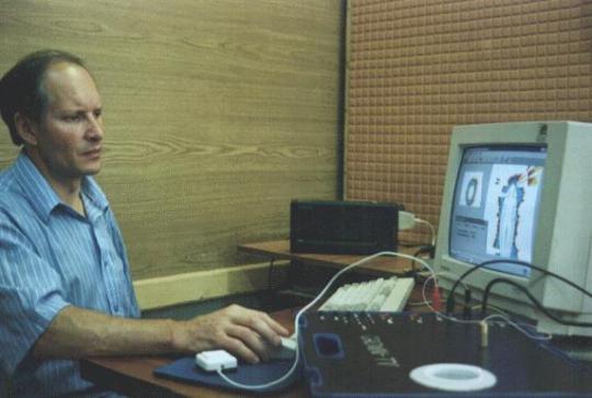
Dr. Konstantin Korotkov and GDVCAMERA 1995 – RUSSIA After many years of researches, he was succeeded to build a new Kirlian Camera which declines the photographic film and puts the GDV image directly at a computer’s screen. It was a new milestone in the History of Bioelectrography. First GDVCAMERA by Dr. Konstantin Korotkov on Saint-Petersburg, Russia created. More details on Bio-Well History. The GDV camera is based on the stimulation of photon and electron emissions from an object when it is placed in an electromagnetic field and subjected to brief electrical pulses. This process is called ‘photo-electron emission’ and has been thoroughly studied with cutting edge electronic techniques. The emitted particles accelerate in the electromagnetic field, generating electronic avalanches on the surface of the dielectric (glass) plate in a process called ‘sliding gas discharge. The discharge causes a glow from the excitement of molecules in the surrounding gas which is constantly measured. Voltage pulses stimulate optoelectronic emissions that are amplified in the gas discharge, and light produced by this process is recorded by a sensitive CCD (charge coupled device) camera that converts it into a colored computer image, or BIO-gram. Data obtained from the fingers of both hands are converted into a Human Energy Field image using proprietary sophisticated software. Russian books about Bioelectrography. 1996 – THE “WAVE” Induced by the success of the researches on Kirlian field, radiotechnicians on all countries around the world started to produce Kirlian Cameras from “backyard”. It was the radiotechnicians worldwide “wave”… As their unique intention was merely commercial, they never were interested in a standard and these “backyard standard” Kirlian Cameras never had a good quality and no stability but were enormously cheap, because they used the worse electronic components and also because anyone of these “producers” never made or had any participation in any kind of scientific experiment on Kirlian field and no one of them had any universitary education and the outsiders of Kirlian researches started to use the Kirlian Camera searching Mysticism and Supernatural exploring this new “commercial mystic field” in Fairs and Parks what was again divulged by International Press with much more sensacionalism and the name “Kirlian” lost credibility. Any samples of “backyard standards” around the world. 1998 – THE INTERNET When the Internet becomes firm and trustable, the informations become much more easy to be interchangeable at world wide level and the Kirlian subject appeared in several homepages. Many of them were only showing interest in commercial sales and another ones, on mysticism and supernaturalism. However, many serious homepages were taking aim at scientific publications or at a simple change of scientific informations were beginning to appear as the case of Prof. Newton Milhomens, in Brazil, Dr. Korotkov, in Russia, Dr. Peter Mandel, in Germany. Dr. Tom Chalko in Australia. IUMAB.org, GDVPLANET.com, GDVCAMERA.com, Kirlian.ru, GDVSOFTWARE.com, GDVsputnik.com and many other homepages from serious researchers around the world. 1998 – NEW ZEALAND Prof. Newton Milhomens was invited by Dr. Robert Beasley (center) to dictate a lecture on Kirliangraphy in a Congress at Auckland, New Zealand, where he knew Dr. Tom Chalko (right) that was researching on Kirliangraphy at Melbourne, Australia. 1998 - IUMAB Review IUMAB Review - Annual Digest Established by Professor Pavel Bundzen and Kirill Korotkov in 1998
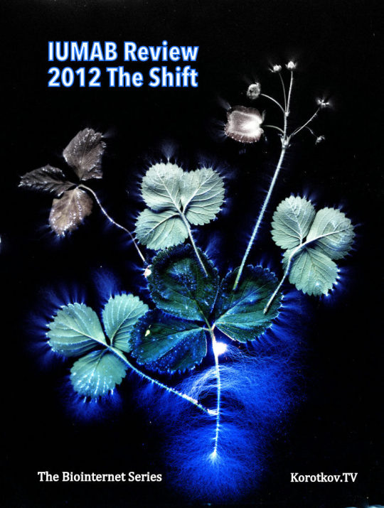
IUMAB Review 2012 The International Union of Medical and Applied Bioelectrography publishes the IUMAB Review one time per year, and is free to all Regular Members. It contains original articles, news and book reviews. Current and past editions of IUMAB Review are ready for download as PDF files for IUMAB Regular Members. 1999 – RUSSIA In the beginning of this year, Ministry of Health, in Russia, declared that Kirliangraphy was Officially Admitted as a Scientific Fact and its main appliance was recommended in Medical Practice. In September, the Russian Academy of Sciences, during a Congress in Moscow, with more than 300 of the most renown russian scientists, officially admitted Kirliangraphy as a SCIENTIFIC FACT. 1999 – BRAZIL In April, at Curitiba (PR), occurred the IVth Brazilian Congress of Kirliangraphy, with the presence of more than 200 persons and the Russian Physicist, Dr. Konstantin Korotkov, PhD, was invited to be the special international invited lecturer. 2000 – RUSSIA Prof. Newton Milhomens was invited as Honor Invited Lecturer to a Congress that occurred at Saint Petersburg. During a certain night, inside a ship on Ladoga Lake, he was honored because he was the first researcher on world to have discovered how to diagnose problems of psychic and organic health using Kirlian Photos. 2000 – BRAZIL Sponsored by IUMAB it was realized, at Curitiba (PR), the Vth Worldwide Conference on Kirliangraphy-2000, with the presence of 350 persons, where 45 of them were europeans. There were 5 lecturers from several European countries and 5 ones from Brazil. During the Vth Worldwide Conference on Kirliangraphy-2000, were presented two new discoveries on organic health area: – The signs that shows Allergic process, by Dra. Celia Cruz, from St Paulo, and the scientific proof of Flower therapy efficiency, by Prof. Rodrigo Campos, from Minas Gerais, a meeting of IUMAB Directory occurred and the executives deliberated that, on all the world, would be valid only 3 (three) OFFICIAL WORLDWIDE STANDARDS of Kirlian Cameras (in alphabetic order): Brazil – Newton Milhomens Standard Germany – Peter Mandel Standard Russia – Konstantin Korotkov Standard

Konstantin Korotkov, Rosemary Steel, Peter Mandel 2000 – BRAZIL (THE 3 OFFICIAL STANDARDS) Left – Camera and Kirlian Photo of “Newton Milhomens Standard” – Brazil Center – Camera and Kirlian Photo of “Peter Mandel Standard” – Germany Right – Camera and Kirlian Image of “Konstantin Korotkov” – Russia During the same Vth Worldwide Conference on Kirliangraphy – 2000’s meeting, Dr. Konstantin Korotkov was elected by unanimous decision the President of IUMAB. Prof. Newton Milhomens was elected the Vice-President of IUMAB on Brazil, and also the IUMAB’s Official Plenipotentiary Representative on Brazil. Yet during the same Vth Worldwide Conference on Kirliangraphy-2000, the Rector of UNESC – Universidade do Extremo Sul de Santa Catarina (University of the Extreme South of Saint Katherine), Brazil, officially informed that Kirliangraphy would be introduced in that University, as official subject Matter in the Courses of Medicine and Psychcology, with official authorization from Brazilian Ministry of Education. Yet during the same Vth Worldwide Conference on Kirliangraphy-2000’s meeting, the IUMAB’s Directory deliberated by unanimous decision to change, from day 12/01/2000 toward, the name Kirliangraphy to Bioelectrography, in honor to Fr. Roberto Landell de Moura, the first researcher on world to use this name and to research on this subject during the period from 1904 to 1912. It happened a ceremony of homage to Fr.Landell symbolized by the delivery of a bust of him to Dr. Korotkov by Profa Vania Abatte, Curator of the Landell de Moura’s Museum, at Porto Alegre (RS), Brazil. 2000 – RUSSIA/BRAZIL The letter from Dr. Korotkov, as IUMAB’s President, confirming to exhibit the bust of Fr. Landell in Kirliangraphy Museum, in Russia, what really came to be true. 2000 – RUSSIA The Fr. Roberto Landell de Moura’s bust is in exhibition in the Kirliangraphy Museum, in Russia, beside the busts of Kirlian couple together a book which tells, in Portuguese, the Biography of this Brazilian eminent scientist and another book, in Russian, with the Biography of the Russian couple. 2001 – BRAZIL The Bioelectrography is taught as official subject of the Course of Holistic Terapies, at University Est?cio de S?, in Rio de Janeiro (RJ), where the first course of How to Interpret Kirlian Photos, was taught in official manner, by Prof. Newton Milhomens, to the first graduating class in that University. 2001/2002 – AROUND THE WORLD Dr. Konstantin Korotkov, PhD During this period, Dr. Korotkov was travelling to U.S.A., Europe and another countries to divulge Bioelectrography. Prof. Newton Milhomens also was traveling to several South America countries and to all the Brazil to do the same. 2002 – BRAZIL Several theses of Graduation and Post-Graduation, including theses of Post-PhD, were presented in several Brazilian Universities, for example, USP, UNICAMP and UFSC, in several Post-Graduation Courses of these Universities, all of them using the Bioelectrography as an auxiliary researches’ instrument. Several Professors from USP and from another Brazilian Universities are purchasing Kirlian Cameras and realizing serious researches, on several areas of human and vegetable health using the Bioelectrography as an auxiliary instrument in their researches. Also many of these Professors are coming to Curitiba (PR) to learn How to Interpret Kirlian Photos with Prof. Newton Milhomens. It was officially and legally founded, at Santo Paulo (SP) , the UBBA – Unio Brasileira de Bioeletrografia Aplicada (Brazilian Union of Applied Bioelectrography) that is recognized by IUMAB and is the greatest directive Oranization in Brazil on the Bioelectrographic field. Its current President of Honor is Prof. Newton Milhomens and the current Executive President is Dr. Celia Cruz. 2003 - BRAZIL Dr. Selma Milhomens Psychologist and wife of Prof.. Newton Milhomens, began teaching in Bioelectrography UNIABEL - Open University of Educational Freedom in Curitiba (PR), as a discipline curriculum for the courses that University ofBiotherapy curitibana. 2004 - COLOMBIA Dr. Edith Torres Noboa, Ecuador Medical, based in Cali, Colombia, degenerative disease specialist and former student of Professor. Newton Milhomens, is using the Bioelectrography as an aid to diagnosis in their patients with great success. RUSSIA – 2002-2012 Dr. Konstantin Korotkov Dr. Korotkov is using GDV CAMERA for sport research. He is Professor, PhD, Deputy Director of Saint-Petersburg Federal Research Institute of Physical Culture. Dr. Korotkov’s Laboratory creates new GDV Software and GDV Cameras. Every year International Bioelectrography Congress collects hundreds researchers from over the World. 2012 - Human Light System Research Project launched IUMAB community are growing. Research project Human Light System brings new Energy to IUMAB. 2012 - Korotkov’s images – Kirlian images downloaded and processed on GDV Software.Korotkov’s images – BEO gram, GDV images, EPI images, Bio-Well images, etc.

GDV basic Korotkov image is a complex 2-D figure with each pixel characterized by its brightness coded by integer in the range of 0 (“black”) to 255 (“white”). Geometrical parameters of GDV-images (e.g., the area defined as a sum of pixels exceeding the specified brightness threshold; fractality coefficient defined as relation of the length of the image perimeter to its average radius multiplied by 2Pi, the breadth of streamers) contain the information about the object’s characteristics. For example, with an increase of ion concentration in liquid the glow increases while the streamer breadth decreases. To include such data into the structure of a complex biophysical experiment quantitative processing of the obtained images is required. GDV chart, GDV Diagnosis Chart, GDV Maps, GDV Tables
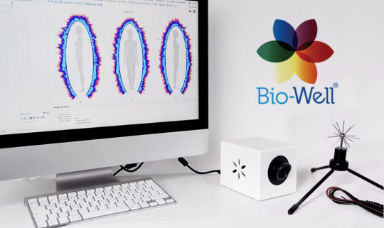
The Energy of Consciousness - new book by Dr. Korotkov 2014 - Bio-Well New model of GDVCAMERA – BIO-WELL opened next level of Electrophotonic Research. GDVCAMERA BIO-WELL has been developed by Dr. Konstantin Korotkov and brings the powerful technology known as Gas Discharge Visualization technique to market in a more accessible way than ever before. The product consists of a desktop camera and accompanying software, which allows a user to quickly and easily conduct human energy scans. Accessory attachments, like GDV SPUTNIK, BIO-COR and other are also available for purchase to conduct environment and object scans.

2014 - GDV Sputnik GDVSPUTNIK is a sensor and attachment system that affixes to the Bio-Well device, allowing for the energy of an environment to be read. BENEFITS of GDVSPUTNIK: Measure the energy of different places of interest during travels. Allows you to see the influence of Moon phases, Sun storms and environmental conditions to space, and, hence, to your wellbeing. Sputnik allows you to find the best position of a bed in your bedroom. Measure response to human emotions, meditations, pray, both individual and collective within an environment. Sputnik may detect the influence of music to the audience. Gas Discharge Visualization (GDV) technology was developed in Russia by the team of Professor Konstantin Korotkov in 1995. The GDV camera is a state-of-the-art computerized system that has superseded traditional Kirlian photography for several reasons. A major difference is that it allows direct, real-time viewing and analysis of changes in human energy fields since the data is quantified and analyzed by sophisticated software. Because the results are obtained so rapidly, it has become an ‘‘express-method’’ not only for diagnosis, but also detecting abnormalities that require more detailed investigation. Most importantly, since this technology and the protocols used are standardized, GDV results obtained by different investigators can be compared with reliability. The results are interpreted based on the energy connections of fingers with different organs and systems via meridians that have been used in acupuncture and traditional Chinese medicine for thousands of years. This technology has extraordinary implications for all health related fields, including conventional as well as complementary or alternative therapies. A comprehensive review of these varied GDV applications can be found in a recent book co-authored with Dr. E. Yakovleva from Moscow Medical University. Research with the GDV device is currently being carried out at universities and research institutes worldwide in medicine, "energy medicine", athletic training, biophysics, parapsychology, and other disciplines (please, see Papers section). GDV has been used in numerous significant research projects that have confirmed its usefulness and reliability and value. GDV technology provides a convenient and user friendly method to assess patients with a wide range of complaints and can also be utilized to assess responses to drugs, meditation, stress reduction therapy or any other interventions.

2014 - Human Light System Online Course Human Light System – Experimental Online 5 years Course. Online lectures, 2-3 times per week. 42 lectures per course. HLS 1.0 – Human Light System devices 2014/2015 Season 2015 - Bio-Well Water tester The Bio-Well Water electrode connects to the Bio-Well Calibration Unit similarly to the Sputnik and allows for the testing of water’s response to environmental stimuli. It is not designed for evaluation of water quality or comparing different types of water from quality standpoint. Bio-Well Water electrode
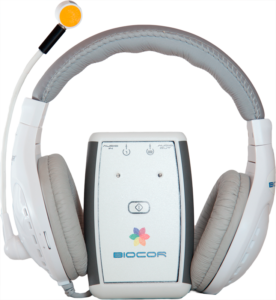
2015 - Biocor BIOCOR is a unique device that aids in shifting and correcting your energy state and balance through the use of high frequencies. This device can be used independently from BioWell or it can be used in conjunction with the Chakra audio setting in the BioWell software. BIOCOR – correction device of human energy field using information extracted fingers with GDV BIO-WELL. These results may help to develop a better quality of life. Biocor uses super Hight Frequency (SHF) in the range Higo-Hertz (4.9 mm (60.12 HHz), 5.6 mm (53,53 HHz) and 7.1 mm (42.19 HHz) of very low intensity (less than 10 mW/cm2). 2015 - Georges Vieilledent and Electrophotonique Ingenierie · France Electrophotonique Ingenierie is an innovative company working on corona effect and development of solutions based on the use of electromagnetic waves. From its basic research, the company has developed a unique patented device to highlight informational fields never identified to date. This device, designed for research laboratories, universities and specialized institutes, opens important perspectives in the healthcare and biotechnology. 2016 - The Biointernet meditation The Biointernet meditation - online experiment with GDV Sputnik. Every Saturday.

The Biointernet Meditation HLS 2.0 Online Course and Human Light System International Workshop, Prague. 2017 - The Energy of Space https://youtu.be/pnXGbwYLlYE The Energy of Space project established.Bio-Well Glove – new version of BioClip that will be called from now on Bio-Well Glove. Our research has shown that blue BioClip sensor had very low sensitivity and was almost useless in experiments.The Energy of Health - new book by Dr. KorotkovThe Energy of Space - new book by Dr. KorotkovHuman Light System Congress Training, Prague 2017 - New Books by Professor Konstantin Korotkov on IUMAB Library

IUMAB Library 2018 - Online Education by Korotkov.TV Bio-Well AnalysisBio-Well Applications Online workshops and trainings with GDVCAMERA BIO-WELL on Human Light System Course 2018 - Bio-Well 2.0 The same quality (Bio-Well 1.0) plus $600! Another one commercial product from Bio-Well company. 2018 - HLS Congress Beach Human Light System Congress on the Beach, Burgas, Bulgaria. HLS Congress Beach - practical 2 weeks the Biointernet training with Translighters and GDVCAMERA. HLS Congress Beach - new system of Education 2018 - GDVCAMERA Crownscopy updated
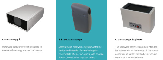

GDV Crownscopy Software 2019 - Bioelectrography Education Inside Human Light System Course: Bio-Well video courseGDV Diagnostics video courseGDV Diagnostics 2 with Kirill Korotkov and Alexander Dvoryanchikov GDV BIOELECTROGRAPHY – PAGES OF HISTORY AND MODERN DEVELOPMENT So where is the similarity in the experiments of Lichtenberg, Tesla and the lightning? In all those cases the gas discharge appears near the earth rod. High field intensity is formed near its sharp end when placed into an electrical field. Electrons, which always exist in the air or are emitted by the bodies, start speeding up in this field and, having picked up necessary speed, ionize air molecules. Those, in their turn, emit photons, mostly in the blue and ultraviolet spectral regions. Here the glow appears. What is more, from the viewpoint of physics both a nail, a tree, a human finger, and a person can be the rod. Everything depends on the scale. Generators used in Bioelectrography have very small power. It means that they can not give high current, even if you lick the electrode with your tongue. In addition, these generators make use of high-frequency voltages and short impulses, and by the laws of physiology such current can not penetrate into the organism, as it slides on the skin surface. In XX'th century many researchers was attracted by Kirlian photography, hundreds books and papers were published, but scientific acceptance of Kirlian photography was rather limited because the quality of equipment used by early investigators varied considerably and results were inconsistent since there was no standardization. Things improved when a multidisciplinary group headed by William Eidson, professor of physics at Drexel University in Philadelphia, showed it was possible to image electrical parameters of a specimen in real time, thus making it possible to map human energy fields and any rapid changes. This six-year project and related research were summarized in a 1976 article in the prestigious journal Science. Bioelectrography patents.

The History of Bioelectrography Light and Love, Dear Friends! Welcome to IUMAB! See more on IUMAB History Research Department Welcome to IUMAB! Be a Member! https://www.gdvplanet.com/product/umab-regular-memberships/ The History of Bioelectrography 2019 - Bio-Well Element - the same Bio-Well device, but GDVCAMERA Bio-Well Element What is the Bio-Well Element?

GDVCAMERA Bio-Well Element Bio-Well Element is a GDVCAMERA (gas discharge visualization) from 4 Generation of GDVCAMERA. It intended for Kirlian photography of various materials and liquids. It has a larger horizontal glass electrode than a GDV Bio-Well camera, which allows the user to lay conductive objects flat on its surface. This is main difference with GDVCAMERA Bio-Well 2.0. Bio-Well Element has the same low quality of images like Bio-Well or Bio-Well 2.0. 2019 Kirlian Photography as Art and Science - online Kirlian museum Kirlian Photography as Art and Science - group on Facebook Read the full article
#B.J.Navratil#Bio-Well#Bio-Well2.0#Bio-WellAnalysis#Bio-WellApplications#Bio-WellCompany#Bio-WellElement#Bioelectrography#Biophotons#Dr.KonstantinKorotkov#ElectroPhotonicImaging#Electrophotonics#GDV#GDV/EPI#GDVCAMERA#HistoryofBioelectrography#IKRA#IUMAB#Kirlian#KirlianResearchAssociation#Korotkov#LandelldeMoura#M.Pogorelski#NewtonMilhomens#NikolaTesla#PeterMandel#SemyonandValentinaKirlian#StanleyKrippner
11 notes
·
View notes
Link
ELECTROPHOTONIC ANALYSIS IN MEDICINE
GDV BIOELECTROGRAPHY RESEARCH
Dr. Ekaterina Yakovleva and
Dr. Konstantin Korotkov
This book is a survey of papers dedicated to Electrophotonic Imaging (EPI) GDVBioelectrography applications in Medicine and Psychology from 2000 to 2012. The most of cited works are presented in proceedings of different conferences, but a lot are published in per-review journals. It is clear that Electrophotonic technique has high potential in analyzing Human Energy Field for health and wellbeing and monitoring the reaction of people to different influences and treatments.
Dr. Ekaterina Yakovleva, M.D., Ph.D., Professor of the Russian National Research Medical University named after N.I. Pirogov, Moscow, Russia, and author of many papers published in per-review journals. From 1999 develops applications of Electtrophotonic Analysis in medicine. She has published 35 papers and a monography on this topic in Russian journals.
Dr. Konstantin Korotkov, Ph.D., Professor of Computer Science and Biophysics at Saint-Petersburg Federal Research University of Informational Technologies, Mechanics and Optics. Deputy Director of Saint-Petersburg Federal Research Institute of Physical Culture and Sport. He has published over 200 papers in leading journals on physics and biology, and he holds 17 patents on biophysics inventions. ISBN 978-1481932981
0 notes
Photo

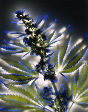
Here is a painting I did of a Leaf and it's electronic energy fields ☇🍁☇ "Kirlian photography is a collection of photographic techniques used to capture the phenomenon of electrical coronal discharges. It is named after Semyon Kirlian, who, in 1939, accidentally discovered that if an object on a photographic plate is connected to a high-voltage source, an image is produced on the photographic plate. The technique has been variously known as "electrography", "electrophotography", "corona discharge photography" (CDP), "bioelectrography", "gas discharge visualization (GDV)", "electrophotonic imaging (EPI)", and, in Russian literature, "Kirlianography". Kirlian photography has been the subject of mainstream scientific research, parapsychology research and art. To a large extent, It has been used in alternative medicine research." #painting #art #energy #field #leaf #vanished98art #photo #electrography #colors #nature #az #artist #kirlian #photography #zen #love #peace (at Phoenix, Arizona)
#energy#peace#electrography#kirlian#photography#photo#colors#leaf#painting#zen#love#field#nature#art#vanished98art#az#artist
0 notes
Text
ANALYSIS OF STRUCTURED LIQUIDS
ANALYSIS OF STRUCTURED LIQUIDS WITH ELECTROPHOTONIC TECHNIQUE Korotkov K, Ph.D., Professor, Orlov D. University of Informational Technologies, Mechanics and Optics, Saint Petersburg, Russia Key words: structured water, Electrophotonic imaging, Gas Discharge Visualization, high dilutions, consciousness influence

ANALYSIS OF STRUCTURED LIQUIDS Abstract Basic principles of Electrophotonics – Gas Discharge Visualization (EPI/GDV) technique – method of analysis of stimulated by electromagnetic field glow of liquids are discussed in the paper. Examples of experimental studies of different samples of water, blood reaction to allergens, low concentrations of different salts are presented. High selectivity and sensitivity of the EPI/GDV approach is proven by publications of different authors. EPI/GDV parameters depend on the chemical composition of a liquid, but most interesting is their dependence from structural properties of water and the possibility of data transfer through water. Measured parameters are defined by the emission activity of surface layer of liquid, which depends on surface-active valency. It is clear that this property is defined by the structure of the near-surface clusters, so the EPI/GDV method may serve as one of the informational methods of study the structural-informational properties of liquids. Developed approach allowed to distinguish the changes of electrophotonic parameters of water under the remote influence of the human consciousness – directed human attention. Introduction Currently considerable attention is being focused on the study of the structural properties of water and the possibility of data transfer through water. A lot of controversial information we may find concerning memory of water (Johansson, 2009; Montagnier, 2009, 2011). According to the viewpoint that has shaped, the phenomena observed during the experiments are determined by the processes of clusters and clathrates formation, mainly at the atoms of admixtures (Del Giudice, Vitiello, 2006). The task of introducing these notions into the scope of contemporary scientific thinking requires, first of all, a set of probative and reproducible experimental facts. Water is a complex subject of study, and its properties depend on a great number of factors; this requires that several independent techniques should be used in parallel, and that new informative methods for study of water properties should be developed and introduced into practice (Voeikov, Del Giudice, 2009). The high degree of informativeness of the Dynamic Electrophotonic Imaging (EPI) analysis based on Gas Discharge Visualization (GDV) method (Korotkov, 2002) applied for studying liquid-phase subjects was first demonstrated during the study of the glow of microbiological cultures (Gudakova et al, 1990), blood of healthy people and cancer patients (Korotkov et al, 1998), reaction of blood to allergens (Sviridov et al, 2003), homeopathic remedies of 30С potency (Bell et al, 2003), and very small concentrations of various salts (Korotkov, Korotkin, 2001). The differences between the glow parameters of the NaCl, KCl, NaNO3 and KNO3, solutions and distilled water were observed until the 2-15 dilution; however, the dynamic trends of the 2-15 dilution and distilled water still had different directions. Great interest has been roused by the studies directed at detecting the differences between the glow of natural and synthetic essential oils with identical chemical composition (Korotkov et al, 2004, Vainshelboim et al, 2004). The oils were analyzed in order to detect possible differences between oils that were obtained by means of natural and synthetic processes, between oils of organic and regular origin; between oils obtained in different climatic conditions and extracted by means of different methods; between oils with different optical activity; between fresh oils and oils that were oxidized by various methods. The combinations of oils under study did not show any statistically significant differences when analyzed by means of the gas chromatography method. Technique Study of Electrophotonic parameters of liquids is based on using commercially produced instrument “GDV Camera”, which is manufactured by KTI company, St. Petersburg (web. Ref 1,2). This instrument is well-known for analyzing stimulated photon emission from human fingers which is being used for health and well-being diagnostics (Measuring 2002), analysis of athletes (Bundzen et al, 2005), altered states of consciousness (Bundzen et al, 2002. Korotkov et al. 2005), influence of music (Gibson, Williams 2005) and Qigong to people (Rubik, Brooks 2005), as well as geo-active zones (Hacker et al, 2005) and minerals (Vainshelboim et al, 2005). When the EPI parameters are measured for liquid subjects, a drop of the liquid is suspended at 2-3 mm distance above the glass surface of the optical window of the device, and the glow from the meniscus of the liquid is registered (Fig.1). The volume of liquid is about 5*10-3 ml. Temperature is kept in the range 22-24 C, the relative humidity is maintained from 42% to 44%. The train of triangular bipolar electrical 10 mcs impulses of amplitude 3 kV at a steep rate of 106 V/s and a repetition frequency of 103 Hz, is applied to the conductive transparent layer at the back side of the quartz electrode thus generating electromagnetic field (EMF) at the surface of the electrode and around the drop. Under the influence of this field, the drop produces a burst of electron-ion emission and optical radiation light quanta in the visual and ultraviolet light regions of the electromagnetic spectrum. These particles and ions initiate electron-ion avalanches, which give rise to the sliding gas discharge along the dielectric surface (Korotkov, Korotkin 2001). A spatial distribution of discharge channels is registered through a glass electrode by the optical system with a charge coupled device TV camera, and then it is digitized in the computer.

ANALYSIS OF STRUCTURED LIQUIDS Full text PDF: 2012 Water EPI
Bioelectrography Water Research
Read the full article
#ANALYSISOFSTRUCTUREDLIQUIDS#Bio-Well#consciousnessinfluence#Electrophotonicimaging#Electrophotonics#GasDischargeVisualization#GasDischargeVisualization(EPI/GDV)#highdilutions#structuredwater
1 note
·
View note
Text
Quality Analysis for Digital Kirlian Effect
Quality Analysis for Digital Kirlian Effect
Enhanced Region-specific Algorithm: Image Quality Analysis for Digital Kirlian Effect Razak Mohd Ali Lee, Janifal Alipal Abstract Research on digital Kirlian effect especially its quality after certain algorithm taken part is overlooked. Thresholding the image in binary form couldn’t give an analysis enough details on its significant features. This study is introducing an Enhanced Region-specific algorithm, ERS to extract the captured digital Kirlian effect as human radiated energy inside an EPI (Electrophotonic Imaging) image. By utilizing image morphology transform, ERS is improving the procedure of blob extraction process by fitting an absolute arithmetic process in-between the gray-level and binary slice of the image. Henceforth, this paper is focusing on the image quality analysis after the process, subsequently offers a new diagnostic information on captured Kirlian effects through an EPI image. This paper present that the quality of processed digital effect under ERS algorithm are in lower MSE and higher PSNR with its correlation coefficient to its original image better than segmented and binary slices. Significant and most-significant details on the image are able to being preserved to its better quality using the proposed algorithm. Keywords Electrophotonic image, EPI Analysis, ERS algorithm, Enhanced Region-specific, Blob extraction, image morphology, Kirlian effect, Human Biofield
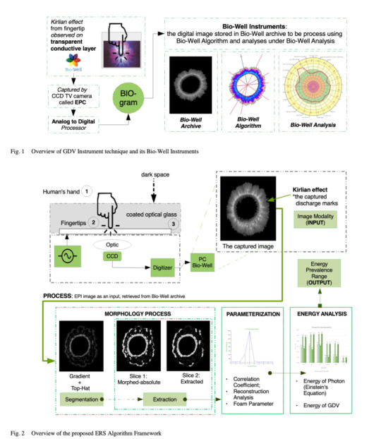
Quality Analysis for Digital Kirlian Effect 2020 Quality Analysis for Digital Kirlian Effect Conference: 2019 IEEE 15th International Colloquium on Signal Processing & Its Applications (CSPA) See also: Qualia and Time Sense, QQQ – Quality, Quantity, Qualia, Qualities and Quantities
Kirlian Photography and Kirlian Effect
Kirlian Photography is a process that uses pulsed high voltage frequencies & electron cascades to take pictures of usually invisible, radiating energy fields that surround us all. Photo techniques.
Kirlian Photography is a high voltage, contact print photography
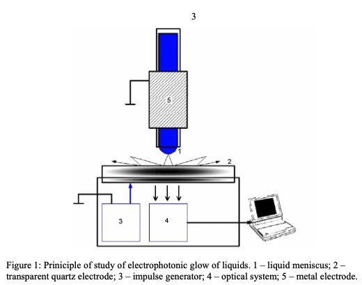
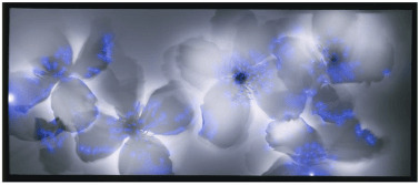
GDV Analysis of Electrophotonic Glow of Liquids Kirlian effect is a visible electro-photonic glow of an object in response to pulsed electrical field excitation. Semyon Kirlian and his wife who first recorded and studied it in detail since 1930s. Kirlian Photography process is simple. Sheet film is placed on top of a metal plate, called the discharge or film plate. The object to photograph is placed on top of the film. High voltage is applied to the plate momentarily to make an exposure. The corona discharge between the object and discharge plate passes through and is recorded onto the film. When the film is developed you have a Kirlian photograph of the object.
Kirlian and Kirlian Photography Kirlian Photography process, being a contact print process, doesn’t require the use of a camera or lens. However when a transparent electrode is substituted for the discharge plate it is possible to use a standard camera (with a bulb setting) or video camera.
Kirlian and Kirlian Photography Kirlian Photography is a simple process which can be even achieved without using a camera. The principle of Kirlian Photography is straight forward: if an object is placed on a photographic plate which is then connected to a source of high-voltage current, an image will appear on the photographic plate. The resulting image appears because of the electrical coronal discharge taking place between the subject and the metal plate.
Kirlian Photography and Kirlian Effect

Kirlian and Kirlian Photography Welcome to Kirlian and Kirlian Photography on IUMAB! Monitoring the Human Health by Measuring the Biofield, “Aura” Korotkov’s images Korotkov’s images, gender and age dependence Post mortem bioelectrographic images Korotkov’s images types GDV Based on Physical Methods and Meridian Analyses Light from the eyes Read the full article
#AnalysisforDigitalKirlianEffect#biowellchakra#Bioelectrography#BioeletrografiaDigital#DigitalKirlianEffect#digitalKirlianimage#DigitalKirlianPhotography#HumanBiofield#KirillKorotkov#Kirlian#KirlianandKirlianPhotography#Kirlianeffect#Kirlianphotography#Kirlian’seffect#KonstantinKorotkov#Korotkov#Korotkov'simages#Korotkov'simages-GDVimages
0 notes
Text
GDVCAMERA
GDVCAMERA by Dr. Korotkov - Gas Discharge Visualization technique
Stress/Energy level measurement and more GDVCAMERA has been developed by Dr. Konstantin Korotkov, Alexander Laptev, Dr. Alexander Kuznetsov, Boris Krylov and Olga Belobaba in 1995 and brings the powerful technology known as Gas Discharge Visualization technique to market in a more accessible way than ever before. The product consists of a desktop camera and accompanying software, which allows a user to quickly and easily conduct energy scans. Accessory attachments for GDVCAMERA are also available for purchase to conduct environment and object scans.

GDVCAMERAS - 1999-2012 GDV Technique is the computer registration and analysis of electro-photonic emissions of biological objects (specifically the human fingers) resulting from placing the object in the high-intensity electromagnetic field on the device lens. When a scan is conducted, a weak electrical current is applied to the fingertips for less than a millisecond. The object’s response to this stimulus is the formation of a variation of an “electron cloud” composed of light energy photons. The electronic “glow” of this discharge, which is invisible to the human eye, is captured by the camera system and then translated and transmitted back in graphical representations to show energy, stress and vitality evaluations. See also: GDV scientific basis Electrophotonics application ABOUT Dr. Konstantin Korotkov

Dr. Konstantin Korotkov is a Professor of Physics at St. Petersburg State Technical University in Russia. He is a leading scientist internationally renowned for his pioneering research on the human energy field. Professor Korotkov developed the Gas Discharge Visualization technique, based on the Kirlian effect. GDV, EPI, etc - please see more Different names of Bioelectrography. Bio-Well – model of GDVCAMERA by Dr. Korotkov GDVCAMERA BIO-WELL - Instrument to reveal Energy Fields of Human and Nature The image (GDV Image, EPI Image, Korotkov's Image, Bio-Well Image), which we create in GDVCAMERA, is based on ideas of Traditional Chinese Medicine and verified by 20 years of clinical experience by hundreds of practitioners with many thousands of patients. The scanning process is quick, easy and non-intrusive. Get real time feedback on what factors - positive and negative - affect your stress state. With the GDV Sputnik measure environment and object energies too. GDVCAMERA is not a medical instrument. It provides an impression of your energy and stress levels and allows users to see their day-to-day transformation and the influence of different situations and stimuli. Friendly software makes data processing simple and convenient for non-experienced users.

Bio-Well by Professor Konstantin Korotkov More about the latest generation of GDVCAMERA: Bio-Well 1.0* Bio-Well 2.0* Bio-Well Reviews Bio-Well Reviews (main page) bio-well practitioner near me, bio well software download, bio well chakra, bio well assessment, bio well training, biowell app, biowell research, gdv camera amazon, bio well assessment, Bio-Well Accessories * Since 2018 Bio-Well company belong to Natalja Romanova Read the full article
0 notes
Text
IUMAB Review

IUMAB Review
Annual Digest Established by Professor Pavel Bundzen and Kirill Korotkov in 1998 The International Union of Medical and Applied Bioelectrography publishes the IR digest one time per year, and is free to all Regular Members. It contains original articles, news and book reviews. Current and past editions of IUMAB Review are ready for download as PDF files for IUMAB Regular Members or view online IR History: 2015 - present days - Korotkov Subscriber / e-mail Event, Science news, links, community, digest, workshops-webinars 2010 - present days - IUMAB Review / PDF digest 2006 - 2009 - IUMAB News / e-mail news, links, community, Science digest 1998 - 2005 - Korrect News / e-mail digest, news

2010

2011

2012 2013, 2014, 2015, 2016, 2017, 2018 Established by Professor Pavel Bundzen Edited by Kirill Korotkov Since 1998 IUMAB Library – books, texts, videos and images about: Bioelectrography, Electrophotonic Imaging, Gas Discharge Visualisation, Kirliangraphy, Kirlian Photography, Electrophotography, GDV, EPC, EPI, etc IUMAB Library Consciousness Research, The Biointernet, Human Light System, Emotions, Yoga and Meditation, New Psychology, Intuitive Information Sight, Distant Influence and more on IUMAB Library Read the full article
#Bio-WellbyDr.Korotkov#Bioelectrography#BioelectrographyPublications#Biophotons#community#digest#Dr.Korotkov#e-mailEvent#HistoryofBioelectrography#IUMABReview#Korotkov#KorotkovSubscriber#KorrectNews#NewtonMilhomens#Sciencedigest#scientificreport
0 notes
Text
Bioelectrography cancer Research
Bioelectrography (EPI/GDV) cancer Research

GDV/EPI software 1. Targeted therapies in gastroesophageal cancer. Kasper S., Schuler M. European Journal of Cancer. 2014:50(7): 1247-1258. 2. Status of cancer care in Russia in 2010 ed. Chissova VI, Starinskiy VV Petrova GV, FGU "MNIOI them. PA Herzen" Health Ministry, 2010. 3 Korotkov KG. Human Energy Field: Study with GDV Bioelectrography. Fair Lawn, NJ: Backbone Publishing Co: 2002. 4. Quantum Events of Biophoton Emission Associated with Complementary and Alternative Medicine Therapies. Hossu M, Rupert R. J Altern Complement Med. 2006, 12(2): 119-124. 5. Bio-electrographic method for preventive health care. Cohly H, Kostyuk N, Isokpehi R, Rajnarayanan R. Biomedical Science & Engineering Conference, First Annual ORNL, 2009,1-4. 6. GDV in Evaluation of Cognitive Functions. Rgeusskaja GV, Listopadov UI. Medical Technology of Electrophotonics J Sci Healing Outcome. 2009, 2(5): 16-19. 7. Biometric Evaluation of Anxiety in Learning English as a Second Language. Kostyuk N, Meghanathan N, Isokpehi R.D., Bell T, Rajnarayanan R, Mahecha1 O, Cohly H., International Computer Science and Network Security, 2010, 10(1): 220-229. 8. Gas Discharge Visualization (GDV) Technique is New in Diagnostics for Patient with Arterial Hypertension. Aleksandrova E, Zarubina T., Kovelkova M., Strychkov P, Yakovleva E. Journal of New Medical Technologies 2010,XVII(1):122-125. 9. The new conceptual approach to the early diagnosis of cancer. Horowitz BL, Krylov BA, Korotkov KG In: From Kirlian effect to Bioelectrography. - St. Petersburg., 1998. - P.125-132. 10. Comparison Bioelektrographic images of cancer patients and healthy subjects. Chowhan R.S., Radzharan P., Rao S. In: From Kirlian effect to Bioelectrography. St. Petersburg., 1998. - P.133-140. 11. GDV in monitoring of lung cancer patient condition during surgical treatment Vepkhvadze R., Gagua R., Korotkov K. et al. Georgian oncology. Tbilisi. 2003. № 1(4). p. 60. 12. The Possibility of Using Electrophotonics in the Early Diagnosis of Polyps and Colon Cancer. Saeidov W. In: Proceedings of XIV International Scientific Congress on Bioelectrography. St Petersburg 2010: 22-23. 13. Identifying Patients with Colon Neoplasias with Gas Discharge Visualization Technique. Yakovleva E G., Buntseva OA, Belonosov SS, Feorov ED, Korotkov KG, and Zarubina TV. The Journal Of Alternative And Complementary Medicine. V.21, N. 11, 2015, pp. 720–724 DOI: 10.1089/acm.2014.0168 14. Engineering Approach To Identifying Patients With Colon Tumors On The Basis Of Electrophotonic Imaging Technique Data. E.G. Yakovleva, K.G. Korotkov, E.D. Fedorov, E.V. Ivanova, R.V. Plahov, S.S. Belonosov The Open Biomedical Engineering J. 2016, 2, 72-80. DOI: 10.2174/1874120701610010072 15. USE OF ELECTROBIOGRAPHIC PHOTO ON COMPARINSON AMONG BREAST CANCER, HEALTHY SEDENTARY, AND HEALTHY RUNNERS WOMEN Kelmerson Henri Buck1,3, Claudio Novelli1,3,8, Fernanda T. Costa1,3, Gustavo C. Martins1,3, Heleise F. R. Oliveira1,3,4, Leandro B. Camargo1,3, Raul Marcel Casagrande1,2,3, Rodrigo R. Dias dos Reis7, Valter R. Moraes1,3, Fabio S. F. Vieira1,3,6, Ricardo Pablo Passos1,3, Guanis de Barros Vilela Junior1,3,5. 16. ONCOLOGICAL KIRLIANGRAPHICAL DIAGNOSIS Published in the “Revista do Hospital das Forças Armadas” (Magazine of the Hospital of the Armed Forces) - (H.F.A.) Brasília (DF) - Brazil Edition of Out/Dec - 1987 Authors: Dr. Hélio Grott Filho, Dr. Júlio Grott 17. Yogic Perspective on Cancer Pradeep B. Deshpande1,* and James P. Kowall2 List of Collaborators 18. THE GDV TECHNIQUE APPLICATION IN ONCOLOGY Gagua PO, Gedevanishvili EG, Georgobiani LG, Kapanadze A., Korotkov KG, Korotkina SA, Achmeteli GG, Kriganivski EV. Read the full article
0 notes
Text
Bioelectrography and Surgery
Bioelectrography and Surgery
For several years research project for evaluation of patients’ condition after surgery was conducted in Saint Petersburg Military Medical Academy. A lot of papers were published in Russian medical journals and two PhD in medicine were awarded. Two papers were published in English . The main conclusions were as follows: 1. There are reliable differences between parameters of GDV-grams of practically healthy people and patients with chronic abdominal surgical pathology. 2. The data obtained indicate that GDI parameters are connected with the functional status of the organism and reflect the severity of the somatic state of patients with abdominal surgical pathology. 3. The most informative parameters are: “integral area of glow JS” in the “GDV Diagram” program; “total” and “normalized area”, “total density”, “average brightness”, as well as “fractality” and “form coefficient” in the “GDV Processor” program. 4. The most informative mode of registration of GDV-grams is the mode “without filter”. On the whole, the application of the filter keeps the trend of changes, but they are often less pronounced and lose statistical significance. 5. The revealed variability of GDV-gram parameters depending on the sex and age of patients indicates that it is necessary to determine their individual norms. 6. The dependence of perioperative dynamics of a number of indices on the severity of the patient's somatic state, the patient’s age and the duration of surgical procedure enables functional monitoring in the postoperative period, and evaluation of operative stress. 7. The parameters of EPI-grams reliably change in response to the operative trauma, and their dynamics depend on the severity of the somatic state of patient, which allows using the technique for functional monitoring of patients in postoperative period, as well as for the assessment of the operative stress. The degree of intensity of changes depended on the volume and character of the undergone physical intervention. Thus, patients who had laparoscopic cholecystectomy showed much smaller changes of bioenergy status in comparison with patients, who had stomach and intestine's operations. 8. The Electrophotonic Imaging technique is mostly advisable for the dynamic assessment of the functional state of patient in perioperative period. Not all the fingers may be used, at that, but only one finger of each hand. For example, the fourth finger, where the changes are the most significant.

Bioelectrography and Surgery Fig. . Dynamics of some GDV indices in perioperative period. Stages of research: 1- before surgery, 2 – 1st hour after surgery, 3 - in 1 day after surgery, 4 – in 2 days after surgery, 5 – in 3 days after surgery, 6 – in 4 days after surgery, 7 – in 5 days after surgery. Dynamics of GDV-gram parameters of systems in perioperative period for 43 people. (0 – before surgery, 1 – first hour after surgery, 2-5 days after surgery). Important approach for monitoring of GDV parameters to predict the development of postoperative delirium was developed by Strukov EU. and Tuzhikova N.V. . 122 people were surveyed using prospective a GDV technique. Electrophotonic Analysis in Medicine Bioelectrography and Surgery Bioelectrography and Surgery GDV technique Three groups were examined. The first (control) were 47 healthy people surveyed by GDV against the background of psycho- emotional well-being. The second group included 50 patients operated on the abdominal organs. GDV-grams were recorded in the group before surgery and during the next five days postoperatively. The third group consisted of 25 patients treated at the clinic of psychiatry with the abstinent syndrome in pre-and delirious state. The most informative was glow area parameter. In series taken in dynamics the most significant changes were revealed. Dynamics of the glow area parameter of patients, whose postoperative period is complicated by the development of delirium, is different from the normal distribution and was characterized by high amplitude of the GDV area. Dynamic changes in the glow area were similar to the dynamics of GDV images of patients with psychiatric profile. However, these changes in the operated patients may be revealed 10-12 hours prior to the development of the clinical picture of delirium. As the delirious syndrome subsided, the parameter of GDV area comes back to the original data and fits into the standard distribution. Such performance of dynamics of the GDV area and its fundamental similarity to that of patients with psychiatric profile allows us to speak with confidence about the possibility of prognosis of delirious syndrome in patients operated on the abdominal organs in the immediate postoperative period, even before the development of complications. Bioelectrography and Surgery Read the full article
#Bioelectrography#BioelectrographyandSurgery#ELECTROPHOTONICANALYSISINMEDICINE#GDV-gram#Korotkov'simages#Surgery
0 notes
Text
Electro-diagnostic techniques
ELECTROPHOTONIC IMAGING (GDV) TECHNIQUE Konstantin Korotkov, PhD, Professor Electro-diagnostic techniques such as Electro-encephalogram and Electro-cardiogram are widely used in medical practices worldwide. A promising method already utilized in sixty-two countries to great success is bioelectrography, based on the Kirlian effect. This effect occurs when an object is placed on a glass plate and stimulated with current; a visible glow occurs, the gas discharge. With EPI/GDV (electro-photon imaging through gaseous discharge visualization) bioelectrography cameras, the Kirlian effect is quantifiable and reproducible for scientific research purposes. Images captured (EPI-grams) of all ten fingers on each human subject provide detailed information on the person’s psycho-somatic and physiological state1. The EPI/GDV camera systems and their accompanying software are currently the most effective and reliable instruments in the field of bioelectrography2,3,4,5. EPI/GDV applications in other areas are being developed as well6,7,8,9,10,11. Through investigating the fluorescent fingertip images, which dynamically change with emotional and health states, one can identify areas of congestion or health in the whole system. The parameters of the image generated from photographing the finger surface under electrical stimulation creates a neurovascular reaction of the skin, influenced by the nervous-humoral status of all organs and systems. Due to this, the images captured on the EPI/GDV register an ever-changing range of states. In addition, most healthy people’s EPI/GDV readings vary only 8- 10% over many years of measurements, indicating a high level of precision in this technique. A specialized software complex registers these readings into parameters which elucidate the person’s state of wellbeing at that time12. The EPC/GDV camera is presently the state-of-the-art in bioelectrography. It utilizes a high frequency (1024 Hz), high-voltage (10 kV) input to the finger (or other object to be measured), which is placed on the electrified glass lens of the EPC camera. Because the electrical current applied to the body is very low, most human subjects do not experience any sensation when exposing their fingertip to the camera. In practice, the applied electric field is pulsed on and off every 10 microseconds, and the fingertip is exposed for only 0.5 seconds. This causes a corona discharge of light-emitting plasma to stream outward from the fingertip. The light emitted from the finger is detected directly by a CCD (charge-coupled detector), which is the state-of-the-art in scientific instruments such as telescopes to measure extremely low-level light. The signal from the CCD is sent directly to a computer, and software analysis is done to calculate a variety of parameters that characterize the pattern of light emitted, including brightness, total area, fractality, and density. The software can also provide color enhancement to enable subtle features such as intensity variations of the image to be perceived. The underlying principle of camera operation is similar to the well-known Kirlian effect13 but modern technology allows reproducible stable data with quantitative computer analysis. Purposeful investigations allowed the discovery of the parameters that are optimal from the point of obtaining critical information on the biological object’s state with the minimum of invasiveness. These findings are described in more than 200 research works in the international scientific literature, 15 patents, 9 books in English, French, German, Italian, Russian, and Spanish. This biophysical concept of the principles of GDV measurements is based on the ideas of quantum biophysics714. This is a further development of well-known ideas of A. Szent-Györgyi concerning the transfer of electron-excited states along the chains of molecular protein complexes815. The EPC technique measures the level of functional energy stored by the particular systems of an organism. This level is defined by the power of the electron-excited states and the character of their transport along the chains of albumin molecules. The level of functional energy is correlated with health status, but it is only one of many of the components that define health. It works together with genetic predisposition, psycho-emotional states, environmental loading (food, water, air, ecology) and other factors. This approach may be associated with the oriental notion of the energy transfer along meridians.

Electro-diagnostic techniques Read the full article
0 notes
Text
GDV/EPI Papers published in 2016-2017
IUMAB Library
IUMAB Library
GDV/EPI Papers published in 2016-2017 From the time of the previous Congress several papers have been published. Herewith we present the list of published papers. Публикации 2016 – 2017 гг Со времени, прошедшего с прошлого Конгресhe list of papersса, у нас вышел целый ряд публикаций, использующих Био-Велл. Приводим их список. Банаян А.А. Грачев А.А. Коротков К.Г., Короткова А.К. Прогноз соревновательной готовности спортсменов-паралимпийцев на базе оценки циркадного ритма на спортивных мероприятиях методом ГРВ. Адаптивная физическая культура. № 2 (66) 2016, 2-5.Korotkov K.G. The Energy of Health. 2017. Amazon.com Publishing.Korotkov K.G. L’Energie de la Sante. 2017. Amazon.it Publishing.E.G. Yakovleva, K.G. Korotkov, E.D. Fedorov, E.V. Ivanova, R.V. Plahov, S.S. Belonosov Engineering Approach To Identifying Patients With Colon Tumors On The Basis Of Electrophotonic Imaging Technique Data. The Open Biomedical Engineering J. 2016, 2, 72-80. DOI: 10.2174/1874120701610010072Kuldeep K. Kushwah, Thaiyar M. Srinivasan, Hongasandra R. Nagendra, Judu V. Ilavarasu. Effect of yoga based techniques on stress and health indices using electrophotonic imaging technique in managers. Journal of Ayurveda and Integrative Medicine 7, 119-123, 2016Kuldeep Kumar Kushwah, Thaiyar M Srinivasan, Hongasandra R Nagendra, and Judu V Ilavarasu. Development of normative data of electro photonic imaging technique for healthy population in India: A normative study. Int J Yoga. JanJun; 9(1): 49–56. 2016, doi: 10.4103/09736131.171713Shiva K.K., Srinivasan TM., Guru Deo, VenkataGirikumar P., Nagendra HR. Electro- photonic imaging for detecting intervention (meditation). Intern J of Current Medical and Pharmaceutical Research. 2, 2016Romesh Kumar Bhat, Guru Deo, Ramesh Mavathur, and Thaiyar Madabusi Srinivasan. Correlation of Electrophotonic Imaging Parameters With Fasting Blood Sugar in Normal, Prediabetic, and Diabetic Study Participants. Journal of Evidence-Based Complementary & Alternative Medicine 1-8, 2016. DOI: 10.1177/2156587216674314Shiva K.K., Srinivasan TM., Nagendra HR., Marimuthu P. Electrophotonic Imaging Based Analysis of Diabetes. Int J of Altern and Complement Medicine. 2016. 4 (5): 134- 137Kelmerson Henri Buck1,3, Claudio Novelli1,3,8, Fernanda T. Costa1,3, Gustavo C. Martins1,3, Heleise F. R. Oliveira1,3,4, Leandro B. Camargo1,3, Raul Marcel Casagrande1,2,3, Rodrigo R. Dias dos Reis7, Valter R. Moraes1,3, Fabio S. F. Vieira1,3,6, Ricardo Pablo Passos1,3, Guanis de Barros Vilela Junior. O uso da bioeletrografia na comparação entre mulheres com câncer de mama, mulheres saudáveis sedentárias e mulheres praticantes de corrida. Centro de Pesquisas Avançadas em Qualidade de Vida | Vol. 8| No. 2 | Ano 2016 | p. 9 GDV/EPI Papers published in 2016-2017 GDV estimation of homeostasis of ontological patients during singlet oxygen therapy rehabilitation after radical methods of therapy E. Gedevanishvili, R. Gagua, I. Rusov, A. Kapanadze, L. Giorgobiani Universal medical center of Georgia (National Cancer Center of Georgia) Association of Oncologists of Georgiaю Association of Medical Physicists of Georgia Association of Integral Medicine of Georgia One of the main problems of oncology is optimization of therapy. That is why development of new methods of evaluation of organism conditions in process of therapy and after it is very important task for the oncology. The aim of this study was as follows: 1. Create of new computed GDV (Gas Discharge Visualization) technology for holistic estimation of functional state of Human organism and its monitoring in the process of therapy of oncology patients; 2. Create of new computed GDV (Gas Discharge Visualization) technology for holistic estimation of compensation ability of Human organism and its monitoring in the process of therapy of oncology patients. There were 20 patients on chemotherapy and 10 patients on radiotherapy with cancer of different localization. After completion of main course of therapy patients received the rehabilitation course of Singlet Oxygen therapy. Our results showed that after main courses of therapy all GDV parameters was worsened and after rehabilitation course the main part of this parameters became better. These results allowed us to conclude that cancer patients need rehabilitation after main courses of radical methods of therapy. To elaborate the technology of GDV monitoring more factual material is needed. ADOPTING ELECTROPHOTONIC IMAGING IN A WESTERN HEALTH PARADIGM: A LITERATURE REVIEW Brennan Carrithers, M.S.1 & Aaron Carrithers, [email protected] of Louisville School of Medicine; 2University of Kansas Medical Center In Western societies there exists an increasing prevalence of chronic diseases and overall decline of population health. The U.S. has recently launched the National Center of Complementary and Integrative Health which supports research that studies integrative medicine (IM) approaches to healing. This effort coincides with a prevention-based medical paradigm. IM aims to heal the patient as aunified system, focusing on the patient’s mind, body, and spirit.Despite the increase of attention, many biomedical (allopathic) physicians remain unconvinced of the significance of integrative health. The large majority of Western-trained physicians discredit IMmodalities because they’re based on the concept of a permeating life force, notable in ancient Eastern medicine. Many conventional scientists argue that there is a lack of objective evidence and scientific understanding supporting the existence of this esoteric bioenergy. Electrophotonic Imaging (EPI) is a technique used to capture and quantify biological energy, offering a bridge between ancient Eastern philosophy and mainstream Western medicine. Although this technique is being used worldwide for many purposes, much of the time, researchers in the U.S. decline opportunities to investigate the clinical utility of EPI due to the lack of data published in peer- reviewed journals. This article reviews existing peer-reviewed evidence and research regarding the clinical capabilities of EPI. Additionally, we offer a biomedical framework for its mechanism of action. Moreover, this article will facilitate the implementation ofEPI’s function of assessing bioenergy, IM therapies, and biopsychosocial wellness in a Western health paradigm. GDV ESTIMATION OF HOMEOSTASIS OF ONTOLOGICAL PATIENTS DURING SINGLET OXYGEN THERAPY REHABILITATION AFTER RADICAL METHODS OF THERAPY E. Gedevanishvili, R. Gagua, I. Rusov, A. Kapanadze, L. Giorgobiani Universal medical center of Georgia (National Cancer Center of Georgia) Association of Oncologists of Georgia Association of Medical Physicists of Georgia Association of Integral Medicine of Georgia One of the main problems of oncology is optimization of therapy. That is why development of new methods of evaluation of organism conditions in process of therapy and after it is very important task for the oncology. The aim of this study was as follows: 1. Create of new computed GDV (Gas Discharge Visualization) technology for holistic estimation of functional state of Human organism and its monitoring in the process of therapy of oncology patients; 2. Create of new computed GDV (Gas Discharge Visualization) technology for holistic estimation of compensation ability of Human organism and its monitoring in the process of therapy of oncology patients. There were 20 patients on chemotherapy and 10 patients on radiotherapy with cancer of different localization. After completion of main course of therapy patients received the rehabilitation course of Singlet Oxygen therapy. Our results showed that after main courses of therapy all GDV parameters was worsened and after rehabilitation course the main part of this parameters became better. These results allowed us to conclude that cancer patients need rehabilitation after main courses of radical methods of therapy. To elaborate the technology of GDV monitoring more factual material is needed. ELECTRO-PHOTONIC IMAGING (BIO-WELL) AS A TRANSLATIONAL TECHNOLOGY WITH YOGA PHILOSOPHY CAN UNVEIL A UNIFIED SYSTEM OF MEDICINE Rajan Narajanan Electro-Photonic Imaging (EPI) measures instant communication within the human system. The nature of this communication seen from the lens of modern anatomy and physiology is not well understood. The objective of this study is to propose a model of Unified System of Medicine (USM) based on yoga philosophy with corroborating observations from life sciences and physical sciences and methods used in yoga and traditional medicine, and suggest that EPI is a translational technology that can help to bridge yoga and traditional medicine with conventional life sciences, and further research and advancement in medicine. Research in the areas of epigenetic factors instantly affecting gene transcription, consistent gene transcription leading toneuroplasticity, functionality of the brain, role of ‘placebo’ and ‘nocebo’ effect’, acupuncture mechanism, electro-dermal response in acupuncture points of Electro-Acupuncture according to Voll (EAV) and EPI previously known as Gas Discharge Visualization (GDV), emerging research in the area of Primo Vascular System (PVS), Marma and Reflexology, cell rhythm of Randoll, role of sleep and immune system, study of monozygotic twins, experience of psychedelic drugs, non-contact healing, coherence in space and water as the basis of communication are reviewed against the philosophy and principles of yoga, and its manifestation in traditional systems of medicine. Then USM is proposed with four sources of input and one source of output in the form of gene expression, and on a moment by moment basis the gene expression combined with the neural system of the person makes the person at the moment. EPI is presented as a translational technology to enable this measurement. Measured Yoga Therapy (MYT) developed by the author with the EPI system Bio-Well is presented as an example of a non-invasive treatment that corroborates EPI as a translational technology while supporting the concept of USM. In conclusion the impact of such a model of the human system on research and the future of medicine is noted. Resonance Acoustics Device (RAD) Acoustic Influence Based On Psycho-Physiological Reactions Of Human Organism (Formed By Evolution Process) With RespectTo «Individual Norm Model». Using Sound And Music ForHealing, Changing The Brain State (Neurological Activity) Negodaylov A.N. Institute of Human BioRestoration, Moscow, Russia [email protected] A new generation acoustic system is proposed — Resonance Acoustic Device (RAD). Acoustic system has the properties of an ideal sound source —pulsing sphere — in best manifestation possible for the material world. RAD was developed as an alternative to traditional acousticcompression type systems. System“s work is based on re-reflected sound, diffuse sound field, compulsory nonlinear amplitude- frequency response, circular beam pattern. RAD system has been tested in several research institutions (National Center for Child Health; St. Petersburg State University; Emergency Medicine Institute; Medical center «Eurika»; Institute of HumanBioRestoration; Psycho-neurological boarding school #18, etc.). Obtained results allow us to make a conclusion that RAD system can be efficiently used for adaptation of human organism in stressful situations, and for minimization of rehabilitation time after the stress. It is recommended for various rehabilitation and adaptation methods. PYRAMIDS AROUND THE WORLD & BOSNIAN PYRAMID HEALING ENERGY Dr. Sam Osmanagich, Ph.D. Discoverer of the Bosnian pyramids Foreign member of Russian Academy of Natural Sciences Director of Center for Archaeology and Anthropology at American University in Bosnia-Herzegovina www.samosmanagich.com Dr. Osmanagich will present his investigation of the pyramids around the world and Bosnian Pyramid discovery that has become the most active archaeological site in the world. He will present scientific, energy, spiritual and healing aspect of the pyramid energy. PYRAMIDS AROUND THE WORLD The world of the past was the world of the pyramids. Author investigates hundreds of pyramids in China, Mauritius, Canary Islands, Bolivia, Peru, Indonesia, Mexico, Egypt, Guatemala, Honduras, El Salvador, Belize, Spain, Italy, Greece, Sudan and US making original conclusions on their origin. LOST PYRAMIDS OF BOSNIA Dr. Sam Osmanagich discovered first pyramids on European soil: Lost pyramids of Bosnia. After 360.000 hours in archaeological digging in a period 2005-2017, sample testing, radiocarbon dating and five scientific conferences, this discovery prove that our view of ancient past has to be changed. Bosnian pyramids are the biggest and oldest pyramidal structures on the Planet, with the best quality concrete and most precise side orientation to the cardinal points. Despite obstacles from cultural, media and political establishments, this project has grown to the most active archaeological site in the World. ENERGY PHENOMENA OF BOSNIAN PYRAMIDS Physicists, electrical and sound engineers detected and measured several artificial energy phenomena on the top of the Bosnian Pyramids: electromagnetic beam of 28 kHz, ultrasound, infrasound and beneficial Schuman resonance of 7.83 Hz. Pioneering steps have been made in the Pyramid Science bringing us step closer in deciphering their true purpose and connection with Tesla’s free energy. MYSTERIOUS LABYRINTH UNDER THE BOSNIAN PYRAMIDS The most extensive underground network of tunnels and chambers has been discovered in the Bosnian Valley of the Pyramids 100 feet below the surface. Tunnels run for dozens of miles. Absence of cosmic radiations and natural radioactivity and high concentration of negative ions make this underground world extremely beneficial to human health. Experiments prove that this complex was used as a place for rejuvenation and self-healing. INFORMATIONAL IMPACT OF MODERN TECHNOLOGIES ON THE ENVIRONMENT, HUMAN BEINGS AND ANIMALS VIA GEOLOGICAL RUPTURES Alexandre Rusanov, SARL TELLUS [email protected] Continuously increasing scale of anthropomorphic influence on our planet Earth, contamination with chemicals and radioactive wastes, and industrial electric currents are causing changes of chemical and physical properties of the lands. It was found, particularly in France, that construction of cellphone base stations, wind generator towers, earthing of the solar panels, in the places of earth ruptures cross-sections, where water flows, causes appearance of new type of irradiation – geotechnogenic or informational. This irradiation is transmitted inside the ruptures for several kilometers and can cause severe problems for the health of human beings and animals that leave right above these ruptures. Examples of such influences on human beings and animals were studied. Special corrective devices were developed and are being used for protection and correction of negative informational influences of ruptures and artificial electromagnetic emissions. Scientific research has been made in order to study the possibilities of application of EPI/GDV method and other methods for measuring and quantifying the influence of the corrective devices (installed in the house) on the persons state. Influences on water of these corrective devices were studied in laboratory (Moscow) by the use of liquid chromatography method, and in-field studies were made on the farm and in the city hall building in France by measuring water’s conductivity withdifferential conductometric method. Measurements and experiments were made in houses, apartments and in factories in France, Germany, Switzerland, Belgium, Bulgaria, Netherlands, Albania, Greece, Morocco, Tunisia, Spain, Russia and Ukraine, on cattle farms in France, Belgium and Netherlands. GDV/EPI Papers published in 2016-2017 From the time of the previous Congress several papers have been published. Herewith we present the list of published papers. Публикации 2016 – 2017 гг Со времени, прошедшего с прошлого Конгресhe list of papersса, у нас вышел целый ряд публикаций, использующих Био-Велл. Приводим их список. Банаян А.А. Грачев А.А. Коротков К.Г., Короткова А.К. Прогноз соревновательной готовности спортсменов-паралимпийцев на базе оценки циркадного ритма на спортивных мероприятиях методом ГРВ. Адаптивная физическая культура. № 2 (66) 2016, 2-5.Korotkov K.G. The Energy of Health. 2017. Amazon.com Publishing.Korotkov K.G. L’Energie de la Sante. 2017. Amazon.it Publishing.E.G. Yakovleva, K.G. Korotkov, E.D. Fedorov, E.V. Ivanova, R.V. Plahov, S.S. Belonosov Engineering Approach To Identifying Patients With Colon Tumors On The Basis Of Electrophotonic Imaging Technique Data. The Open Biomedical Engineering J. 2016, 2, 72-80. DOI: 10.2174/1874120701610010072Kuldeep K. Kushwah, Thaiyar M. Srinivasan, Hongasandra R. Nagendra, Judu V. Ilavarasu. Effect of yoga based techniques on stress and health indices using electrophotonic imaging technique in managers. Journal of Ayurveda and Integrative Medicine 7, 119-123, 2016Kuldeep Kumar Kushwah, Thaiyar M Srinivasan, Hongasandra R Nagendra, and Judu V Ilavarasu. Development of normative data of electro photonic imaging technique for healthy population in India: A normative study. Int J Yoga. JanJun; 9(1): 49–56. 2016, doi: 10.4103/09736131.171713Shiva K.K., Srinivasan TM., Guru Deo, VenkataGirikumar P., Nagendra HR. Electro- photonic imaging for detecting intervention (meditation). Intern J of Current Medical and Pharmaceutical Research. 2, 2016Romesh Kumar Bhat, Guru Deo, Ramesh Mavathur, and Thaiyar Madabusi Srinivasan. Correlation of Electrophotonic Imaging Parameters With Fasting Blood Sugar in Normal, Prediabetic, and Diabetic Study Participants. Journal of Evidence-Based Complementary & Alternative Medicine 1-8, 2016. DOI: 10.1177/2156587216674314Shiva K.K., Srinivasan TM., Nagendra HR., Marimuthu P. Electrophotonic Imaging Based Analysis of Diabetes. Int J of Altern and Complement Medicine. 2016. 4 (5): 134- 137Kelmerson Henri Buck1,3, Claudio Novelli1,3,8, Fernanda T. Costa1,3, Gustavo C. Martins1,3, Heleise F. R. Oliveira1,3,4, Leandro B. Camargo1,3, Raul Marcel Casagrande1,2,3, Rodrigo R. Dias dos Reis7, Valter R. Moraes1,3, Fabio S. F. Vieira1,3,6, Ricardo Pablo Passos1,3, Guanis de Barros Vilela Junior. O uso da bioeletrografia na comparação entre mulheres com câncer de mama, mulheres saudáveis sedentárias e mulheres praticantes de corrida. Centro de Pesquisas Avançadas em Qualidade de Vida | Vol. 8| No. 2 | Ano 2016 | p. 9 GDV/EPI Papers published in 2016-2017 GDV estimation of homeostasis of ontological patients during singlet oxygen therapy rehabilitation after radical methods of therapy E. Gedevanishvili, R. Gagua, I. Rusov, A. Kapanadze, L. Giorgobiani Universal medical center of Georgia (National Cancer Center of Georgia) Association of Oncologists of Georgiaю Association of Medical Physicists of Georgia Association of Integral Medicine of Georgia One of the main problems of oncology is optimization of therapy. That is why development of new methods of evaluation of organism conditions in process of therapy and after it is very important task for the oncology. The aim of this study was as follows: 1. Create of new computed GDV (Gas Discharge Visualization) technology for holistic estimation of functional state of Human organism and its monitoring in the process of therapy of oncology patients; 2. Create of new computed GDV (Gas Discharge Visualization) technology for holistic estimation of compensation ability of Human organism and its monitoring in the process of therapy of oncology patients. There were 20 patients on chemotherapy and 10 patients on radiotherapy with cancer of different localization. After completion of main course of therapy patients received the rehabilitation course of Singlet Oxygen therapy. Our results showed that after main courses of therapy all GDV parameters was worsened and after rehabilitation course the main part of this parameters became better. These results allowed us to conclude that cancer patients need rehabilitation after main courses of radical methods of therapy. To elaborate the technology of GDV monitoring more factual material is needed. ADOPTING ELECTROPHOTONIC IMAGING IN A WESTERN HEALTH PARADIGM: A LITERATURE REVIEW Brennan Carrithers, M.S.1 & Aaron Carrithers, [email protected] of Louisville School of Medicine; 2University of Kansas Medical Center In Western societies there exists an increasing prevalence of chronic diseases and overall decline of population health. The U.S. has recently launched the National Center of Complementary and Integrative Health which supports research that studies integrative medicine (IM) approaches to healing. This effort coincides with a prevention-based medical paradigm. IM aims to heal the patient as aunified system, focusing on the patient’s mind, body, and spirit.Despite the increase of attention, many biomedical (allopathic) physicians remain unconvinced of the significance of integrative health. The large majority of Western-trained physicians discredit IMmodalities because they’re based on the concept of a permeating life force, notable in ancient Eastern medicine. Many conventional scientists argue that there is a lack of objective evidence and scientific understanding supporting the existence of this esoteric bioenergy. Electrophotonic Imaging (EPI) is a technique used to capture and quantify biological energy, offering a bridge between ancient Eastern philosophy and mainstream Western medicine. Although this technique is being used worldwide for many purposes, much of the time, researchers in the U.S. decline opportunities to investigate the clinical utility of EPI due to the lack of data published in peer- reviewed journals. This article reviews existing peer-reviewed evidence and research regarding the clinical capabilities of EPI. Additionally, we offer a biomedical framework for its mechanism of action. Moreover, this article will facilitate the implementation ofEPI’s function of assessing bioenergy, IM therapies, and biopsychosocial wellness in a Western health paradigm. GDV ESTIMATION OF HOMEOSTASIS OF ONTOLOGICAL PATIENTS DURING SINGLET OXYGEN THERAPY REHABILITATION AFTER RADICAL METHODS OF THERAPY E. Gedevanishvili, R. Gagua, I. Rusov, A. Kapanadze, L. Giorgobiani Universal medical center of Georgia (National Cancer Center of Georgia) Association of Oncologists of Georgia Association of Medical Physicists of Georgia Association of Integral Medicine of Georgia One of the main problems of oncology is optimization of therapy. That is why development of new methods of evaluation of organism conditions in process of therapy and after it is very important task for the oncology. The aim of this study was as follows: 1. Create of new computed GDV (Gas Discharge Visualization) technology for holistic estimation of functional state of Human organism and its monitoring in the process of therapy of oncology patients; 2. Create of new computed GDV (Gas Discharge Visualization) technology for holistic estimation of compensation ability of Human organism and its monitoring in the process of therapy of oncology patients. There were 20 patients on chemotherapy and 10 patients on radiotherapy with cancer of different localization. After completion of main course of therapy patients received the rehabilitation course of Singlet Oxygen therapy. Our results showed that after main courses of therapy all GDV parameters was worsened and after rehabilitation course the main part of this parameters became better. These results allowed us to conclude that cancer patients need rehabilitation after main courses of radical methods of therapy. To elaborate the technology of GDV monitoring more factual material is needed. ELECTRO-PHOTONIC IMAGING (BIO-WELL) AS A TRANSLATIONAL TECHNOLOGY WITH YOGA PHILOSOPHY CAN UNVEIL A UNIFIED SYSTEM OF MEDICINE Rajan Narajanan Electro-Photonic Imaging (EPI) measures instant communication within the human system. The nature of this communication seen from the lens of modern anatomy and physiology is not well understood. The objective of this study is to propose a model of Unified System of Medicine (USM) based on yoga philosophy with corroborating observations from life sciences and physical sciences and methods used in yoga and traditional medicine, and suggest that EPI is a translational technology that can help to bridge yoga and traditional medicine with conventional life sciences, and further research and advancement in medicine. Research in the areas of epigenetic factors instantly affecting gene transcription, consistent gene transcription leading toneuroplasticity, functionality of the brain, role of ‘placebo’ and ‘nocebo’ effect’, acupuncture mechanism, electro-dermal response in acupuncture points of Electro-Acupuncture according to Voll (EAV) and EPI previously known as Gas Discharge Visualization (GDV), emerging research in the area of Primo Vascular System (PVS), Marma and Reflexology, cell rhythm of Randoll, role of sleep and immune system, study of monozygotic twins, experience of psychedelic drugs, non-contact healing, coherence in space and water as the basis of communication are reviewed against the philosophy and principles of yoga, and its manifestation in traditional systems of medicine. Then USM is proposed with four sources of input and one source of output in the form of gene expression, and on a moment by moment basis the gene expression combined with the neural system of the person makes the person at the moment. EPI is presented as a translational technology to enable this measurement. Measured Yoga Therapy (MYT) developed by the author with the EPI system Bio-Well is presented as an example of a non-invasive treatment that corroborates EPI as a translational technology while supporting the concept of USM. In conclusion the impact of such a model of the human system on research and the future of medicine is noted. Resonance Acoustics Device (RAD) Acoustic Influence Based On Psycho-Physiological Reactions Of Human Organism (Formed By Evolution Process) With RespectTo «Individual Norm Model». Using Sound And Music ForHealing, Changing The Brain State (Neurological Activity) Negodaylov A.N. Institute of Human BioRestoration, Moscow, Russia [email protected] A new generation acoustic system is proposed — Resonance Acoustic Device (RAD). Acoustic system has the properties of an ideal sound source —pulsing sphere — in best manifestation possible for the material world. RAD was developed as an alternative to traditional acousticcompression type systems. System“s work is based on re-reflected sound, diffuse sound field, compulsory nonlinear amplitude- frequency response, circular beam pattern. RAD system has been tested in several research institutions (National Center for Child Health; St. Petersburg State University; Emergency Medicine Institute; Medical center «Eurika»; Institute of HumanBioRestoration; Psycho-neurological boarding school #18, etc.). Obtained results allow us to make a conclusion that RAD system can be efficiently used for adaptation of human organism in stressful situations, and for minimization of rehabilitation time after the stress. It is recommended for various rehabilitation and adaptation methods. PYRAMIDS AROUND THE WORLD & BOSNIAN PYRAMID HEALING ENERGY Dr. Sam Osmanagich, Ph.D. Discoverer of the Bosnian pyramids Foreign member of Russian Academy of Natural Sciences Director of Center for Archaeology and Anthropology at American University in Bosnia-Herzegovina www.samosmanagich.com Dr. Osmanagich will present his investigation of the pyramids around the world and Bosnian Pyramid discovery that has become the most active archaeological site in the world. He will present scientific, energy, spiritual and healing aspect of the pyramid energy. PYRAMIDS AROUND THE WORLD The world of the past was the world of the pyramids. Author investigates hundreds of pyramids in China, Mauritius, Canary Islands, Bolivia, Peru, Indonesia, Mexico, Egypt, Guatemala, Honduras, El Salvador, Belize, Spain, Italy, Greece, Sudan and US making original conclusions on their origin. LOST PYRAMIDS OF BOSNIA Dr. Sam Osmanagich discovered first pyramids on European soil: Lost pyramids of Bosnia. After 360.000 hours in archaeological digging in a period 2005-2017, sample testing, radiocarbon dating and five scientific conferences, this discovery prove that our view of ancient past has to be changed. Bosnian pyramids are the biggest and oldest pyramidal structures on the Planet, with the best quality concrete and most precise side orientation to the cardinal points. Despite obstacles from cultural, media and po ENERGY PHENOMENA OF BOSNIAN PYRAMIDS Physicists, electrical and sound engineers detected and measured several artificial energy phenomena on the top of the Bosnian Pyramids: electromagnetic beam of 28 kHz, ultrasound, infrasound and beneficial Schuman resonance of 7.83 Hz. Pioneering steps have been made in the Pyramid Science bringing us step closer in deciphering their true purpose and connection with Tesla’s free energy. MYSTERIOUS LABYRINTH UNDER THE BOSNIAN PYRAMIDS The most extensive underground network of tunnels and chambers has been discovered in the Bosnian Valley of the Pyramids 100 feet below the surface. Tunnels run for dozens of miles. Absence of cosmic radiations and natural radioactivity and high concentration of negative ions make this underground world extremely beneficial to human health. Experiments prove that this complex was used as a place for rejuvenation and self-healing.

Magic stone Shungite See also: Measuring Sacred Sites in Egypt by GDV Great Pyramid, Human Aura and the Chakra System Influence of Geopathic Zones on the Human Body Mexico, Scientific Experiments by the End of the World Read the full article
0 notes
Text
Kirlian Photography Publications
Bioelectrography publications
Kirlian Photography Publications
IUMAB Library – books, texts and videos about: Bioelectrography, Electrophotonic Imaging, Gas Discharge Visualisation, Kirliangraphy, Kirlian Photography, Electrophotography, GDV, EPC, EPI, etc 1) Boyers, D G & Tiller, W A (1973). Corona discharge photography. Journal of Applied Physics, 44, 3102-3112. 2) Dakin, H S (1975). High voltage photography. San Francisco: Dakin & Co. 3) Dumitrescu, I & Kenyon, J N (1983). Electrographic imaging in medicine and biology. Sudbury: Spearman. 4) Ebrahim, H & Williams, R. (1982). Kirlian photography – an appraisal. Journal of Audiovisual Media in Medicine, 5, 84-91. 5) Ellison, A J (1962). Some recent experiments in psychic perceptivity. Journal of the Society for Psychical Research, 41, 355-364. 6) Ellison A J (1967). Review of the book “The human aura” by W J Kilner. Journal of the Society for Psychical Research, 44, 27-34. 7) Ellison, A J (1981). The Unexplained, 630-634 and 650-653. 8) Kilner, W J (1920/1965). The human aura (2nd edition, reprint). New ork: University Books. 9) Konikiewics, L W (1977). Kirlian photography in theory and clinical application. Journal of the Biological Photographic Association, 45, 115-134. 10) Leadbeater, C W (1902/1969). Man visible and invisible. Wheaton, IL: Quest. 11) Neher, A (1980). The psychology of transcendence (pp. 186-191). Englewood Cliffs, NJ: Spectrum. 12) Omni, June 1981, p48. 13) Pehek, J O and others (1976). Science, 194, 63-270. 14) Tart, C T (1972). Concerning the scientific study of the human aura. Journal of the Society for Psychical Research, 46, 1-21. 15) Tiller, W A (1974). New Scientist, 25 April, 160-163.

Kirlian Photography Publications Kirlian Bioelectrography publications
Kirlian Photography Publications

Kirlian Photography Publications Welcome to
KIRLIAN PHOTOGRAPHY AS ART AND SCIENCE
Newton Milhomens books Books by DrK on Professor Konstantin Korotkov website Dr. Konstantin Korotkov, Dr. Piter Mandel, Prof. Newton Milhomens, Dr. Thelma Moss, Semyon Kirlian, Dr. Krishna Madappa, Dr. Stanley Krippner, Professor Pavel Bundzen and other researchers, inventors and scientists on IUMAB Library Read the full article
#BioelectrographyPublications#Dr.KonstantinKorotkov#Dr.KrishnaMadappa#Dr.StanleyKrippner#Dr.ThelmaMoss#IUMABLibrary#Kirlianphotography#KirlianPhotographyLibrary#KirlianPhotographyPublications#Prof.NewtonMilhomens#ProfessorPavelBundzen#SemyonKirlian
0 notes
Text
GDV/EPI publications in 2008-2018
GDV/EPI papers on medicine and psychophysiology published in 2008-2018 In 2010 we published review of papers on application of GDV/EPI technology in medicine and psychophysiology published before 2007 . Herewith we present list of papers published in 2008-2018. Full list of Bioelectrography publications here

GDV Software
Welcome to IUMAB Library!
IUMAB Library – books, texts, videos and images about: Bioelectrography, Electrophotonic Imaging, Gas Discharge Visualisation, Kirliangraphy, Kirlian Photography, Electrophotography, GDV, EPC, EPI, etc 1 Korotkov K.G., Matravers P, Orlov D.V., Williams B.O. Application of Electrophoton Capture (EPC) Analysis Based on Gas Discharge Visualization (GDV) Technique in Medicine: A Systematic Review. The J of Alternative and Complementary Medicine. January 2010, 16(1): 13-25. 2 Aleksandrova EV., Kovelkova TN. , Strychkov P V., Yakovleva E G. , Korotkov K G.. Electrophotonic Analysis of Arterial Hypertension. J of Science of Healing Outcome. V.7, N 28, pp 4-12. 2015. 3 Augner Chr., Hacker G.W., Schwarzenbacher S., Pauser G.: Gas Discharge Visualization (GDV): Eine auf physikalischen Methoden und Meridiananalysen basierende Technik zur Untersuchung von Stressreaktionen und energetischen Schwachstellen – Zwischenbericht laufender Forschung. (Gas Discharge Visualization (GDV): A Technique Based on Physical Methods and Meridian Analyses to Detect Stress Reactions and Energetic Weaknesses – Report of Ongoing Research.) Dt. Ztschr. f. Akup. (DZA) – German Journal of Acupuncture & Related Techniques 53, 14-20 (2010). 4 Banupriya D. A Randomised, Blinded, Placebo-Controlled, Three Armed Parallel Study On Electrophotonic Image Changes During Homoeopathic Pathogenetic Trial Using Molecular And Ultra-Molecular Doses. PhD thesis, National Institute of Homoeopathy, India, 2018. 5 Berne S. Electrophotonic Imaging: Measuring Human Consciousness. J of Optometric Phototherapy. 3, 9-15, 2010. 6 Bhargav H, Srinivasan TM, Varambally S, Gangadhar BN, Koka P. Effect of mobile phone induced electromagnetic field on brain haemo-dynamics and human stem cell functioning.. possible mechanism link to cancer risk and early diagnostic values of electrophotonic imaging. J Stem cells, 2015; 10(4) 287.94. 7 Bhargav P, SureshV, Hankey A, Bharga H. Application of Gas Discharge Visualization Technique for Assessing Effects of Mobile Phone-induced Electromagnetic Field on Subtle Energy Levels of Teenagers and Protective Value of Yoga Intervention. www.ijoyppp.org on Wednesday, November 1, 2017, 8 Bhat RK, Guru Deo, Mavathur R, and Srinivasan TM. Correlation of Electrophotonic Imaging Parameters With Fasting Blood Sugar in Normal, Prediabetic, and Diabetic Study Participants. Journal of Evidence-Based Complementary & Alternative Medicine 1-8, 2016. 9 Boulter C. The Affect Of The Great Pyramid On The Human Aura And The Chakra System. In: Proceedings of XVI International Scientific Congress on Bioelectrography. St Petersburg 2012: 2-8 10 Buck KH, Novelli C, Costa FT, Martins GC, Oliveira HFR, Camargo LB, Casagrande RM, Dias dos Reis RR, Moraes VR, Vieira FSF, Passos RP, de Barros Vilela Junior G. O uso da bioeletrografia na comparação entre mulheres com câncer de mama, mulheres saudáveis sedentárias e mulheres praticantes de corrida. Centro de Pesquisas Avançadas em Qualidade de Vida | Vol. 8| No. 2 | Ano 2016 | p. 9 11 Bulatova TE. Dynamics of GDV indexes for school children. . In: Proceedings of XII International Scientific Congress on Bioelectrography. St Petersburg 2011: 42-45. 12 Ciesielska I.L., Masajtis J. The preliminary studies of influence of garments on human beings' corona discharge.International Journal of Clothing Science and Technology. 2008. 20. 5. 299 – 316 13 Ciesielska I.L. Images of corona discharges as a source of information about the influence of textiles on humans. Autex research journal, 2009,.9, 1, 36-41. 14 Ciesielska I.L. The precursory analysis of the influence of garments on corona discharge created around a human fingertip. Textile research journal, 2010; v. 80: pp. 216 - 225. 15 Ciesielska LL, Masajtis J. The Influence of Textiles on Corona Discharge Created Around a Human Fingertip. FIBRES & TEXTILES in Eastern Europe 2007, 15, 5 – 6: 64 – 65. 16 Cohly H.H., Kostyuk N, Rajnarayanan R, Isokpehi D. Bio-Electrographic Method For Preventive Health Care. In: Proceedings of XIV International Scientific Congress on Bioelectrography., St. Petersburg, 2009, 113-116 17 Deo G., Kumar S. K., Srinivasan TM, Kushwah KK. Cumulative effect of short-term and long-term meditation practice in men and women on psychophysiological parameters of electrophotonic imaging: a cross-sectional study. J Complement Integr Med. 2016; 13(1): 73–82. 18 Deo G., Kumar S. K., Srinivasan TM, Kushwah KK. Changes in electrophotonic imaging parameters associated with long term meditators and naive meditators in older adults practicing meditation. European Journal of Integrative Medicine. 7. 663-668. 2015. 19 Deshpande P. B., Korotkov K., and Kowall J. P., Bioenergy Measurements for Predictive Medical Diagnosis, Journal of Consciousness Exploration and Research, 7, 2, 2016. pp. 126-136. 20 Dobson P, O’Keeffe E. Cognition as a moderator of GDV emission: past research, a current explanation and some ideas for the future. In: Korotkov K.G. Energy fields Electrophotonic analysis in humans and nature. 2012. 240 p. e- book: Amazon.com 21 Dobson P., O’Keefe E. Measuring Human Personality by Machine: Could it be true? In: Proceedings of International Scientific Congress on Bioelectrography. St Petersburg 2010: 14-17. 22 Drozdovski A, Gromova I, Korotkov K, Shelkov O, Akinnagbe F. Express-evaluation of the psycho-physiological condition of Paralympic athletes. Open Access Journal of Sports Medicine. Journal of Sports Medicine, No 3, 2012, p. 215-222. 23 Erdentuja C., Battulga.M, Umsuran I., Navchaa C. The GDV Analysis of the Environment Impact on the Psychophysiological Condition of People In Mongolia. In: Proceedings of International Scientific Congress on Bioelectrography. St Petersburg 2016: 81-83. 24 Gagua R, Osmanova V, Gedevanishvili E.G, Kapanadze A.G., Giorgobiani L.E. New Radiobiological Concept Of Urine Droplet Gas Discharge Visualization (Gdv) In Cancer Patients. In: Proceedings of International Scientific Congress on Bioelectrography. St Petersburg 2010: 66 25 Garinov G., Korotkov K. Prostate Cancer Groups Statistics Pilot Study. In: Proceedings of XVI International Scientific Congress on Bioelectrography. St Petersburg 2012:56-57. 26 Gedevanishvili E.G, Kapanadze A.G., Giorgobiani L.E., Rusov I.P. Application of the GDV method in oncology. In: Proceedings of International Scientific Congress on Bioelectrography. St Petersburg 2015: 36-45. 27 Gedevanishvili E.G, Gagua I., Kapanadze A.G., Giorgobiani L.E., Rusov I.P. GDV estimation of homeostasis of ontological patients during singlet oxygen therapy rehabilitation after radical methods of therapy. In: Proceedings of International Scientific Congress on Bioelectrography. St Petersburg 2017: 32-33. 28 Gimbut VS, Chernositov AV, Kostrikina EV. GDV parameters of woman in phase dynamics of menstrual cycle. In: Proceedings of International Scientific Congresses on Bioelectrography. St Petersburg 2000: 16-19 and 2004:80-82. 29 Hassan M. Measuring The Influence of the Sacred Sites’ Electromagnetic Energy on the Human Biofield Using GDV Technology. An Observational Study in Egypt. In: Proceedings of International Scientific Congress on Bioelectrography. St Petersburg 2017: 34-35. 30 Kolosova O.S. Psychophysiological correlates of life values of students. . In: Proceedings of International Scientific Congress on Bioelectrography. St Petersburg 2010: 59-61. 31 Korobka I.E., Yakovleva T.G., Korotkov K.G. , Belonosov S.S. , Kolesnichenko T.V. Electrophotonic Imaging technology in the diagnosis of autonomic nervous system in patients with arterial hypertension. J Appl Biotechnology and Bioengineering 2018, 5(1): 00112 32 Korobka I.E., Yakovleva T.G., Belonosov S.S. , Korotkov K.G. , Zarubina T.V. Gender Differences in the Activity of the Autonomic Nervous Systems of Healthy and Hypertensive Patients in Russia. J of Appl Biotechnology and Bioengineering. 3 (6): 84-87. 2017 33 Korotkov K (Ed). Science Confirms Reconnective Healing. Amazon Publishing, 2011. 34 Korotkov K., Korotkova A. Influence of Massage with Essential Oils t Human Energy. Open Access Journal of Biomedical Engineering and its Applications. 2/2, 2018. 35 Korotkov K., De Vito D., Arem K., Madappa K., Williams B., Wisneski L. Healing Experiments Assessed with Electrophotonic Camera. Subtle Energies & Energy Medicine • V 20, N 3, pp 1- 15, 2010 36 Korotkov K G.. Electrophotonic Analysis of Complex Parameters of the Environment and Psycho-Emotional State of a Person, WISE Journal. V 4, N 3, 2015, pp 49-56. 37 Korotkov K.G. Recent Advances in Electrophotonic Image Processing. Recent Patents and Topics on Imaging, 5, 1- 5, 2015 38 Korotkov K, Shelkov O, Shevtsov A, Mohov D, Paoletti S, Mirosnichenko D, Labkovskaya E, Robertson L. Stress Reduction with Osteopathy assessed with GDV Electro-Photonic Imaging: Effects of Osteopathy Treatment. J Alt Compl Med 18, 3: 251-257, 2012. 39 Kostyuk N, Ayensu WK, Isokpehi RD and Cohly HP. Therapeutic Evaluation Of Soqi (Solar Energy) Utilizing GDV. In: Proceedings of International Scientific Congress on Bioelectrography. St Petersburg 2010: 8-9. 40 Kostyuk N, Rajnarayanan R, Isokpehi D. and Cohly H.H., Autism from a Biometric Perspective. Int. J. Environ. Res. Public Health, 7, 1984-1995, 2010 41 Kostyuk N, Meghanathan N, Isokpehi R.D., Hacker Hacker T, Rajnarayanan R, Mahecha1 O, Cohly H., Biometric Evaluation of Anxiety in Learning English as a Second Language. International Journal of Computer Science and Network Security, V.10 No.1, pp 220-229, 2010 42 Krashenyuk A.I., Korotkov K.G ., Kuryleva N.A. Study of the Influence of Diagnostic Ultrasound on the Human Aqua- System with Bio-Well Device. J of Science of Healing Outcome. 2017, 9 (36) 5-15. 43 Kumar S.K., Srinivasan TM., Nagendra HR., Marimuthu P. Electrophotonic Imaging Based Analysis of Diabetes. Int J of Altern and Complement Medicine. 2016. 4 (5): 134-137 44 Kumar S. K., Srinivasan TM., Guru Deo, Venkata G. P., Nagendra HR. Electro-photonic imaging for detecting intervention (meditation). Intern J of Current Medical and Pharmaceutical Research. 2, 2016 45 Kumar S.K., Srinivasan TM., Nagendra HR. Neural Network Based Analysis of Electro Photonic Data for Disease Diagnosis and Intervention Recognition. PhD thesis. University Bengaluru, India 2017. 46 Kushwah K K, Nagendra H R, Srinivasan TM. Effect of Integrated Yoga Program on Energy Outcomes as a Measure of Preventive Health Care in Healthy People. Central European Journal of Sport Sciences and Medicine. 12 (4): 61– 71, 2015 47 Kushwah KK, Srinivasan TM, Nagendra HR, Ilavarasu JV. Effect of yoga based techniques on stress and health indices using electro photonic imaging technique in managers. Journal of Ayurveda and Integrative Medicine 7, 119- 123, 2016 48 Kushwah KK. Efficacy Of Integrated Yoga Practices On Healthy People Using Electro Photonic Imaging Technique. PhD Thesis. Swami Vivekananda Yoga Anusandhana Samsthana (SVYASA). 2016. 49 Kushwah KK, Srinivasan TM, Nagendra HR, Ilavarasu JV. Development of normative data of electro photonic imaging technique for healthy population in India: A normative study. Int J Yoga. Jan-Jun; 9(1): 49–56. 2016, 50 Narajanan R. Understanding Diabetes from the Perspective of Electro-Photonic Imaging (Bio-Well) and Proposing Yoga Therapy for Reversing Type-2 Diabetes. In: Proceedings of International Scientific Congress on Bioelectrography. St Petersburg 2017: 36. 51 Naranjan R. EPI readings of type II diabetes. In: Proceedings of International Scientific Congress on Bioelectrography. St Petersburg 2018: 16-23. 52 Naranjan R. EPI readings of pain and other conditions. In: Proceedings of International Scientific Congress on Bioelectrography. St Petersburg 2018: 24-27. 53 Narayanan RC, Korotkov K, Srinivasan TM. Bioenergy and its Implication For Yoga Therapy. International Journal of Yoga 2018, 11, (2) 157-165 54 Osmanagich S. Bosnian Pyramid Healing Energy. In: Proceedings of International Scientific Congress on Bioelectrography. St Petersburg 2017: 37-40. 55 Pesotskaya LA, Goncharenko VI. Application of the GDV technique for the evaluation of children treatment. In: Proceedings of XIV International Scientific Congress on Bioelectrography. 2010: 16-18. 56 Pesotskaya L.A., Kulikovich J.N. , Braga E.F., Danilova O.V. Application Kirlianografii In The Diagnosis Of Urological Disorders. In: Proceedings of International Scientific Congress on Bioelectrography. St Petersburg 2011: 16.-21. 57 Polushin J, Levshankov A, Shirokov D, Korotkov K. Monitoring Energy Levels during treatment with GDV Technique. J of Science of Healing Outcome. 2:5. 5-15, 2009. 58 Rabe L. Evaluation of Training Sessions for the EMF Balancing Technique using THE GDV/EPI Measurement Technology. In: Proceedings of XIV International Scientific Congress on Bioelectrography.,St. Petersburg, 2009, 140- 144. 59 Rao T.I. & Nagendra H.R.. The Effect of Active and Silent Music Interventions on Patients with Type 2 Diabetes Measured with Electron Photonic Imaging Technique. International Journal HumanitiesAnd Social Sciences (Ijhss)Issn(P): 2319-393x; Issn(E): 2319-3948. 3, (5):7-14, 2014 60 Rao T.I. Kushwah K.K., Srinivasan T.M. Effect of Indian Devotional Music on Students and Performers Measured with Electron Photonic Imaging. Online International Interdisciplinary Research Journal, {Bi-Monthly}, IV, (IV), 2014. 61 Rao T.J., Kumar I.R. Kushwah K.K., Srinivasan T.M. Effect of anapanasati meditation technique through electrophotonic imaging parameters: A pilot study. International J of Yoga. 12. 117-121. 2015. 62 Rgeusskaja G.V., Listopadov U.I. Medical Technology of Electrophotonics – Gas Discharge Visualization - in Evaluation of Cognitive Functions. J of Science of Healing Outcome. V.2, N 5, pp.15-17, 2009. 63 Semenichin EE., Geltjakove IN, Geltjakova UA. Correlations between GDV indexes and data of psychological testing. In: Proceedings of International Scientific Congress on Bioelectrography. St Petersburg 2011: 56-59. 64 Semenikhin E.E., Zeltyakova I.N., Kozlov A.V., Kozlova N.V. Assessment Of Individual Influence Of The Music Therapy By Means Of GDV-Technic. In: Proceedings of International Scientific Congress on Bioelectrography. St Petersburg 2010: 5-58. 65 Sharma B., Hankey A, Nagendra H R. Gas Discharge Visualization Characteristics of an Indian Diabetes Population. Voice of Research 2 (4):28-33, 2014 66 Sorokin O.V. V. S. Druzhinin, V. G. Efimenko, M. E. Golubkov, K. V. Popov, A. D. Kuimov, K. G. Korotkov, V. Y. Kulikov. The Nature Of The Relationship Between Photoelectron Emission And Autonomic Regulation Of Cardiac Rhythm In Patients With Ischemic Heart Disease. Medicine and Education in Siberia. 4. 23-27. 2009. 67 Sorokin O.V., Godunov A.I. , Korotkov K.G., Kulikov V.Y. Photoelectron (GDV) Emission as a Reflection of Microvascular Fluctuations. Medicine and Education in Siberia. 4. 28-32. 2009. 68 Strukov EU. Tuzhikova N.V. Monitoring of GDV Parameters To Predict The Development of Postoperative Delirium. . In: Proceedings of XIV International Scientific Congress on Bioelectrography. St Petersburg 2010: 24-26. 69 Sushrutha S, Hegde M, Nagendra HR, Srinivasan TM. Comparative study of Influence of Yajña and Yogāsana onstress level as Measured by Electron Photonic Imaging (EPI) Technique. International Journal of Science and Research (IJSR) ISSN (Online): 2319-7064. 3 (8):1402-1406, 2014 70 Sushrutha S , Madappa K, Nagendra HR. Effect of Bhaishajya Maha Yajna on Human Energy Field and Environment. International Journal of Innovative Research in Science & Engineering. ISSN (Online) 2347-3207. 2015 71 Tumanova A.L. Information risk factors in early diagnosis and prognosis of thalassemia with GDV. In: Proceedings of International Scientific Congress on Bioelectrography. St Petersburg 2015: 46-49. 72 Usubov R, Sherbakov DB, Fesenko MU. GDV Application in Pediatrics. In: Proceedings of International Scientific Congress on Bioelectrography. St Petersburg 2009: 26-28. 73 Vasilenko SV, Kozik SV, Karnatovskaya NI. Evaluation of Psychological State by Gas Discharge Visualization. Proceedings of the International Conference “Ecology and Health” Kaliningrad, 2012:69-71. 74 Yakovleva E G., Buntseva OA, Belonosov SS, Feorov ED, Korotkov KG, and Zarubina TV. Identifying Patients with Colon Neoplasias with Gas Discharge Visualization Technique. J of Alternative Complementary Medicine. 2015;21(11):720–724 75 Yakovleva E.G., Korotkov K.G., Fedorov E.D., Ivanova E.V., Plahov R.V., Belonosov S.S. Engineering Approach To Identifying Patients With Colon Tumors On The Basis Of Electrophotonic Imaging Technique Data. The Open Biomedical Engineering J. 2016, 2, 72-80. Read the full article
#Bioelectrografia#books#GDV#GDV/EPIpublications#GDVCAMERA#IUMABLibrary#medicineandpsychophysiology#ProfessorKonstantinKorotkov#psychophysiology
0 notes
Text
Ultrasound influence on the Human
STUDY OF THE INFLUENCE OF DIAGNOSTIC ULTRASOUND ON THE HUMAN AQUA-SYSTEM WITH BIO-WELL DEVICE
Krashenyuk A.I.1, Korotkov K.G 2., Kuryleva N.A.1
1. Academy of Hirudotherapy, St. Petersburg, Russia 2. St. Petersburg Federal University of Information Technologies, Mechanics and Optics, St. Petersburg, Russia Abstract Objective: To evaluate the influence of diagnostic ultrasound onto the condition of people using different several technologies. Materials and Methods: The methods of heart rate variability (HRV), Electrophotonic Imaging (or gas discharge visualization EPI/GDV), and Akabane acupuncture test were used to assess subjects. Analysis of data from 138 apparently healthy people aged between from 14 and to 63 years, was carried out. Results: In the Akabane test, after exposure to diagnostic ultrasound, imbalance in some channels was observed in all subjects. Analysis of the HRV data showed a shift of indexes to the pronounced sympathicotonia after the ultrasound exposure. Statistically significant changes were found in of the Electrophotonic EPI/GDV parameters before and 40 minutes after the ultrasound exposure. Conclusions: Our experimental results prove that diagnostic ultrasound had a pronounced impact on a person's aquatic systems. The EPI/GDV method using the Bio- well device allows the detection of the effect of diagnostic ultrasound on the human aquatic systems, allowing us to recommend this device for the evaluation of such effects on the human body. This opens up perspectives for the use using of the Bio-Well device to assess the impact of various medical technologies, both diagnostic and therapeutic, on the human body. Keywords: diagnostic ultrasound, heart rate variability, Electrophotonic Imaging.

Introduction The term "aqua-system" in relation to the human body was first proposed in1, as the description of the aqua systems of cells, tissues, and organs. We propose a broader definition of aqua system of the body by including all fluids, such as sweat, blood, lymph, cerebrospinal fluid, saliva, urine, and intracellular and intercellular water of all tissues and organs. The dose of ultrasound is difficult to quantify. The action of the vibration stops the moment the ultrasound is turned off. The time required to restore the normal functioning of the cells may be up to tens of minutes2. A number of methodological issues arise while assessing the impact of diagnostic ultrasound on a person. Owing to the nonlinear effects of ultrasound on aquatic systems, there is no equivalent parameter for predicting the effects of ultrasound, in particularly long-term effects. Ultrasound is not perceived directly by human senses, it is not possible to determine the level of energy received by a patient, as it depends on the type of apparatus and diagnostic modality method, and the doctor's qualifications and experience3. The effect of ultrasound on the operators of ultrasound devices has not been physically explored. The most studied physical effects of ultrasound that cause unwanted effects are the mechanical and thermal effects4. There is a cavitation effect when the ultrasound pressure exceeds a certain limit. An essential aspect is that the level of ultrasonic power needed for heating tissue by 1°C depends on the type of tissue2. The consequences of the above-mentioned effects of ultrasound exposure are uncontrolled chemical reactions in the area of wave propagation that may influence chemical processes in the body. Ultrasound increases cell membrane permeability and accelerates diffusion processes, the hydrogen-ion concentration changes in tissues, causing a cleavage of high-molecular compounds, thus accelerating metabolism5. In addition to the release of mechanical energy, the formation of cavitation bubbles is accompanied by the appearance of electric charges on the boundary surfaces, causing luminescence and ionization of water molecules, the formation of free radicals, and hydroxyl radicals. Chemically, the decaying products of ionized water molecules in the tissues of the body are extremely active. This high activity leads to a number of biological effects under the influence of ultrasound5. For example, the ultrasound increases nitrogen solubility in water by 12%6, which can affect the dynamic network of hydrogen bonds among water molecules. Polymorphic changes in almost all tissues, organs, and systems of the human body under the influence of high-frequency ultrasound have been mentioned in several studies1-4. Ultrasound may have a negative effect on the health of doctors. For example, in the Chuvash State University, in Russia, the health of 85 doctors of ultrasonic diagnostics was studied: many violations were revealed, including those in the cardiovascular, nervous system in the form of dysfunction of vegetative centers dysfunction, and the changes in the macro- and microelement composition of blood serum in the form of reduced iron, phosphorus, calcium and chloride7. The aim of our work was to study possible effects of diagnostic ultrasound on the state of meridians of traditional Chinese medicine (TCM), the state of the autonomic nervous system, and the aqua system of people. Materials and methods To assess the influence of diagnostic ultrasound the following methods were used: 1. Ultrasonography with ultrasound scanner DP-9900 Plus Mindray (Korea); 2. Akabane test (thermo acupuncture) with “Reflexomaster PM-01” device (Russia); 3. Cardiorhythmography with “Expert-01” device (Russia); 4. Electrophotonic imaging (EPI/GDV) with Bio-Well device (USA); The Akabane test consists of measuring the heat sensitivity of the end points of different meridians, situated on at the hands of a person, and reflects the asymmetry of the temperature sensitivity of the right and left fingers, related to the particular meridian8. The result depends on the subjective response of the patient, like "no feelings– I feel" during the test. In the Akabane test, only the time until the “I feel” response in the symmetric points is compared. There is an imbalance in the channels in which the response time of the left and right sides differ more than twofold. In the HRV method, the heart rhythm is recorded, followed by with a subsequent mathematical analysis of its structure. The main HRV indexes are discussed in9. Electrophotonic Imaging EPI (GDV bioelectrography) is already in use in 62 countries, with great success in many fields10-13. This effect occurs when an object is placed on a glass plate and stimulated with a high-intensity electrical field, resulting in a visible glow produced by the gas discharge. This glow is detected by a sensitive CCD camera and processed in the computer as a digital image. In the EPI technology, images captured of all 10 fingers of each human subject provide a set of quantitative parameters, which may be used for statistical analysis and practical applications. EPI applications are being developed10-20 in different areas. The parameters of the image generated from photographing the finger surface under electrical stimulation create a neurovascular reaction on a part of the skin, influenced by the nervous-humoral status of organs and systems. Owing to this, the images captured on the GDV/EPI register an ever-changing range of states. In addition, the EPI readings of most healthy people vary only 8–10% over many days of measurements, indicating a high level of precision in this technique.10-13. A total of 134 people aged between from 14 and 65 years were surveyed— all of them by the EPI method; 51 people with the Akabane test (45 women and 6 men), and 15 people (of 22 to 46 years, 14 women and 1 man) with the HRV test. The distribution by sex and age is presented in Table 1. All people were apparently healthy, without serious chronic problems and health complaints; arterial pressure, measured before the test, was within the normal range for all the participants. All participants were explained the test procedure and they signed an informed consensus. The protocol of the experiment was approved by the ethics committee of the Academy of Hirudotherapy. People were advised not to eat, smoke, drink coffee or strong tea 1.5–-2 hours before the examination and not to take a deep breathe, cough or swallow during the HRV measurement. A survey of patients was conducted during the first part of the day in a darkened room by eliminating emotional arousal factors, including conversation and phone calls. Women were measured in the inter-menstrual period. Before the measurement, patients rested for 5–-10 minutes in a horizontal position. The HRV study included a 5-minute ECG recording (no less than 300 cardiocycles) in the supine position with quiet breathing. After this, GDV testing was performed, lasting for about 3 minutes. During ultrasound test patients lay on the their stomach, the ultrasound sensor was positioned on the back in the lumbar area, and kept there for the duration of the entire experiment. After initial 10 minutes, the ultrasound signal was turned on for 10 minutes and then turned off. Patients were measured before, immediately after, and 40–-60 minutes after the application of ultrasound. Results As a result of experiments using the Akabane test, imbalance in some channels was observed among all subjects after exposure to diagnostic ultrasound — the most pronounced changes being seen in the meridians of the bladder (V), kidney (R), spleen- pancreas (RP), stomach (E), large intestine (GI) and lungs (P) (Fig.1). Changes in the meridian system returned to its original state in most cases within one hour. With these results, a method was developed to determine the impact of diagnostic ultrasound on the body in real time21. Analysis of HRV data demonstrated a shift in indexes to the pronounced sympathicotonia after the ultrasound exposure: SDNN — standard deviation of all NN intervals — decreased; PAPR — adequacy of regulation processes –increased; SI — the index of regulatory systems tension — grew; AMo — the number of RR intervals in the unit range — was enhanced; and there was a rigidity of rhythm and reduced heart rate variability. It was shown that after the work with ultrasound, the rigidity of the cardiac rhythm was much greater expressed among the operators of the ultrasound device than among for patients subjected to ultrasonic examination. Parameters began returning to the initial values after about one hour. The detected changes were seen as a presence of a large number of rigid and the increased amount of short chains of shock waves of the first, second, and third orders. Statistically significant changes of in the Electrophotonic EPI/GDV parameters before and 40 minutes after the ultrasound exposure are presented in the Tables 2. A significant difference was found in the GDV-grams of different fingers for the following indexes: area, normalized area, energy, intensity, and internal noise. (description of the parameters is given in10-13). For most participants, they increased after the exposure, while the stress index and form factor decreased. This can be seen as a sign of activation of a person's aquatic system due to the impact of low-intensity ultrasound. Changes in these indexes for different sectors of the fingers, corresponding to different organs and systems of the body in accordance with the data of Bio-Well software, are presented in Table 3. We may conclude from the results shown in Table 3 that the areas of the body, in which statistically significant differences before and after exposure to diagnostic ultrasound was found, were situated in the projection of the bladder channel. Of all the channels, the bladder meridian (V) has the largest length. It originates at the inner corner of the eye and ends on the foot. According to the concepts of traditional Chinese medicine22, Channel V is functionally connected to the channel of the kidneys; so, we may conclude that it reflects the influence of the ultrasound on all body fluids. Electrophotonic technique enables the tracking of the changes of in EPI area index over time. This signal is taken from the standard cardio electrode positioned on the right wrist of the patient. Fig. 2 demonstrates examples of time dynamics of the EPI area index before, during, and after the application of the low-intensity ultrasound. As we see from Fig. 2, reaction to the ultrasound was different: in most of the of people, the EPI area index increased while in some it people it decreased. No dependence on age or gender was found. Controlled experiments, imitating the process of measurements, but without application of the ultrasound, demonstrated no significant changes in the measured parameters for all applied technologies. Discussion As can be seen from all this data, a person’s aquatic systems demonstrate pronounced response to exposure to ultrasound. The nature of the response suggests the occurrence of a wave process: during the first hour of observation, we could noted the appearance of four to five peaks in the time dynamics of the luminescence area of people subjected to ultrasound. We can assume that active generation of free radicals in the aquatic systems takes place in the contact zone of the ultrasound sensor with tissues. Given that the whole aquatic system is joined united by a network of hydrogen bonds, it can be assumed that sweat on the surface of terminal phalanges of fingers react to the impact in the lumbar region. As was shown in11 , Electrophotonic signal strongly depends on microcirculation of fingers and, in particular, on the level of perspiration. This was confirmed by the experiments in the Russian institute of Hydrodynamics. The authors conducted model experiments of corona discharges stimulation according to the EPI/GDV method on multiple artificial capillaries made of polymethylmethacrylate with an inner diameter of 50 μm filled with an aqueous solution of NaCl. Photos of glow on the fingertips and on artificial capillaries looked similar23. The authors concluded that the observed glow on the contour of the fingers of a person was strongly depended on corona discharge in the open pores of the sweat glands. In24 , a group of volunteers drank water with a high negative value of the redox potential (RP), up to hundreds of millivolts. This water in a closed container retains its properties, including the negative RP value in the range -400/-200 mV for at least 6 months. This resulted in the highly reliable differences in the EPI/GDV parameters before and after drinking this water. For all volunteers, an increase in the luminescence area was observed. The detected increase was tracked for 14 days for eight of the 20 subjects (40%) who took this water daily. All these results confirm the idea of the importance of the human aqua system for body functioning. Water is sensitive to the acoustic and electromagnetic effects, which may affect the cluster structure of intracellular water, and water entering the structure of meridians. It is shown that the acoustic signal can cause a wave resonance in a person's the aquatic systems. Our results illustrate the need for more careful handling of diagnostic ultrasound, especially in the early stages of pregnancy. It is known that organogenesis occurs in 6–-8 weeks of pregnancy. Strong external influence at this stage may cause teratogenic effects. It should be noted that the risk of ultrasound in early pregnancy has been demonstrated in experiments on animals such as pregnant mice and chimpanzees25,26 Conclusions 1. The results of our experiments prove that diagnostic ultrasound has a pronounced impact on the aquatic systems of a person. This study is especially relevant for assessing the risk involved in ultrasonography for the fetus in early pregnancy and for assuming that it is it as one of the causes of occupational diseases of doctors using ultrasound diagnostic and therapy. 2. The EPI/GDV method using the Bio-Well device allows detection of the effects of diagnostic ultrasound on the human aquatic systems, which allows us to recommend this device for the evaluation of such effects on the human body. 3. The obtained results allow a significantly extension the diagnostic interpretation of the Electrophotonic information, particularly in monitoring of the impact of any influences on the human body, both therapeutic and professional. This opens up perspectives for the use of the Bio-Well device to assess the impact of various medical technologies on the human body. 4. The obtained results should be considered as preliminary and need confirmation at the next level of experiments. Acknowledgments The authors thank the management of St. Petersburg "Yunionmed" Center for providing assistance and support in conducting the experiments. References 1. Slesarev VI, Danilov AD. Water and the phenomenon of "Aqua-communication" - the physical-chemical basis of aqua paradigm in medicine. Proceedings of the Congress "Neurobiotelecom 2012", Saint-Petersburg p. 84-86. 2. Akopyan VB, Erschov YA. Fundamentals of ultrasound interaction with biological subjects. Moscow: MGTU Publishing house, 2005. 4. The safe use of ultrasound in medical diagnosis. Editor Gail ter Haar. The British Institute of Radiology, 2012. 5. Makino K, Mossoba MM, Riesz P. Chemical effects of ultrasound on aqueous solutions. Formation of hydroxyl radicals and hydrogen atoms, J. Phys. Chem, Apr 14;87(8):1369-1377, 1983 г. 6. Gerasimov VD, Suchkova EP. Investigation of methods of water treatment and water components of the restored dairy products and their influence on the activity index of the water. University ITMO Scientific journal. 2013(2): 16-19. 9. McCraty R, Shaffer F. Heart Rate Variability: New Perspectives on Physiological Mechanisms, Assessment of Self-regulatory Capacity, and Health Risk. Global Advances in Health Medicine 2015, 4(1): 46-61. 10. Yakovleva E G., Buntseva OA, Belonosov SS, Feorov ED, Korotkov KG, and Zarubina TV. Identifying Patients with Colon Neoplasias with Gas Discharge Visualization Technique. J Altern Complementary Med. 2015, 21(11):720–724 11. Korotkov KG. Energy Fields Electrophotonic analysis in humans and nature. e- book: Amazon.com Publishing. 2012. 12. Jakovleva E, Korotkov KG. Electrophotonic applications in medicine. EPI bioelectrography research. Amazon.com Publishing. 2013. 13. Korotkov KG, Matravers P, Orlov DV, Williams BO. Application of Electrophoton Capture Analysis Based on Gas Discharge Visualization Technique in Medicine: A Systematic Review. J Altern Complement Med. 2010, 161: 13-25. 14. Hossu M, Rupert R. Quantum Events of Biophoton Emission Associated with Complementary and Alternative Medicine Therapies. J Altern Complement Med. 2006, 12(2) :119-124. 15. Cohly H, Kostyuk N, Isokpehi R, Rajnarayanan R. Bioelectrographic method for preventive health care. Biomedical Science & Engineering Conference, First Annual ORNL, 2009, 1-4. Bulavin L. The use of ultrasound in medicine: Physical foundations. CRC Press. 2007 Suvorova NB. Hygienic studies of working conditions and health of doctors of ultrasound diagnostics. PhD Thesis Abstract, Kazan, 2007 Mugikov VG. Theory and practice of termopunktur channel diagnosis and treatment. St. Petersburg, 2000 16. Rgeusskaja GV, Listopadov UI. Medical Technology of Electrophotonics in Evaluation of
Cognitive Functions. J Sci Healing Outcome. 2009, 2(5): 16-19. 17. Szadkowska I, Masajtis J, Gosh JH. Images of corona discharges in patients with cardiovascular diseases as a preliminary analysis for research of the influence of textiles on
images of corona discharges in textiles' users. Autex research journal, 2010, 1(10):26-30. 18. Alexandrova R, Fedoseev B, Korotkov KG, et al. Analysis of the Bioelectrograms of bronchial asthma patients. In: Proceedings of conference “Measuring the human energy field: State of the science”. National Institute of Health.
Baltimore, MD: 2003. 70-81. 19. Korotkin DA, Korotkov KG, Concentration dependence of gas discharge around drops of inorganic electrolytes. J Applied Physics. 2001, 89(9): 4732-4736. 20. Korotkov K, Shelkov O, Shevtsov A, et.al. Stress Reduction with Osteopathy assessed with EPI Electro-Photonic Imaging: Effects of Osteopathy Treatment. J Altern Complement Med. 2012, 18(3): 251-257. 21. Krashenyuk AI, N. Kuryleva NA, Danilov AD. A method for assessing the influence of diagnostic ultrasound on humans. A positive decision on the application for the patent (No. 2014133022/14 (053179) on 25.06.2015. 22. Yanchi Liu. The essential book of traditional Chinese medicine. Columbia University press. 1995. 23. Sankin GN, Teslenko VS. Study of electrical discharge in air with an electric capillary electrode. J Technical Physics letters, 1996,24: 49-53. 24. Volkov AV, Telesheva TY, Postnov SE. Registration of action of electrochemically polarized water (A-water) on the human body by the GDV-method. Proceedings of the XI international Congress on GDV Bioelectrography. St. Petersburg, 2007, 76- 77. 25. McClintic AM, King BH, Webb SJ, Mourad PD. Mice Exposed to Diagnostic Ultrasound In Utero Are Less Social and More Active in Social Situations Relative to Controls. Autism Research, 2014,7(3): 295-304. 26. Tarantal AF, O'Brien WD Jr, Hendrickx AG. Evaluation of the bioeffects of prenatal ultrasound exposure in the cynomolgus macaque (Macaca fascicularis). Teratology. 1993,47(2): 159-170. Full text PDF: INFLUENCE OF DIAGNOSTIC ULTRASOUND ON THE HUMAN Read the full article
#Akabanetest#Aqua-communication#aqua-system#Bio-Well#Bio-Welldevice#DIAGNOSTICULTRASOUND#doseofultrasound#ECG#GDV#HRVstudy#Materialsandmethods#Ultrasound#UltrasoundinfluenceontheHuman
0 notes
Link
Kirlian Photography Publications
IUMAB Library – books, texts and videos about: Bioelectrography, Electrophotonic Imaging, Gas Discharge Visualisation, Kirliangraphy, Kirlian Photography, Electrophotography, GDV, EPC, EPI, etc
0 notes