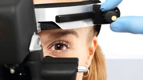
#Direct Indirect Ophthalmoscope
Explore tagged Tumblr posts
Visit Tumblr Blog
Explore Tumblr blogs with no restrictions, modern design and the best experience.
Last Seen Tumblr Blogs


Fun Fact
There were a total of 171.5 billion posts on Tumblr in 2019.
Text
Direct and Indirect Ophthalmoscope
Ophthalmology is a field of medicine that deals with the diagnosis and treatment of eye-related problems. As an ophthalmoscope supplier, you play an important role in providing ophthalmologists with the tools they need to diagnose and treat their patients. Two essential tools in any ophthalmologist's toolkit are the direct and indirect ophthalmoscope.
Direct Ophthalmoscopes
A direct ophthalmoscope is a handheld device that allows ophthalmologists to examine the interior of the eye, including the retina, optic disc, and blood vessels. This instrument works by illuminating the eye with a bright light and magnifying the image of the retina. Direct ophthalmoscopes are especially useful for detecting conditions such as macular degeneration, diabetic retinopathy, and glaucoma.
When selecting a direct ophthalmoscope to supply, it's important to consider features such as the size and weight of the device, the strength of the light source, and the magnification power. Some ophthalmologists prefer ophthalmoscopes with rechargeable batteries or LED lights, while others prefer disposable devices that can be used once and then discarded.
Indirect Ophthalmoscopes
An indirect ophthalmoscope is a more complex device that uses a special lens to provide a wider view of the retina and other structures inside the eye. This instrument is mounted on a headband and is typically used in combination with a slit lamp or other examination equipment. Indirect ophthalmoscopes are especially useful for detecting conditions such as retinal detachments, macular holes, and vitreous hemorrhages.
When selecting an indirect ophthalmoscope to supply, it's important to consider features such as the quality and clarity of the lens, the adjustability of the headband, and the compatibility of the device with other equipment used in ophthalmic examinations.
Benefits of Working with an Ophthalmoscope Supplier
As an ophthalmoscope supplier, we play a vital role in providing ophthalmologists with the tools they need to diagnose and treat eye-related conditions. By working as reputable supplier, we ensure that our customers have access to the latest and most advanced ophthalmic instruments, as well as reliable customer support and technical assistance.
In addition to supplying direct and indirect ophthalmoscopes, we also provide a range of other ophthalmic instruments, such as tonometers, refractometers, and slit lamps. By offering a comprehensive range of products, you can become a trusted partner for ophthalmologists and other eye care professionals.
Conclusion
Direct and indirect ophthalmoscopes are essential tools in the field of ophthalmology. As a supplier of these instruments, we understand the needs of our customers and provide them with high-quality, reliable products that meet their specific requirements. By working closely with ophthalmologists and other eye care professionals, you can build a reputation as a trusted supplier and help to improve the quality of eye care around the world.
Get More Product Details @ https://nationalindco.com/category/direct-and-indirect-ophthalmoscopes-24.html
1 note
·
View note
Text
Ophthalmoscopes: A Vital Medical Device for Eye Examinations

The history of the ophthalmoscope dates back to the 1850s when Hermann von Helmholtz, a German physician and physicist, invented the first basic version to examine the interior of the human eye. Since then, the device has undergone many innovations and advancements. In 1890, American ophthalmologist Henry Gradle developed an improved version of the direct device which allowed for better retinal examinations. In the early 1900s, indirect ones became available which enabled eye inspection without contact with the patient's eyeball making examinations more comfortable. In the following decades, power sources were introduced to provide brighter illumination for viewing the fundus and photo capabilities were integrated to document findings. Modern ones today are highly portable, lightweight digital devices offering advanced imaging, magnification and illumination functions. Workings
Ophthalmoscopes utilize a combination of lenses and light sources to view the interior structures of the eye. Direct ones hold a lens near the patient's eye which reflects and magnifies the retinal image while the examiner views through an eyepiece. Indirect scopes project an inverted magnified image of the retina onto a lens placed on the patient's forehead, allowing the examiner to observe from a short distance. Both types focus a small bright light beam on the retina using lenses or an optic fiber bundle. Dioptic lenses enable viewing of both the illuminated retina and immediate surroundings simultaneously. Powered ones contain a battery that powers high-intensity LEDs or halogen bulbs. Digital models have cameras to photograph the retina electronically. Applications in Medical Screening and Diagnosis Routine eye examinations conventionally involve visualizing the retina, optic disc and macula using an ophthalmoscope. This helps detect signs of numerous common conditions. Diabetic retinopathy screenings rely heavily on retinal visualization via ophthalmoscopy. Changes indicative of the disease such as microaneurysms, hemorrhages and exudates can be spotted. These are indispensable for hypertensive retinopathy evaluation where they reveal arteriolar narrowing, silver wiring of vessels and optic disc edema. Papilledema, a swelling of the optic disc, is a key sign often picked up on ophthalmoscopy in conditions like brain tumors. Retinal detachments, tumors and occlusions can also be promptly diagnosed with these instruments. Ophthalmoscopic findings are regularly recorded to monitor progression or response to treatment over time. Applications in Research and Telemedicine Ophthalmoscopy makes significant contributions beyond clinical practice. Research in ocular diseases extensively utilizes information gathered through standardized retinal examinations using scopes. Digital retinal images obtained are stored in databases for large population studies analyzing risk factors. Technology partnerships have enabled “eyecams” that can tele-ophthalmoscopically transmit retinal photos from remote locations to expert reading centers, expanding access to specialty eye care. The US military uses tele-ophthalmoscopy to remotely screen troops for eye and systemic issues like hypertension. These are pivotal research tools, aiding discovery of pathological changes, disease mechanisms and treatment responses. Advancing scope technology itself drives retinal imaging research contributing to medicine.
0 notes
Text
Exploring the Synergy of Indirect Ophthalmoscopes and Soft Noncontact Tonometers
Technological developments in the dynamic field of ophthalmology are constantly improving the ability to diagnose, ensuring less intrusive and more accurate eye examinations. The soft noncontact tonometer and the indirect ophthalmoscope are two essential tools that have completely changed how eye care is provided. Combined these tools provide a thorough and patient-centered method of eye exams.

Indirect Ophthalmoscopes: A Closer Look
In the toolbox of an eye care specialist, an indirect ophthalmoscope is an essential tool. With a broad field of view of the retina, indirect ophthalmoscopes allow for a detailed study of the internal tissues of the eye, in contrast to direct ophthalmoscopes, which have a narrow field of view. An enlarged, stereoscopic image of the retina is created by this device using a light source and several lenses.
An indirect ophthalmoscope has numerous advantages. It enables the detection of retinal conditions like retinal detachment, diabetic retinopathy, and macular degeneration. Its large field of vision and improved picture quality allow for a more complete examination, making it a vital instrument for diagnosing and ongoing observation of eye problems.
Soft Noncontact Tonometers: Gentle Yet Accurate
However, a soft noncontact tonometer is made to measure intraocular pressure (IOP) without coming into contact with the eye, which allows patients to feel more at ease during the procedure. Traditional tonometry techniques frequently call for direct contact with the cornea, which can be painful and call for applying anaesthetic drops. On the other hand, noncontact tonometers examine the cornea's resistance by using a little puff of air to calculate the intraocular pressure.
One of the main causes of blindness, glaucoma, can be prevented by maintaining an ideal intraocular pressure. The soft noncontact tonometer offers a rapid, precise, and easy-to-use method for screening and keeping track of this condition. Particularly for young patients and those with a high blink reflex, its noninvasiveness lowers the risk of infection and improves patient compliance.

The Synergy of Indirect Ophthalmoscopes and Soft Noncontact Tonometers
Examining the eyes with both an indirect ophthalmoscope and a soft noncontact tonometer can greatly increase the effectiveness of the examination. Here’s how:
Comprehensive Diagnosis: The soft noncontact tonometer enables accurate intraocular pressure measurements, while the indirect ophthalmoscope offers a thorough view of the retina. When combined, they provide a comprehensive evaluation of eye health, assisting in the early diagnosis and management of several disorders.
Patient Comfort: The patients will have a comfortable experience because both equipment are non-invasive. For young people, the elderly, and those who are anxious about eye exams, this is especially crucial. More patient compliance and accurate outcomes are the direct result of increased comfort.
Enhanced Monitoring: Regular monitoring is essential for patients with long-term eye problems such as diabetic retinopathy or glaucoma. Together, these two tools provide comprehensive and reliable monitoring, which makes it possible to act quickly and effectively to treat these disorders.
A breakthrough in ophthalmic diagnostics has been made with the merging of soft noncontact tonometers and indirect ophthalmoscopes. Together, these instruments offer a thorough, effective, and patient-centered method of eye treatment. As technology develops further, these tools working together will surely improve the skills of eye care providers and benefit patients all around the world. Accepting these advancements brings us one step closer to a time when eye conditions are identified early, treated successfully, and handled with the highest care.
#Ophthalmology#EyeCare#IndirectOphthalmoscope#NoncontactTonometer#EyeHealth#MedicalTechnology#Optometry#VisionCare#PatientComfort#GlaucomaPrevention#RetinaHealth#IntraocularPressure#EyeExams#DiagnosticTools#InnovativeHealthcare#AdvancedOphthalmology
1 note
·
View note
Text
The Importance Of Routine Retina Tests: When Should You Get Yours?
The retina serves as the eye's equivalent of a camera, capturing images essential for clear vision. Regular retina tests are vital for maintaining eye health by detecting any potential issues early on. In this article, we'll delve into understanding the retina, the procedure of a retina test, and the critical times when such examinations are paramount.
Understanding the Retina
The retina, a delicate tissue layer at the eye's rear, comprises millions of photoreceptor cells known as rods and cones. These cells detect light and transmit signals to the brain via the optic nerve, enabling visual perception. Additionally, the retina houses supporting cell layers crucial for photoreceptor function and overall retinal health.
What Constitutes a Retina Test?
A Retina Test, also termed Retinal Examination or Retinal Imaging, is a diagnostic process conducted by eye care professionals to evaluate retina health. Various methods are employed for this examination:
Direct Ophthalmoscopy: Involves illuminating the eye and using an ophthalmoscope to examine the retina.
Indirect Ophthalmoscopy: Utilizes a special lens and bright light to view the retina through a dilated pupil.
Optical Coherence Tomography (OCT): A non-invasive imaging technique generating detailed cross-sectional images of the retina for thorough examination.
Significance of Routine Retina Tests
Routine retina tests hold several crucial purposes:
Early Detection: They detect eye diseases like diabetic retinopathy and macular degeneration in their initial stages, before symptoms manifest.
Monitoring: Vital for individuals with existing eye conditions, aiding in the prevention of complications.
Treatment Evaluation: Assesses the efficacy of treatments like intravitreal injections or laser therapy.
Overall Health Assessment: Changes in the retina can signal underlying systemic health issues, necessitating further evaluation if indicated.
When is a Retina Check-Up Crucial?
While the frequency of retina tests varies based on individual factors, certain scenarios necessitate prioritizing a retina check-up:
Vision Symptoms: Sudden changes in vision, floaters, or flashes of light warrant prompt evaluation and may necessitate a retina check-up.
Diabetes: Individuals with diabetes require regular retina check-ups due to the heightened risk of diabetic retinopathy.
Family History: Those with a family history of eye diseases should prioritize regular retina exams for early detection and management.
Age-related Changes: Adults aged 50 and above should undergo routine retina tests to monitor for age-related eye conditions.
Regular eye tests are imperative for maintaining eye health, detecting issues early, and ensuring effective treatment. Protect your vision and overall well-being by scheduling regular eye check-ups. If you have concerns or a predisposition to eye problems, don't hesitate to book an appointment with us at 'Dr Ruchika Eye Clinic'. Your eyes are precious, and regular tests can help preserve your vision for years to come. Dr. Ruchika Kedia, a distinguished Ophthalmologist in Thane West, specializes in comprehensive eye care. With years of experience and expertise, she is committed to providing exceptional eye care services. As a proficient Eye Specialist Doctor in Thane West, Dr. Ruchika Kedia offers a wide range of treatments for various eye conditions. Whether it's Cataract, Glaucoma, Diabetic Retinopathy, Macular Degeneration, Dry Eye, Refractive errors, or other eye problems, Dr. Ruchika Kedia is the top choice when seeking an Eye Doctor in Thane West. For advanced eye care services, consider visiting Dr. Ruchika Eye Clinic in Thane West. Dr. Ruchika Kedia, an accomplished Cataract Surgeon in Thane West, provides top-notch Cataract Surgery. Specializing in Lasik Surgery, she is recognized as one of the eminent Lasik Eye Surgeons in Thane West, with expertise in Laser Lasik Eye Surgery. If you reside in or near Thane East, Thane West, Ghodbandar Road, Hiranandani Estate, VasantVihar, Brahmand, Anand Nagar, Ovala, Majiwada, Waghbil, Gaimukh, Kolshet, Bhiwandi, Kalwa, Upvan, Pokaran Road, Shastri Nagar, Balkum, Shivai Nagar, or Lakmanya Nagar, consult Dr. Ruchika Kedia for world-class eye care services at Dr. Ruchika Eye Clinic in Thane West.
0 notes
Text
Fundus Examination: A Comprehensive Insight
Fundus examination is a fundamental aspect of ophthalmology and plays a crucial role in diagnosing various eye diseases and systemic conditions. This article aims to delve into the significance of fundus examination, the procedure involved, equipment used, common findings, indications, challenges, and future trends associated with this diagnostic tool.
Introduction to Fundus Examination
The fundus of the eye refers to the interior surface of the eye, including the retina, optic disc, macula, and blood vessels. Fundus examination involves the evaluation of these structures to assess ocular health and detect abnormalities.
Importance of Fundus Examination
Fundus examination is vital for diagnosing and managing a wide range of ocular conditions, including diabetic retinopathy, glaucoma, macular degeneration, and hypertensive retinopathy. It also provides valuable insights into systemic diseases such as hypertension, diabetes, and cardiovascular disorders.
Equipment Used in Fundus Examination
Direct Ophthalmoscope
A direct ophthalmoscope is a handheld device used to examine the fundus by illuminating and magnifying the structures of the eye. It allows for a direct view of the retina and optic nerve.
Indirect Ophthalmoscope
An indirect ophthalmoscope is a binocular instrument equipped with a light source and a condensing lens. It provides a wider field of view and allows for a more comprehensive examination of the fundus.
Procedure of Fundus Examination
Preparing the Patient
Before performing a fundus examination, it is essential to obtain informed consent from the patient and explain the procedure. Dilating eye drops may be administered to dilate the pupil for better visualization of the fundus.
Positioning and Lighting
The patient is typically seated comfortably in a dimly lit room. The examiner adjusts the lighting and positions the patient's head to achieve optimal visualization of the fundus.
Using the Ophthalmoscope
The examiner holds the ophthalmoscope in one hand and uses the other hand to stabilize the patient's head. By adjusting the focus and aperture settings, the examiner directs the light beam onto the fundus and systematically examines its various structures.
Common Findings in Fundus Examination
Fundus examination may reveal abnormalities such as retinal hemorrhages, exudates, cotton-wool spots, optic disc edema, and macular degeneration. These findings provide valuable diagnostic clues for identifying underlying eye diseases and systemic conditions.
Indications for Fundus Examination
Fundus examination is indicated in various clinical scenarios, including routine eye examinations, diabetic screening, hypertensive evaluations, preoperative assessments, and monitoring of ocular diseases.
Role of Fundus Examination in Various Medical Conditions
Fundus examination plays a pivotal role in the diagnosis and management of conditions such as diabetic retinopathy, hypertensive retinopathy, retinal vein occlusion, age-related macular degeneration, and glaucoma.
Fundus Examination in Ophthalmology Practice
In ophthalmology practice, fundus examination is an essential diagnostic tool used by ophthalmologists to evaluate patients with visual disturbances, ocular trauma, and systemic diseases affecting the eye.
Fundus Examination in Non-Ophthalmology Settings
Fundus examination is also performed by non-ophthalmic healthcare providers, including primary care physicians, internists, and emergency room physicians, to screen for eye diseases and assess systemic health.
Fundus Examination in Diabetic Patients
Regular fundus examination is recommended for diabetic patients to detect and monitor diabetic retinopathy, a leading cause of blindness worldwide. Early detection and timely intervention can help prevent vision loss in these patients.
Challenges and Limitations of Fundus Examination
Despite its utility, fundus examination may be challenging in patients with small pupils, media opacities, or uncooperative behavior. Limited access to specialized equipment and trained personnel can also hinder its widespread implementation.
Future Trends in Fundus Examination Technology
Advancements in imaging technology, such as optical coherence tomography (OCT) and fundus photography, are revolutionizing fundus examination by providing high-resolution images and quantitative data. These technologies hold promise for early disease detection and personalized treatment approaches.
Training and Education for Fundus Examination
Proper training and education are essential for healthcare providers performing fundus examination to ensure proficiency and accuracy in interpreting findings. Continuous medical education and hands-on training programs help enhance skills and knowledge in this specialized area.
Ethical Considerations in Fundus Examination
Ethical considerations in fundus examination include patient autonomy, informed consent, confidentiality, and equitable access to healthcare services. Healthcare providers must uphold ethical standards and respect patients' rights throughout the examination process.
Conclusion
Fundus examination is a valuable diagnostic tool that provides crucial insights into ocular and systemic health. By understanding its significance, mastering the procedural techniques, and staying abreast of technological advancements, healthcare providers can optimize patient care and improve clinical outcomes.
FAQs
1. How often should I undergo a fundus examination?
It depends on your age, medical history, and risk factors. Your eye care provider can recommend the appropriate frequency based on your individual needs.
2. Does fundus examination require dilating eye drops?
In many cases, dilating eye drops are used to dilate the pupil for better visualization of the fundus. However, not all examinations require pupil dilation.
3. Can fundus examination detect systemic diseases?
Yes, fundus examination can detect signs of systemic diseases such as diabetes, hypertension, and cardiovascular disorders through changes in the retinal blood vessels and other structures.
4. Is fundus examination painful?
No, fundus examination is a painless procedure. You may experience slight discomfort from the bright light and dilation drops, but it is generally well-tolerated.
5. Who performs fundus examination?
Fundus examination can be performed by various healthcare providers, including ophthalmologists, optometrists, and primary care physicians, depending on their training and scope of practice.
#choroida#fundus examination#eyes#health#medical devices#medical care#eyecare#eye health#medical equipment#choroid
0 notes
Text
Global Laser Indirect Ophthalmoscope Market Is Estimated To Witness High Growth Owing To Increasing Demand for Advanced Ophthalmic Equipment
The Global Laser Indirect Ophthalmoscope Market is estimated to be valued at US$ 89.5 million in 2017 and is expected to exhibit a CAGR of 4.2% over the forecast period 2018-2026, according to a new report published by Coherent Market Insights. Market Overview: Laser indirect ophthalmoscope is a medical device used by ophthalmologists to examine the retina and diagnose various eye disorders. It provides a wider view of the retina compared to direct ophthalmoscopy, allowing for better visualization of eye conditions such as retinal detachment, macular degeneration, diabetic retinopathy, and glaucoma. Laser indirect ophthalmoscopes have gained popularity due to their advantages such as non-contact examination, high-resolution imaging, and ease of use. The increasing prevalence of eye diseases and the growing geriatric population are driving the demand for advanced ophthalmic equipment, including laser indirect ophthalmoscopes. Market Key Trends: One key trend in the laser indirect ophthalmoscope market is the increasing adoption of digital imaging technology. Digital imaging provides a detailed and accurate representation of the retina, allowing for better diagnosis and monitoring of eye conditions. It eliminates the need for traditional film-based imaging, reducing costs and improving workflow efficiencies. For example, digital imaging systems like fundus cameras and confocal scanning laser ophthalmoscopes are increasingly being integrated with laser indirect ophthalmoscopes, enabling ophthalmologists to capture, analyze, and store retinal images digitally. PEST Analysis: - Political: Government initiatives promoting access to eye care services and investments in healthcare infrastructure are driving the market growth. - Economic: Increasing healthcare spending and rising disposable income of individuals are contributing to market growth. - Social: Growing awareness about eye health and the importance of early detection and treatment of eye disorders are fueling the demand for ophthalmic equipment. - Technological: Advancements in technology, such as the integration of artificial intelligence and machine learning algorithms in ophthalmic devices, are improving the accuracy and efficiency of diagnosis and treatment. Key Takeaways: - The Laser Indirect Ophthalmoscope Market Size is expected to witness high growth, exhibiting a CAGR of 4.2% over the forecast period, due to increasing demand for advanced ophthalmic equipment. - North America is the fastest growing and dominating region in the market, driven by favorable reimbursement policies, high healthcare expenditure, and the presence of key market players. - Key players operating in the global laser indirect ophthalmoscope market include Ellex Medical Lasers Ltd, Iridex Corporation, Alcon, Carl Zeiss Meditec AG, Lumenis Ltd., Topcon Medical Laser System, Nidek Co. Inc., Ziemer Ophthalmic Systems AG, Heine Optotechnik GmbH, and Keeler Ltd. These companies focus on product innovation, mergers and acquisitions, and collaborations to strengthen their market position. In conclusion, the global laser indirect ophthalmoscope market is expected to grow significantly in the coming years due to the increasing demand for advanced ophthalmic equipment. The adoption of digital imaging technology and advancements in technology are key trends driving market growth. Furthermore, favorable political, economic, and social factors, along with advanced technological developments, are expected to propel the market further. North America is anticipated to be the fastest growing region in the market. Key players in the industry are taking strategic initiatives to enhance their market presence and cater to the growing demand for laser indirect ophthalmoscopes.
#Laser Indirect Ophthalmoscope Market#Laser Indirect Ophthalmoscope Market Insights#Laser Indirect Ophthalmoscope Market Value#Laser Indirect Ophthalmoscope Market Forecast#Coherent Market Insights
0 notes
Text
Physical Examination of the Eye in Veterinary Medicine
A thorough ocular examination is an important diagnostic procedure in veterinary medicine. Primary and secondary ocular disease may occur. The changes seen in the eye may be related to another disease process, directing you to further diagnostics.
Before being able to perform a proper ocular examination, you must understand the anatomy of the species that you are examining. This post will largely focus on mammalian eyes.
Basic ocular anatomy includes: The cornea - clear tissue covering the front of the eye. The uvea (ciliary body, choroid, and iris) - vascular tissue of the eye. The iris is the coloured section surrounding the pupil. The lens - clear disc within the eye that is used to focus light. The retina - photoreceptors are present here. Light is converted into a signal for the brain to interpret as a visual. The sclera - the white connective tissue surrounding the globe. The optic nerve - carries nervous information to the brain.
Related structures include: The third eyelid (nictitating membrane). The eyelids and conjunctiva. The lacrimal glands - produce the watery portion of tears. The meibomian glands - produce the oily portion of tears.
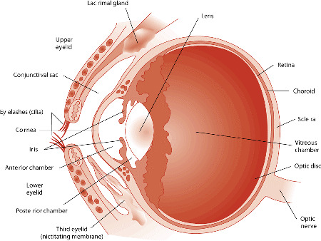
Image source: https://www.merckvetmanual.com/dog-owners/eye-disorders-of-dogs/eye-structure-and-function-in-dogs
Vision
The patient should be observed as they move around the examination room. Signs that the patient may be experiencing difficulty in seeing include reluctance to move, running into things, and high stepping.
Menace response: This test is performed by performing a threatening hand gesture heading towards the eye. The contralateral eye should be covered when performing this test. It is important to avoid pushing air/creating a breeze toward the patient as air stimulus alone may cause the patient to blink. You must avoid contact with the whiskers. A positive menace response is achieved when the patient blinks. The menace response is a learned response and is not present consistently in cats and dogs under the age of 3-4 months. Tests: peripheral and central visual pathways, visual cortex, CN7.
Pupillary light reflex: This test does NOT test vision as it is a subcortical reflex. This may be normal in a cortically blind animal. This test is performed by shining a light into the eye and observing the response of pupil size. This should be done with a bright light in a dimly lit room. Both direct (the eye in which light is being shined) and indirect (the contralateral eye) PLRs are observed. Both direct and indirect should be present when light is shined into either eye. Birds and reptiles do not have a PLR. They are able to control the constriction and dilation of their pupils independent of light.
Dazzle: This test does NOT test vision as it is a subcortical reflex. This test is performed by intensely illuminating the eye. A positive dazzle response is achieved when the patient reacts to the light. This reaction can be a blink, third eyelid protrusion, head movement, or globe retraction. Tests: retina, CN2, CN7.
Periocular
Evaluate the orbits (bone surrounding the globe) for symmetry, deformity, or enlargement. Breed variations are important to understand during this portion of the examination. The orbital rim should be palpated to ensure no irregularities are missed.
The position and general movement of the eyes should be observed: Strabismus - abnormal alignment of the eyes. Nystagmus - rapid involuntary movement of the eyes.
Eyelids
Blink (palpebral) reflex: This test is performed by touching the skin near both corners of the eye. This should produce a blink response. The finger should be gently dragged across the closed eye to ensure that the patient is capable of holding their eye closed. Tests: CV7, orbicularis oculi muscle, CN2, CN5.
The eyelids should be observed for any abnormalities. Abnormalities may include: Masses or cysts. Entropion - rolling in of the eyelid. Ectropion - rolling out of the eyelid. Ectopic cilia - eyelashes in abnormal locations.
Conjunctiva
The upper and lower eyelids should be manually everted. This allows for evaluation of the tissue. Abnormalities may include: Increased vascularity. Lacerations or foreign bodies. Growths. Chemosis - edema.
Third eyelid
The third eyelid can often be extruded by placing gentle pressure on the tissues near the inner corner of the eye. Abnormalities may include: Prolapse of the gland - cherry eye. Conjunctivitis. Enlarged glands or lymphoid tissue.
Sclera
The sclera should be observed for changes in vasculature, colour, masses, or lacerations.
Cornea
The cornea should be avascular and unpigmented. It should be smooth. The cornea should be examined for change in transparency/opacity, vascularization, pigmentation, laceration/ulceration, masses, and foreign bodies.
Fluorescein stain: This stain is used to confirm the absence or presence of superficial ulcers. No corneal examination is complete without this test. The dye does not stick to the outer layer of the cornea as it is lipid selective. The corneal stroma, when exposed, allows rapid diffusion of the dye. An area of fluorescein retention is indicative of an epithelial defect such as an ulcer or erosion.
Anterior Chamber
A focal light source should be shined through the anterior chamber of the eye to examine the aqueous humor. When increased protein is present, the beam of light should appear to be passing through smoke. This is called aqueous flare and is indicative of uveitis.
Lens
The lens should be transparent and avascular. The lens should be observed with an ophthalmoscope for opacities, position, and size.
Cataracts: Focal - localized within various parts of the lens. Nuclear cataracts are usually stationary. Equatorial cataracts are usually progressive. Immature cataract - involves more of the lens and causes blurred vision. Mature cataract - entire lens affected, vision loss.
Lenticular sclerosis: Begins to develop in dogs around 6 years of age. This is seen as a blue zone in the nucleus of the lens that does not impair visualization of the fundus.
Fundus
An ophthalmoscope is required for examination of the fundus. An examination can be performed without drug induced mydriasis (dilation of the iris) though it assists in complete examination.
Abnormalities may include: Vascular patterns. Retinal detachment. Congestion or hemorrhage. Changes in pigmentation.
Tear Production
Tear film is essential in maintaining normal corneal health. When the cornea is not appropriately moisturized, ulcers and other pathology can result.
The schirmer tear test is used to quantify the amount of aqueous tear portion present in each eye. This test does not evaluate the quality or make-up of the tears. The test strip is placed in the lacrimal lake at the junction of the lateral and middle thirds of the lower eyelid. It is held in place for 60 seconds while the animal typically holds their eye closed. The test is not linear and therefore you cannot measure for 30 seconds and simply multiply by two. Normal values are > 15mm for cats and dogs. Other species may have species-specific values published.
Intraocular Pressure
This is measured with tonometry. Applanation tonometers, such as the tonopen, are very accurate. Normal IOP is 16.8 ± 4.0 mm Hg in dogs; 20.2 ± 5.5 in cats. Low IOP indicates uveitis while high IOP indicates glaucoma.
#veterinarian#veterinary medicine#veterinary#vet med#vet school#vet student#ophthalmology#vet ophthalmology#vetblr
56 notes
·
View notes
Text
Top 7 Ophthalmic Equipment You Must Have in Your Clinic
If you're a practicing ophthalmologist, it's important to have the right equipment in your clinic to provide the best possible care for your patients. Here are seven pieces of ophthalmic equipment that you should definitely have in your clinic.
1. Slit lamp
The slit lamp is a common ophthalmoscope used to examine the interior of the eye. It consists of a low-power microscope with a slit in the middle of the eyepiece, allowing the examiner to see a narrow strip of tissue. The lamp is placed close to the patient's eye, and the examiner looks through the eyepiece to view the tissue. The slit lamp is used to examine the posterior segment of the eye, including the retina, optic nerve, and posterior chamber. It can also be used to measure intraocular pressure.
2. Indirect ophthalmoscope
An indirect ophthalmoscope is a ophthalmic equipment that is used to examine the interior of the eye. This device consists of a telescope that is attached to a light source. The light is directed into the eye and the image is projected onto a screen. This device can be used to diagnose a variety of eye conditions.
3. Fundus camera

A fundus camera is a medical device that is used to photograph the inside of the eye. It is a small, handheld device that is placed close to the eye. The camera takes a picture of the back of the eye, which is called the fundus. This image can be used to check for signs of eye disease.
4. Tonometer
A tonometer is a ophthalmic equipment used to measure the intraocular pressure (IOP) of an eye. There are a few different types of tonometers, but the most common is the Goldmann applanation tonometer. This device uses a small metal disk that is placed on the surface of the cornea. The disk is then pressed against the eye by a small plunger, which measures the IOP.
5. Phoropter
The phoropter is an important ophthalmic equipment used by optometrists and ophthalmologists. It is used to measure the refractive error of the eye. The phoropter contains a number of different lenses that are used to correct vision.
6. Autorefractor
An autorefractor is a device used to measure the prescription of eyeglasses. The autorefractor is a small, hand-held device that is placed in front of the patient's eyes. The patient looks into the autorefractor, and the device measures the prescription and provides a reading.
7. Keratometer
A keratometer is a device used to measure the curvature of the cornea. It is a common tool used in eye exams to help determine the prescription for eyeglasses. A keratometer works by shining a light into the eye and then measuring the angle of reflection. This angle is then used to calculate the curvature of the cornea.
With the right equipment in your clinic, you can provide your patients with the best possible care. Make sure to include these seven pieces of ophthalmic equipment in your clinic to ensure that you're providing the best possible care for your patients.
0 notes
Text
Multifunctional otoscope inspection glasses set
How to apply the ophthalmoscope
When examining the rest of the fundus, the subject should be able to rotate the eye to cooperate with the examination, and the examiner moves the position around the subject's head, and the handheld LCD Display Digital Chargeable Otoscope Ophthalmoscope Set and the examiner's head move with it. The images examined are opposite up and down, and also opposite left and right. To examine the peripheral part of the fundus, such as the 6 o'clock orientation, the examiner is positioned at the top of the subject's head and the affected eye is made to look down at the 6 o'clock orientation. The metal scleral compressor is worn on the middle finger or index finger of the examiner's right hand, and the head of the compressor is placed outside the corresponding eyelid of the examined eye, and if necessary, the conjunctival sac is examined after epiretinal anesthesia. During the examination, attention should be paid to the patient at any time to close the eyelid to moisten the cornea, when suspected of intraocular occupying lesions, do not compress the examination.
Examination methods of ophthalmoscopy
Direct examination method
The fundus image can be magnified about 15-16 times, and the image seen is positive, the fundus can be seen in a small range, but more detailed and detailed, and can also be used to examine the refractive interstitium of the eye conveniently. The LCD Display Digital Chargeable Otoscope Ophthalmoscope Set, which comes with its own light source, can be used to correct the refractive error of the examiner and the subject during the examination.
Indirect Examination Method
The indirect ophthalmoscope can magnify the fundus by 4.5 times, and the image seen is an inverted real image, which can be seen in a wide range, up to 25°~60° at a time, with a strong sense of stereo and a wide depth of field. With the scleral compressor, the most peripheral parts of the fundus such as the serrated edge and even the flattened part of the ciliary body can be seen. The ophthalmoscope is equipped with a translucent, semi-reflective lateral mirror that can be used for teaching purposes.
The LCD Display Digital Chargeable Otoscope Ophthalmoscope Set is equipped with a strong light source and a focusing adjustment system, so that the projected light can be close to the left and right eye line of sight of the examinee, so that the examinee can observe with both eyes.
During the examination, the examinee takes a sitting or lying position, the examination distance is about 50cm, the examiner holds the +13D - 28D lens with the thumb and index finger, the ring finger and pinky finger lean on the forehead of the examinee as a support, and lift the upper lid, the lens moves in front of the examinee within 4-9cm until the fundus image is seen.
The LCD display digital rechargeable otoscope ophthalmoscope set manufactured by Sintrue medical instrument in Ningbo is the preferred choice for hospital medical clinics.
0 notes
Text
Ophthalmoscopes Market Analysis
Technological advancements in the disease diagnostic field has led to advanced product development. Key manufacturers are focused on developing new ophthalmoscopes for unmet need of ophthalmic diagnostic practices. The increasing product launches by key players is expected to drive growth of the market over the forecast period. For instance, in 2019, Nidek Co. Ltd launched Mirante Scanning Laser Ophthalmoscope in Japan. The device is a multimodal fundus imaging platform, which combines high definition scanning laser ophthalmoscope and optical coherence tomography. Moreover, such product launches are expected to boost growth of the global ophthalmoscopes market.
Global Ophthalmoscopes Market - Impact of Coronavirus (Covid-19) Pandemic
The Covid – 19 pandemic is expected to drive growth of the global ophthalmoscopes market over the forecast period. Since December 2019, a novel coronavirus spread throughout China and across the world, causing a continuous increase in confirmed cases within a short period of time. The disease has spread to more than 100 countries across the globe and the World Health Organization declared it as a public health emergency. For instance, according to the World Health Organization Coronavirus Disease (COVID-19) Dashboard (WHO) report, the manifestation of the coronavirus disease (COVID-19) has resulted in more than 11 million infected individuals worldwide as of September 2020. This outbreak of corona expected to hinder countries overall growth.
The U.S has the highest number of corona patients which is expected to impact the country’s economic condition and government healthcare spending. Similarly, the countries include Italy, Spain, Russia, India, Brazil and many other economies are also suffering from Covid 19 attack that is expected to impact on lowering the medical devices market growth. Furthermore, due to COVID-19 pandemic, hospitals are closed in containment zones, that has slowed sale of medical devices like Ophthalmoscopes and similar devices.
Global ophthalmoscopes market is estimated to be valued at US$ 216.8 million in 2020, and is expected to exhibit a CAGR of 4.3% over the forecast period (2020-2027).
Figure 1: Global Ophthalmoscopes Market Share (%) Analysis, By Product Type, 2019

Global Ophthalmoscopes Market- Regional Insights
On the basis of region, the global ophthalmoscopes market is segmented into North America, Latin America, Europe, Asia Pacific, Middle East, and Africa. North America is expected to be the most lucrative region for growth of the ophthalmoscopes market over the forecast period, owing to increasing incidence of ophthalmic disease in the U.S. For instance, according to the Centers for Disease Control and Prevention (CDC), in 2015, around 100 million U.S. population aged 18 years and older were diagnosed with diabetes or pre-diabetic. In 2015, in the U.S. prevalence of retinopathy in all adults with diabetes those are aged 40 and above was around 28.5%. In 2015, the estimated rate of prevalence is around 4.4% (0.7 million) of people for vision-threatening diabetic retinopathy in the U.S. Moreover, according to the Glaucoma Research Foundation, in 2018, over 120,000 (9-12% of total blindness cases) population in the U.S were blind due to glaucoma disease. Such high prevalence of eye diseases is expected to drive growth of the market in North America region.
Ophthalmoscopes Market Report Coverage
Report Coverage
Details
Base Year:
2019
Market Size in 2019:
US$ 216.8 Mn
Historical Data for:
2017 to 2019
Forecast Period:
2020 to 2027
Forecast Period 2020 to 2027 CAGR:
4.3%
2027 Value Projection:
US$ 289 Mn
Geographies covered:
§ North America: U.S. and Canada
§ Latin America: Brazil, Argentina, Mexico, and Rest of Latin America
§ Europe: Germany, U.K., Spain, France, Italy, Russia, and Rest of Europe
§ Asia Pacific: China, India, Japan, Australia, South Korea, ASEAN, and Rest of Asia Pacific
§ Middle East: GCC, Israel, Rest of Middle East
§ Africa: South Africa, North Africa, and Central Africa
Segments covered:
§ By Product Type: Direct Inspection, Indirect Inspection.
§ By End User: Hospitals, Eye Clinics, Ambulatory Surgical Centers.
Companies covered:
Medline Industries, Inc., Heine USA Ltd., US Ophthalmic, Welch Allyn, Dino-Lite Europe, HONSUN Group, Rudolf Riester GmbH, IRIDEX Corporation, Zumax Medical Co., Ltd., Oscar Boscarol S.r.l., Suzhou Kangjie Medical Inc., NIDEK Inc., and KIRCHNER & WILHELM GmbH Co. KG
Growth Drivers:
§ Rising incidence of ophthalmic disease across the globe
Restraints & Challenges:
§ Technological advancement in ophthalmoscopes
Asia Pacific ophthalmoscopes market is expected to witness significant growth due to growing healthcare infrastructure, high prevalence of eye diseases, and increasing engagement of government bodies in preventing the cataract and blindness cases among the population. For instance, according to the data published by the Indian Journal of Ophthalmic, in September 2018, cataract was responsible for 50-80% of bilateral blindness in India. Moreover, government initiatives such as the National Program for Control of Blindness (NPCB) and Vision 2020 aims to eliminate preventable blindness at the national level. Such initiatives are expected to boost demand for ophthalmoscopes and further expected to drive growth of the market.
Figure 2: Global Ophthalmoscopes Market, Value (US$ Mn) & Y-o-Y Growth (%), 2016-2027

Request Free Sample: https://www.coherentmarketinsights.com/insight/request-sample/4157
Download PDF brochure: https://www.coherentmarketinsights.com/insight/request-pdf/4157
Global Ophthalmoscopes Market- Competitive Landscape
Major players operating in the global ophthalmoscopes market include Medline Industries, Inc., Heine USA Ltd., US Ophthalmic, Welch Allyn, Dino-Lite Europe, HONSUN Group, Rudolf Riester GmbH, IRIDEX Corporation, Zumax Medical Co., Ltd., Oscar Boscarol S.r.l., Suzhou Kangjie Medical Inc., NIDEK Inc., and KIRCHNER & WILHELM GmbH + Co. KG
About Us:
Coherent Market Insights is a global market intelligence and consulting organization focused on assisting our plethora of clients achieve transformational growth by helping them make critical business decisions.
What we provide:
• Customized Market Research Services
• Industry Analysis Services
• Business Consulting Services
• Market Intelligence Services
• Long term Engagement Model
• Country Specific Analysis
Contact Us:
Mr. Shah
Coherent Market Insights Pvt. Ltd.
Address: 1001 4th ave, #3200 Seattle, WA 98154, U.S.
Phone: +1-206-701-6702
Email: [email protected]
Source:https://www.coherentmarketinsights.com/market-insight/ophthalmoscopes-market-4157
0 notes
Text
Trust Indira Medical Tourism for Your Eye Cure & Opthalmology.

Indira Medical Tourism Delhi –the best medical tourism company in delhi has always been committed to provide the highest quality of eye care. Over the years, state of the art equipment and the best of doctors have helped us in establishing our institution as the leading hospital for Ophthalmology not just in Kolkata, but in Eastern India. Our objective has always been to provide the best diagnosis and treatment of all complex problems across our different units.
The Eye department at Indira Medical Tourism Delhi functions with facilities for retina check-up, Glaucoma check-up besides vision screening and refractions. It is equipped with Auto refractometer, a High-Tec slit lamp with Haag-streit Applanation tonometer, indirect and direct ophthalmoscopes and various diagnostics lenses. A few facilities at this unit include routine eye check-ups, complete solution for cataract treatment and a surgical retina treatment centre.
The Bhubaneswar unit is another renowned hospital for Ophthalmologythat provides comprehensive eye care and services for all eye and vision problems under one roof. We have exclusive clinics for Refractive error and contact lenses, Cataract, Glaucoma, Cornea, Paediatric Ophthalmology, Vitreo-Retina, Diabetic Retinopathy, ROP, ARMD, Oculoplasty, Ocular Oncology, Ocular trauma, Uveitis, Neuro-ophthalmology, Computer vision syndrome etc.
The Ophthalmology department at Indira Medical Tourism Delhi some of the most advanced equipment and the best medical personnel that makes it a one stop centre for eye treatment and care. The unit is renowned to be the first hospital for ophthalmology in Eastern India to be equipped with state-of-the-art Oertli “Faros” phaco and posterior segment system. This equipment delivers the highest quality of surgical tissue handling that only a few can match, both for routine as well as complicated surgeries.
The department at Indira Medical Tourism Delhi was initially set up with the primary objective being able to cater to the basic and routine needs of a super-specialty hospital. Over the years Indira Medical Tourism Delhi has developed into one of the best lasik eye surgery in delhi. The new department at the annex –I building of AMRI Salt Lake providesall the world class facilities like:
Routine eye check-ups
A complete medical retina clinic with angiography and lasers
A hi-tech glaucoma clinic with Applanation tonometry, perimetry and 90 D examination
A complete end to end solution for cataract treatment
A one stop surgical retina treatment centre
AMRI Salt Lake is widely regarded as one of the most advanced eye-treatment centres in Eastern India. The department in this unit boasts of:
A topcon digital fluorescein angiography (latest TRC 50Dx) to deliver crisp clean images of the retina via the Image net (patented) software, imported from Japan
A Humphrey auto perimeter which has become the gold standard in diagnosis and follow up of glaucoma, from Carl Zeiss, a world leader in this technology.
A blue green laser from Ellex corp, Australia complete with indirect ophthalmoscope delivery system (first of its kind in eastern India) for highly accurate and effective retinal photocoagulation
A sonomed biometry machine which gives quick and highly accurate acquisition of data for lens power calculation in cataract surgery
The Moller Wedel microscope, imported from Germany is one of a kind in the industry, with no contemporaries in the stereo base setup.
A dedicated Fabius Anaesthesia machine capable of handling long duration surgeries in, backed by the best monitors and lifesaving equipment.
FOR MORE UPDATES VISIT TO OUR OFFICIAL WEBSITE:- https://www.indiramedicaltourism.net/
FOLLOW US ON FACEBOOK:- https://www.facebook.com/pg/indiramedicaltourism/about/
VISIT TO OUR OFFICE:- https://goo.gl/maps/pM7iyd2n8cAWMCbj8
0 notes
Text
Indirect Fundoscopy
What is indirect fundoscopy, and how does it differ from direct fundoscopy?
Indirect fundoscopy involves using a condensing lens and a light source to view the fundus, creating an inverted and magnified image. In contrast, direct fundoscopy uses a handheld ophthalmoscope for a non-inverted, but less magnified view. Indirect fundoscopy allows for a more comprehensive examination of the peripheral retina.
Is indirect fundoscopy more commonly used in specific eye conditions?
Indirect fundoscopy is often preferred in cases where a more extensive view of the retina is required, such as in the assessment of peripheral retinal tears, detachments, or during surgical procedures. It provides a wider field of view compared to direct fundoscopy.
#health#eyes#eyecare#medical care#medical devices#choroid#Ophthalmic#eye health#medical equipment#Ophthalmology Devices#ophthalmology#Ophthalmoscope#retinal disease#fundus examination#choroida
0 notes
Text
Consumer Electronics to Dominate India Ophthalmoscope Market through FY2027 | TechSci Research
Increase in number of ophthalmic clinics and advancements in laser technology is expected to drive the India ophthalmoscope market for the forecast period.

According to TechSci Research report, “India Ophthalmoscope Market By Product (Direct Ophthalmoscope v/s Indirect Ophthalmoscope) By Light Source (Halogen Illumination Ophthalmoscope, Xenon Illumination Ophthalmoscope, LED illumination Ophthalmoscope) By Application (Glaucoma, Lattice Degeneration, Diabetic Retinopathy, Others) By End User (Hospitals & Clinics, Eye Care Centers, Ambulatory Care Centers, Others) By Company, By Region, Forecast & Opportunities, FY2027”, India ophthalmoscope market is expected to witness steady growth for the next five years. Ophthalmoscopy is the process for conducting test for eye examination using an ophthalmoscope to examine inside the fundus of eye. Ophthalmoscope helps in detection of abnormalities of retina, vitreous humor, optic disk, and lens and focuses a small beam of light though the pupil of patient’s eye. The ophthalmoscope devise comprises of eye piece, light illuminating source, battery handle and an aperture selector. India healthcare industry is developing at the rapid rate and there is high prevalence of eye diseases. Increase in the government initiatives to prevent blindness and cataract among the population such as National Program for Control of Blindness (NPCB) is another factor responsible for the growth of the ophthalmoscope market. Surge in healthcare expenditures for treatment of blindness, sight loss and sight-threatening conditions are expected to boost the demand for eye care and examination market which is further expected to drive the demand of ophthalmoscope market. Rise in occurrence of chronic diseases such as diabetes and hypertension can lead to retinopathy which is a significant factor accelerating the ophthalmoscope market growth.
Due to the ongoing pandemic COVID-19, lockdown restrictions were imposed all over the country. Complete shutdown was observed, and people started practicing social distancing and following the precautionary guidelines. Imposition of strict regulations and policies by leading authorities led to huge losses suffered by businesses. Also, the demand for medical devices have gone down. Manufacturing units were shut down and loss of availability of skilled labors led to market decline. Number of units imported or exported to other countries also suffered a setback. After the lifting of lockdown restrictions, companies are again operational and producing with lower volume and the market is expected to pick up the pace eventually.
However, lack of skilled healthcare professionals and eye surgeons may hamper the growth of ophthalmoscope market for the forecast period.
Browse XX Figures spread through XX Pages and an in-depth TOC on “India Ophthalmoscope Market”.
https://www.techsciresearch.com/report/india-ophthalmoscope-market/7529.html
India ophthalmoscope market is segmented into product, light source, application, end user, regional distribution, and company. Based on application, market can be further divided into glaucoma, lattice degeneration, diabetic retinopathy, and others. The diabetic retinopathy segment is expected to account for major market share for the forecast period, FY2023-FY2027. Diabetic retinopathy is a diabetes complication which effects the eye adversely by damages the blood vessel in the tissue at the back of the eye. Diabetic retinopathy is treated with the help of laser technology fueling the growth of ophthalmoscope market. Sometimes, glaucoma leads to diabetic retinopathy which is another factor responsible for high demand of ophthalmoscope market. Based on product, market can be divided into direct ophthalmoscope and indirect ophthalmoscope. Direct ophthalmoscopy is expected to witness growth for the next five years as it has an advantage of capturing erect view of fundus with moderate to high resolution. Indirect ophthalmoscopy in examination of retinal health and it can be carried out without giving anaesthesia to patients.
Hill-Rom India Private Limited, Riester-Halma India, Scorpia India Medicare Pvt. Ltd. (HEINE Optotechnik), Prkamya Visions, Carl Zeiss India Pvt. Ltd., Medline Industries India Private Limited, Easilens Healthcare Computers Limited, United Optical Company are the leading players operating in India ophthalmoscope market. Manufacturers are increasingly focusing on research and development process to fuel higher growth in the market. To meet evolving customer demand with respect to better efficiency and durability, several ophthalmoscope manufacturers are coming up with their technologically advanced offerings.
Download Sample Report @ https://www.techsciresearch.com/sample-report.aspx?cid=7529
Customers can also request for 10% free customization on this report.
“Advancements in technology to upgrade diagnostic procedures such as introduction of ophthalmoscopes having multimodal fundus imaging platform which combines the high-definition scanning laser ophthalmoscope and optical coherence tomography. is driving the demand of the market. Launch of novel products by key players to meet the unmet needs of ophthalmic procedures is fostering the growth of ophthalmoscope market. Increase in number of geriatric population and occurrence of eye-related disorders is expected to accelerate the ophthalmoscope market growth till FY2027” said Mr. Karan Chechi, Research Director with TechSci Research, a research-based global management consulting firm.
“India Ophthalmoscope Market By Product (Direct Ophthalmoscope v/s Indirect Ophthalmoscope) By Light Source (Halogen Illumination Ophthalmoscope, Xenon Illumination Ophthalmoscope, LED illumination Ophthalmoscope) By Application (Glaucoma, Lattice Degeneration, Diabetic Retinopathy, Others) By End User (Hospitals & Clinics, Eye Care Centers, Ambulatory Care Centers, Others) By Company, By Region, Forecast & Opportunities, FY2027” has evaluated the future growth potential of India ophthalmoscope market and provided statistics & information on market size, shares, structure and future market growth. The report intends to provide cutting-edge market intelligence and help decision makers take sound investment decisions. Besides, the report also identifies and analyzes the emerging trends along with essential drivers, challenges, and opportunities in the of India ophthalmoscope market.
Browse Related Reports:
Global Ophthalmic Equipment Market By Product (Vision Care Products, Ophthalmology Surgical Devices, Diagnostic and Monitoring Devices, Others) By Application (Glaucoma, Amblyopia, Cataract, Retinal Detachment, Others) By End User (Hospitals & Clinics, Ambulatory Care Centers, Others) By Region, Competition Forecast & Opportunities, 2026
https://www.techsciresearch.com/report/ophthalmic-equipment-market/7363.html
Global Eye Care Market By Product Type (Eyeglasses, Eye Drops, Contact Lens, Intraocular Lens, Eye Vitamins, Others), By Eye Drops (Prescription v/s Over-The-Counter), By Coating (Anti-Glare, UV Coating, Others), By Lens Material (Polycarbonate, Normal Glass, Trivex, Others), By Distribution Channel (Retail Stores, E-commerce, Clinics, Hospitals), By Region, Competition Forecast & Opportunities, 2026
https://www.techsciresearch.com/report/eye-care-market/4879.html
Contact
Mr. Ken Mathews
708 Third Avenue,
Manhattan, NY,
New York – 10017
Tel: +1-646-360-1656
Email: [email protected]
Website: https://www.techsciresearch.com/
For More Market Research Blogs Visit: https://techsciblog.com/
#TechSci#Market Research Reports#India Ophthalmoscope Market#Healthcare#Ophthalmoscope Market#India Ophthalmoscope Market Size#Medical Devices#India Ophthalmoscope Market Forecast
0 notes
Text
LCD Display Digital Chargeable Otoscope Ophthalmoscope Set
Types of otoscope
otoscope can be divided into two kinds of direct otoscope and indirect otoscope.
Direct ophthalmoscopy
Direct ophthalmoscopy can directly examine the fundus, without dilating the pupil, in a dark room for examination, the examiner's eye must be close to the patient's eye, with the right eye to examine the patient's right eye, the right hand holding the ophthalmoscope, sitting or standing on the right side of the patient, the left eye is the other way around, the doctor's other hand to open the patient's eyelid, first placed the ophthalmoscope in front of the patient about 20cm, with +10D lens to check whether the patient's refractive interstitial transparency If the doctor is orthoptotic or has corrective lenses, the refractive power used to see the fundus indicates the refractive condition of the examined eye. Generally, the eye is first made to look straight ahead to examine the optic papilla, then along the retinal vessels to examine the superior and inferior temporal, superior and inferior nasal quadrants, and finally the eye is made to look to the temporal side to examine the macula. The size of the fundus lesion is expressed by the diameter of the optic papilla, and the degree of concavity of the lesion is measured by the refractive index of the lens, which is equivalent to 1mm in 3D. Some examining glasses have a green filter for better observation of the optic nerve fibers and macula.
Indirect otoscope
Indirect otoscope must be fully dilated pupil, in the dark room, the doctor turn on the power, adjust the distance and the position of the reflector, start with a weaker light observation, see the corneal, crystal and vitreous turbidity, and then the light directly into the pupil of the examined eye, and let the examined eye look at the light source, generally with +20D objective lens placed in front of the examined eye 5cm, the convex side of the objective lens to the examiner, the examiner to The objective lens is placed at 5 cm in front of the examined eye, with the convexity of the objective lens facing the examiner, the examiner holds the objective lens in his left hand and fixes it at the orbital rim of the patient, the examined eye, the objective lens and the examiner's head are fixed, when the optic papilla and macula are seen, the LCD Display Digital Chargeable Otoscope Ophthalmoscope Set is moved toward the examiner, and the stereo image of the optic papilla and macula can be clearly seen at 5 cm in front of the examined eye. The inverted image of the optic papilla and macula can be clearly seen at 5cm in front of the examined eye.
0 notes
Text
code General Medical General Medical Equipment Tender G01/BED/01 Manual inpatient bed G01/BED/02 Electric inPatient Bed G01/BED/03 ICU Electric Bed G01/BED/04 Hydraulic Patient Trolley G01/BED/05 Patient lifter G01/BED/06 Bed side cabinet G01/BED/07 Overbed table G01/BED/08 Baby Cot G01/BED/09 Examination couch OPD G01/BED/10 Screen for examination couch OPD G01/BED/11 Gynachology examination couch G01/BED/12 burn bed (shower trolly) G01/BED/13 Elehtric Chair for dialysis machine G01/BED/14 Air Mattress G01/BED/15 Sequential compression device (DVT) General Medical FURNITURE Tender G02/FUR/01 Foot stool G02/FUR/02 Kick bucket G02/FUR/03 Double Bowel stand G02/FUR/04 Mayo cart G02/FUR/05 Adjustable Stool G02/FUR/06 St St Shelving unit G02/FUR/07 Drip stand with 5 wheel G02/FUR/08 Wheel Chair G02/FUR/09 Fileing trolly G02/FUR/10 St St instrment cupboard G02/FUR/11 Galvanized St instrment cupboard G02/FUR/12 Dressing cart 70x50 (2 shelves) G02/FUR/13 Dressing cart 60x90 (2 shelves) G02/FUR/14 Dressing cart (120 x 80) ( 2 shleves) G02/FUR/15 Anesthesia Cart G02/FUR/16 Dressing cart (180 x 80) (2 shelves) G02/FUR/17 St. St. Hamper linen G02/FUR/18 Instrument cabinet G02/FUR/19 Medication Trolley G02/FUR/20 Crush Cart General Medical Accessories Tender G03/ACC/01 Adult Sphygmomanometer G03/ACC/02 Pediatric Sphygmomanometer G03/ACC/03 Adult Sphygmomanometer mobile stand G03/ACC/04 Stethoscope G03/ACC/05 Pediatric Stethoscope G03/ACC/06 Cardiac Stethoscope G03/ACC/07 Otoscope G03/ACC/08 Laryngoscope adult G03/ACC/09 Laryngoscope pediatric G03/ACC/10 Mackoy laryngoscope G03/ACC/11 Head Light G03/ACC/12 Vein Vewar G03/ACC/13 Digital Thermometer G03/ACC/14 Patella hammer G03/ACC/15 Tuning fork 128Hz G03/ACC/16 X-ray film viewer G03/ACC/17 Scale Infant G03/ACC/18 Scale adult G03/ACC/19 Dipper scale Infusion Tender G04/Infusion/01 Syringe Pump G04/Infusion/02 Infusion Pump G04/Infusion/03 Docking Station G04/Infusion/04 Auto Transfusion Device (Cell Saver)- TRANSFUSION SET Warming Tender G05/WARMING/01 Fluid Warmer Cabinet G05/WARMING/02 Blood Warming System G05/WARMING/03 Warming mattress Radiology Tender Radiology/US Tender G06/RAD-US/01 2D Color Doppler Ultrasound( GYNACOLOGY) G06/RAD-US/02 2D Color Doppler Ultrasound( Anesthesia) G06/RAD-US/03 2D Color Doppler Ultrasound( NEONATE) G06/RAD-US/04 Middle range color Doppler G06/RAD-US/05 Echo Ultrasound + Convex (ICU) G06/RAD-US/06 Color Doppler Echocardiography System ( ECHO Clinic) G06/RAD-US/07 High End 4D Color Doppler G06/RAD-US/08 4D GYNACHOLOGY G06/RAD-US/09 4D Cardiac Ultrasound with TEE Radiology/x-ray Tender G06/RAD-XRAY/01 Conventional x-ray G06/RAD-XRAY/02 Mobile X-Ray G06/RAD-XRAY/03 CR System G06/RAD-XRAY/04 x-ray System with DR G06/RAD-XRAY/05 x-ray System with DR U ARM G06/RAD-XRAY/06 Digital Fluoroscopy G06/RAD-XRAY/07 x-ray Bone Densitometer G06/RAD-XRAY/08 Digital Mobile X-Ray G06/RAD-XRAY/09 Digital Mammography System - G06/RAD-XRAY/10 x-ray Bone Densitometer G06/RAD-XRAY/11 Shock Wave Lithotripsy Radiology/CT Tender G06/RAD-CT/01 Multi-Slice CT 16 Slice G06/RAD-CT/02 Multi-Slice CT 128 Slice G06/RAD-CT/03 Multi-Slice CT Advanced (more than160 slice) G06/RAD-CT/04 Mobile CT Complete with Vehicle Radiology/MRI Tender G06/RAD-MRI/01 1.5 T Closed MRI 60 cm G06/RAD-MRI/02 1.5 T Closed MRI 70 cm G06/RAD-MRI/03 3 T Closed MRI G06/RAD-MRI/04 Open MRI Radiology/CARM Tender G06/RAD-CARM/01 C-Arm G06/RAD-CARM/02 High Power Cardiovascular C-arm G06/RAD-CARM/03 High Power Cardiovascular C-arm flat detector Radiology/SW Tender G06/RAD-SW/01 PACS G06/RAD-SW/02 VNA for PACS Radiology / Intervential Tender G06/RAD-IV/01 Microwave G06/RAD-IV/02 RF ablator Radiology / Safety Tender G06/RAD-SAFETY/01 Dosimeter G06/RAD-SAFETY/02 Apron CATHLAB Tender G06/RAD-CATH/01 Angiography System(small detector) G06/RAD-CATH/02 Angiography System(med detector) G06/RAD-CATH/03 Angiography System Advanced G06/RAD-CATH/04 Angiography System (Biplane) G06/RAD-CATH/05 Hyprid Room G06/RAD-CATH/06 Advanced 3D cardiac mapping System G06/RAD-CATH/07 Cryoablation system G06/RAD-CATH/08 Angiojet G06/RAD-CATH/09 IntraVascular Ultrasound ONCOLOGY Tender G07/ONC/01 CT Simulator G07/ONC/02 Gamma Camera G07/ONC/03 SPECT CT G07/ONC/04 PET CT G07/ONC/05 PET MRI G07/ONC/06 Digital PET CT G07/ONC/07 LINAC BASIC G07/ONC/08 LINAC ADVANCED G07/ONC/09 Brachytherapy system DIAGNOSTIC Tender G08/DIAG/01 Fetal doppler Phono G08/DIAG/02 CTG G08/DIAG/03 12-Channel ECG G08/DIAG/04 Stress Test System G08/DIAG/05 Holter ECG System G08/DIAG/06 Holter NIBP System G08/DIAG/07 EEG G08/DIAG/08 EMG With Magnetic Stimulator G08/DIAG/09 EEG Holter with 3 recorder G08/DIAG/10 Sleep Study Laboratory G08/DIAG/11 Palmonary function G08/DIAG/12 Spirometer DERMAOLOGY Tender G09/DERMA/01 Magnifing glass G09/DERMA/02 Cryo with tank G09/DERMA/03 high forecator G09/DERMA/04 Dermojet G09/DERMA/05 Dermoscope G09/DERMA/06 Wood s lamp G09/DERMA/07 Narrow band UVB G09/DERMA/08 PUVA Cabinet G09/DERMA/09 Ultra violet A1 G09/DERMA/10 IPL with Multi- Source laser G09/DERMA/11 CO2 Laser G09/DERMA/12 Q-switched laser G09/DERMA/13 Eximmer Light G09/DERMA/14 Pulsed Dye Laser G09/DERMA/15 Nd YAG laser for dermatology AUDIOLOGY Tender G10/AUDIOLOGY/01 Tympanometry G10/AUDIOLOGY/02 Otoacoustic Emission with ABR G10/AUDIOLOGY/03 Play Audiometry G10/AUDIOLOGY/04 Audiometer (Single Channel) G10/AUDIOLOGY/05 Clinical Audiometer (Dual channel) G10/AUDIOLOGY/06 Sound proof Room G10/AUDIOLOGY/07 Evoked Potential with speech ABR & Oto acoustic Emission with Suppression G10/AUDIOLOGY/08 Sound proof Room G10/AUDIOLOGY/09 Stroboscope with voice analysis & swallowing system G10/AUDIOLOGY/10 Video nsytagmograph “VNG” with rotatory chair & Video Head impulse Test “VHIT” OPHTHALMOLOGY Tender G11/OPHT/01 Optical Coherence Tomography G11/OPHT/02 Computerized Lens Meter G11/OPHT/03 Autorefractometer with Electric Table G11/OPHT/04 Chart Projector G11/OPHT/05 Slit Lamp with Electric Table G11/OPHT/06 Golden applanationTonometer G11/OPHT/07 Portable Slit Lamp + Tonometer G11/OPHT/08 Direct Ophthalmoscope G11/OPHT/09 Indirect ophthalmoscope G11/OPHT/10 Lens set + Trial Frame G11/OPHT/11 Retinoscope G11/OPHT/12 Visual Field Analyzer G11/OPHT/13 Fundus camera G11/OPHT/14 Argon laser (Retinal photocoagulation laser) G11/OPHT/15 Yag laser G11/OPHT/16 Ocular U.S (A&B scan) G11/OPHT/17 Ophthalmology unit (without equipment) G11/OPHT/18 Computerized non contact tonometer G11/OPHT/19 Optical Biometry G11/OPHT/20 Corneal topography & tomography G11/OPHT/21 Phaco machine G11/OPHT/22 Phaco Machines & Vitrectomy G11/OPHT/23 Ophthlalmlic Diodr Laser G11/OPHT/24 BIOM System Anesthesia & Ventilator Tender G12/ANATH-VENT/01 Adult-Pediatric Ventilator G12/ANATH-VENT/02 Neonatal Ventilator G12/ANATH-VENT/03 Portable Ventilator G12/ANATH-VENT/04 HFO G12/ANATH-VENT/05 Neonatal CPAP G12/ANATH-VENT/06 Bipap G12/ANATH-VENT/07 Ultrasonic Nebulizer G12/ANATH-VENT/08 Advanced Anesthesia Machine G12/ANATH-VENT/09 Basic Anesthesia Machine G12/ANATH-VENT/10 MRI Anesthesia Machine G12/ANATH-VENT/11 Measuring Patient Awareness Device G12/ANATH-VENT/12 Bi-spectral Index (BIS) G12/ANATH-VENT/13 Cerebral Oximetry G12/ANATH-VENT/14 Nerve Stimulator G12/ANATH-VENT/15 Jet Ventillation System DEFIRILLATOR Tender G13/DEFIB/01 Defibrillator with Crush Cart G13/DEFIB/02 Automated External Defibrillator (AED) MONITORING Tender G14/MONIT/01 Portable Vital Sign Monitors With Stand G14/MONIT/02 Compact Monitors G14/MONIT/03 Central Station for Compact Monitor G14/MONIT/04 Modular Monitor G14/MONIT/05 Central Station for Modular Monitor G14/MONIT/06 Portable Monitor G14/MONIT/07 Pulse Oximeter NEONATE Tender G15/NEONATE/01 Neonatal Incubator G15/NEONATE/02 Portable Baby Incubator G15/NEONATE/03 Portable Baby Incubator with ventilator G15/NEONATE/04 Portable Baby Incubator with ventilator(AEROPLANE) G15/NEONATE/05 Servo Resuscitation Unit G15/NEONATE/06 Non Invasive Jaundice Meter G15/NEONATE/07 Phototherapy Unit G15/NEONATE/08 Radient warmer G15/NEONATE/09 Phototherapy bed G15/NEONATE/10 Total Body cooling unit HEMODIALYSIS Tender G16/HEMO/01 Dialysis machine G16/HEMO/02 Dialysis machine HDF G16/HEMO/03 Media supply Unit G16/HEMO/04 Water Treatment Station Endoscope Tender Flexescope Tender G16/scope-flex/01 Neonatal Intubation Scope G16/scope-flex/02 Intubation Scope G16/scope-flex/03 CCD Video Laryngoscope System G16/scope-flex/04 Intubation System With Tower G16/scope-flex/05 Endoscopy Tower G16/scope-flex/06 Gastroscope G16/scope-flex/07 Gastroscope pediatric G16/scope-flex/08 Gastroscope neonate G16/scope-flex/09 Colonoscope G16/scope-flex/10 Colonoscope pediatric G16/scope-flex/11 Duodenoscope (ERCP) G16/scope-flex/12 Double Channel Gastroscope G16/scope-flex/13 Endoscope U.S System G16/scope-flex/14 Bronchoscope Therapeutic + Tower G16/scope-flex/15 flexable uretroscope G16/scope-flex/16 flexable cystoscope G16/scope-flex/17 Disinfection Unit Rigid-scope Tender G16/scope-rigid/01 High END Vedio Chledocoscope G16/scope-rigid/02 Endo urology tower with instrument G16/scope-rigid/03 Laparoscopic Tower with its instrument Set G16/scope-rigid/04 Video Thoracoscope with its instrument Set G16/scope-rigid/05 Rigid bronchoscope with tower G16/scope-rigid/06 Arthroscope G16/scope-rigid/07 ENT Tower and instrument G16/scope-rigid/08 ENT Cabinet G16/scope-rigid/09 Neuro Tower and instrument G16/scope-rigid/10 Colposcope G16/scope-rigid/11 Laparoscopic Tower with hestroscope OPREATING ROOM TENDER Prefabricated Tender G17/OR-fab/01 OR Capsule Complete With Laminar Flow G17/OR-fab/02 OR Capsule Complete With HEPA Filter
source https://www.medical-2019.cf/2020/06/code-general-medical-general-medical.html
0 notes