Don't wanna be here? Send us removal request.
Text
Enhancing Glaucoma Diagnosis and Management with the Humphrey 750 Perimeter
Glaucoma, a leading cause of irreversible blindness worldwide, underscores the critical importance of early detection and precise monitoring in preserving vision. In this context, the Humphrey 750 Perimeter emerges as a pivotal tool in the armamentarium of eye care professionals. This article delves into the significance of the Humphrey 750 Perimeter, exploring its features, benefits, and impact on glaucoma diagnosis and management.
Understanding the Humphrey 750 Perimeter:
The Humphrey 750 Perimeter is a state-of-the-art device designed to assess visual field function with exceptional accuracy and reliability. By employing sophisticated testing algorithms and advanced psychophysical techniques, this instrument enables clinicians to detect and monitor subtle changes in visual function associated with glaucoma and other optic nerve disorders.
Key Features and Functionality:
At the heart of the Humphrey 750 Perimeter lies its cutting-edge technology, which includes a comprehensive array of test patterns and stimulus parameters tailored to evaluate various aspects of visual field function. From standard automated perimetry to advanced testing protocols such as threshold testing and short-wavelength automated perimetry (SWAP), this device offers versatility and precision in assessing visual function across different disease stages.
Advantages for Practitioners:
For eye care professionals, the Humphrey 750 Perimeter offers several advantages. Its intuitive interface and user-friendly software streamline the testing process, enhancing workflow efficiency and reducing examination time. Moreover, the device's ability to generate detailed and customizable reports facilitates communication with patients and colleagues, enabling informed decision-making regarding treatment and management strategies.
Empowering Glaucoma Management:
In the context of glaucoma management, the Humphrey 750 Perimeter plays a crucial role in disease monitoring and progression assessment. By accurately measuring visual field defects and detecting changes over time, this device enables clinicians to tailor treatment strategies and optimize patient care. Furthermore, its ability to quantify disease severity and functional impairment aids in risk stratification and prognosis determination, guiding therapeutic decisions and improving long-term outcomes for patients.
Patient Experience and Compliance:
From the patient's perspective, undergoing visual field testing with the Humphrey 750 Perimeter is generally well-tolerated and non-invasive. The device's automated testing protocols and comfortable testing environment minimize patient discomfort and anxiety, promoting greater compliance and adherence to follow-up appointments. Additionally, the availability of normative data and visual aids enhances patient understanding of their condition, fostering active engagement in their care and treatment decisions.
Challenges and Future Directions:
Despite its numerous benefits, the integration of the Humphrey 750 Perimeter into clinical practice may pose certain challenges, including cost considerations and training requirements. However, ongoing advancements in technology and increased accessibility are poised to overcome these obstacles, paving the way for broader adoption and utilization of this indispensable diagnostic tool in the management of glaucoma and other optic nerve disorders.
0 notes
Text
Unveiling the Power of Precision Imaging: The Heidelberg Spectralis
In the realm of ophthalmology, the ability to visualize and assess ocular structures with precision is paramount for accurate diagnosis and effective management of eye conditions. Enter the Heidelberg Spectralis, a cutting-edge imaging system that has revolutionized retinal imaging and diagnosis. This article delves into the significance of the Heidelberg Spectralis, exploring its features, benefits, and impact on modern eye care practices.
Understanding the Heidelberg Spectralis:
The Heidelberg Spectralis is an advanced imaging platform designed to capture high-resolution, cross-sectional images of the retina, optic nerve, and anterior segment of the eye. Utilizing spectral domain optical coherence tomography (SD-OCT) technology, this device offers unparalleled visualization of ocular structures with micron-level resolution and depth.
Key Features and Functionality:
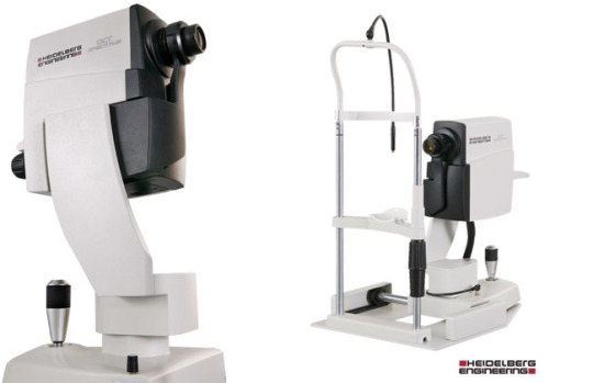
At the core of the Heidelberg Spectralis lies its sophisticated imaging capabilities, which include multiple modalities such as SD-OCT, confocal scanning laser ophthalmoscopy (cSLO), and fundus autofluorescence (FAF) imaging. This integration of modalities allows for comprehensive assessment of retinal morphology, function, and pathology, enabling clinicians to obtain detailed insights into various ocular conditions.
Advantages for Practitioners:
For eye care professionals, the Heidelberg Spectralis offers numerous advantages. Its ability to capture high-quality images in a non-invasive manner facilitates early detection and monitoring of retinal diseases, including age-related macular degeneration, diabetic retinopathy, and glaucoma. Moreover, the device's intuitive software and customizable imaging protocols enhance workflow efficiency, enabling clinicians to make informed decisions regarding patient management and treatment strategies.
Empowering Patient Care:
From the patient's perspective, the introduction of the Heidelberg Spectralis signifies a significant advancement in eye care technology. Its non-invasive imaging procedures and quick scan times ensure a comfortable and efficient experience during retinal evaluations. Furthermore, by enabling early detection and monitoring of ocular pathology, this technology offers the promise of preserving vision and improving long-term outcomes for patients.
Applications Across Ophthalmic Subspecialties:
The versatility of the Heidelberg Spectralis extends beyond retinal imaging, encompassing a wide range of ophthalmic subspecialties. In the field of glaucoma, for example, the device's ability to assess optic nerve head morphology and retinal nerve fiber layer thickness aids in the diagnosis and management of this sight-threatening condition. Similarly, in corneal imaging, the device's anterior segment module provides detailed visualization of corneal layers and pathology, facilitating accurate diagnosis and treatment planning for conditions such as keratoconus and corneal dystrophies.
Challenges and Future Directions:
Despite its remarkable capabilities, the integration of the Heidelberg Spectralis into clinical practice may pose certain challenges, including cost considerations and training requirements. However, ongoing advancements in technology and increased accessibility are poised to overcome these obstacles, paving the way for broader adoption and utilization of this transformative imaging platform in the years to come.
Conclusion:
In conclusion, the Heidelberg Spectralis represents a paradigm shift in the field of retinal imaging and diagnosis, offering clinicians unprecedented insights into ocular structures and pathology. With its advanced imaging modalities, user-friendly interface, and precise measurement capabilities, this device empowers eye care professionals to deliver personalized and effective patient care.
0 notes
Text
Mobilizing Vision Care: Exploring the Benefits of DGH Portable Ultrasound
In the realm of ophthalmology, diagnostic tools that are portable yet powerful hold immense value, especially in settings where access to traditional imaging equipment may be limited. The DGH portable ultrasound stands out as a versatile device that offers eye care professionals the ability to conduct comprehensive ocular assessments in diverse clinical environments. This article delves into the significance of the DGH portable ultrasound, exploring its features, benefits, and impact on modern vision care practices.
Understanding the DGH Portable Ultrasound:
The DGH portable ultrasound is a compact and lightweight imaging device designed to provide detailed imaging of ocular structures, including the cornea, anterior chamber, lens, and posterior segment. Unlike traditional ultrasound machines that are bulky and stationary, this portable device offers flexibility and convenience without compromising imaging quality or diagnostic accuracy.
Key Features and Functionality:
At the heart of the DGH portable ultrasound lies its advanced ultrasound technology, which utilizes high-frequency sound waves to produce real-time images of the eye's internal structures. The device is equipped with a range of transducers and imaging modes, allowing clinicians to perform various ocular assessments, such as pachymetry, biometry, and anterior and posterior segment imaging, with precision and efficiency.
Empowering Patient Care:
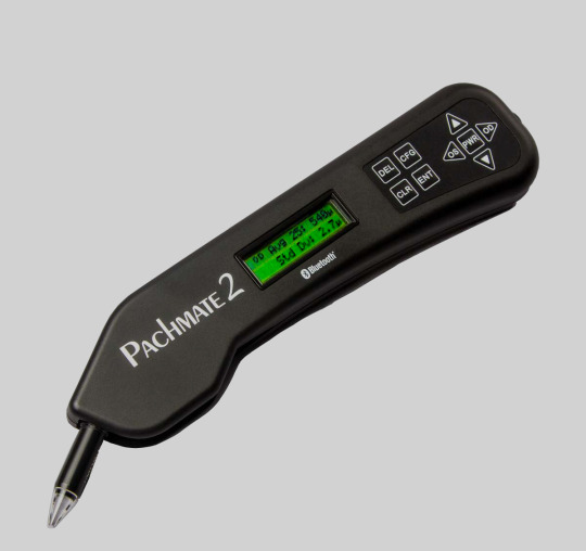
From the patient's perspective, the introduction of the DGH portable ultrasound represents a significant advancement in vision care accessibility and convenience. Its non-invasive imaging procedures and quick scan times ensure a comfortable and efficient experience during ocular evaluations. Furthermore, by facilitating early detection and monitoring of ocular pathology, this technology offers the promise of preserving vision and improving long-term outcomes for patients, especially in underserved or remote communities.
Applications Across Ophthalmic Specialties:
The versatility of the DGH portable ultrasound extends across various ophthalmic specialties, making it a valuable tool for comprehensive eye care. In corneal imaging, for example, the device can assess corneal thickness and detect abnormalities such as edema or dystrophy. In glaucoma management, it can evaluate anterior chamber depth and angle structures, aiding in the diagnosis and monitoring of disease progression.
Challenges and Future Directions:
Despite its numerous benefits, the integration of the DGH portable ultrasound into clinical practice may pose certain challenges, including training requirements and cost considerations. However, ongoing advancements in technology and increased accessibility are poised to overcome these obstacles, paving the way for broader adoption and utilization of this invaluable imaging tool in the years to come.
Conclusion:
In conclusion, the DGH portable ultrasound represents a significant advancement in the field of ocular imaging and diagnosis, offering eye care professionals the ability to conduct comprehensive assessments with flexibility and convenience. With its compact design, advanced ultrasound technology, and wide range of applications, this device empowers clinicians to deliver personalized and effective patient care, even in resource-limited settings.
0 notes
Text
Unlocking Vision Clarity with Zeiss Cirrus 4000 OCT: A New Era in Eye Health
In the ever-evolving landscape of eye care, technology continues to redefine the standards of precision and accuracy. Among the latest innovations, the Zeiss Cirrus 4000 OCT (Optical Coherence Tomography) emerges as a beacon of advancement, offering unparalleled insights into retinal health. This blog explores the transformative potential of the Zeiss Cirrus 4000 OCT, shedding light on its features, benefits, and impact on modern ophthalmic practice.
Exploring Zeiss Cirrus 4000 OCT:
The Zeiss Cirrus 4000 OCT represents a cutting-edge imaging system engineered to deliver detailed, cross-sectional images of the retina. By harnessing the power of light waves, this device enables clinicians to visualize retinal structures with exceptional clarity and precision, revolutionizing the diagnostic and monitoring capabilities in eye care.
Delving into Technology:

At the heart of the Zeiss Cirrus 4000 OCT lies its sophisticated technology, which utilizes a non-invasive imaging technique based on light-wave interference. This approach allows the device to capture high-resolution, three-dimensional scans of the retina, providing comprehensive insights into its anatomical layers and microstructures.
Advantages for Practitioners:
For eye care professionals, the Zeiss Cirrus 4000 OCT offers a myriad of benefits. Its ability to detect subtle changes in retinal thickness and morphology facilitates early diagnosis and monitoring of various ocular pathologies, including macular degeneration, diabetic retinopathy, and glaucoma. Moreover, the device's user-friendly interface and rapid imaging capabilities enhance workflow efficiency, empowering clinicians to deliver personalized care to their patients.
Empowering Patient Care:
From the patient's perspective, the introduction of Zeiss Cirrus 4000 OCT signifies a significant advancement in vision care. Its non-invasive nature and quick scan times ensure a comfortable and hassle-free experience during retinal evaluations. Furthermore, by enabling early detection of retinal abnormalities, this technology offers the promise of preserving vision and improving long-term outcomes for patients.
Transforming Retinal Health:
The widespread adoption of Zeiss Cirrus 4000 OCT holds profound implications for the field of retinal health. By providing clinicians with detailed insights into retinal anatomy and pathology, this technology facilitates timely intervention and personalized treatment strategies. Additionally, its ability to track disease progression over time enables practitioners to monitor therapeutic efficacy and adjust management plans accordingly, ultimately enhancing patient care and quality of life.
Conclusion:
In conclusion, the Zeiss Cirrus 4000 OCT represents a significant milestone in the evolution of eye care, offering clinicians unprecedented insights into retinal health. With its advanced imaging capabilities and user-friendly design, this technology has the potential to revolutionize how ocular conditions are diagnosed, monitored, and managed. As we continue to embrace innovation in ophthalmology, Zeiss Cirrus 4000 OCT stands as a testament to progress, ushering in a new era of clarity and precision in vision care.
0 notes
Text
Revolutionizing Eye Care: The Advantages of the ICARE TA01 Tonometer
In the realm of ophthalmology, precision and accuracy are paramount. The ICARE TA01 Tonometer emerges as a pioneering tool, reshaping the landscape of eye care diagnostics. With its innovative technology and user-friendly design, this device offers a multitude of advantages, revolutionizing the way intraocular pressure (IOP) is measured.
Unparalleled Precision:
The cornerstone of the ICARE TA01 Tonometer lies in its ability to deliver precise and reliable measurements of intraocular pressure. The ICARE TA01 utilizes advanced rebound technology. This method ensures consistent and accurate readings, even in challenging clinical environments. By eliminating the variability associated with other tonometry techniques, the ICARE TA01 provides clinicians with confidence in their diagnostic assessments.
Patient Comfort and Safety:
One of the most significant advantages of the ICARE TA01 Tonometer is its non-invasive and gentle approach to measuring intraocular pressure. Patients often experience anxiety or discomfort during traditional tonometry procedures, which involve direct contact with the cornea. In contrast, the ICARE TA01 utilizes a soft-tipped probe that makes brief, painless contact with the corneal surface. This approach not only enhances patient comfort but also minimizes the risk of corneal abrasions or infections, ensuring a safer and more pleasant experience for the individual undergoing examination.
Enhanced Efficiency:
Efficiency is a hallmark of the ICARE TA01 Tonometer, streamlining the diagnostic process for both clinicians and patients alike. With its rapid measurement capabilities, this device significantly reduces examination time compared to conventional tonometry methods. The straightforward operation and intuitive interface further enhance workflow efficiency, allowing ophthalmic professionals to focus their time and attention where it matters most – patient care.
Portability and Versatility:
The compact and portable design of the ICARE TA01 Tonometer extends its utility beyond traditional clinical settings. Whether in a hospital, private practice, or remote outreach program, this device offers unparalleled versatility. Its lightweight construction and battery-powered operation make it ideal for mobile clinics or fieldwork, enabling ophthalmic professionals to reach underserved communities with ease.
Data Integration and Connectivity:
In the era of digital healthcare, seamless data integration, and connectivity are essential features for medical devices. The ICARE TA01 Tonometer rises to meet this demand with its compatibility with electronic health record (EHR) systems and connectivity options. By seamlessly transferring measurement data to patient records, this device facilitates streamlined documentation and enhances collaboration among healthcare providers.

The ICARE TA01 Tonometer represents a paradigm shift in the field of ophthalmic diagnostics, offering unparalleled precision, patient comfort, and efficiency. Its innovative technology and user-friendly design empower clinicians to deliver high-quality eye care services with confidence and convenience. As the demand for reliable intraocular pressure measurements continues to grow, the ICARE TA01 stands as a beacon of progress, reshaping the way we approach the diagnosis and management of ocular conditions. Top of Form
0 notes
Text
Advantages of the Zeiss IOL Master 500 in Precise Cataract Surgery Planning
In the realm of cataract surgery, precision is paramount. Surgeons rely on advanced tools and technologies to ensure optimal outcomes for their patients. Among these tools, the Zeiss IOL Master 500 stands out as a cornerstone in preoperative planning. With its cutting-edge features and unmatched accuracy, this device revolutionizes the way surgeons approach cataract procedures.
Unrivaled Precision in Biometry Measurements
The Zeiss IOL Master 500 is renowned for its unparalleled precision in biometry measurements. By utilizing partial coherence interferometry (PCI) technology, it achieves precise axial length measurements, corneal curvature assessments, and anterior chamber depth calculations. These accurate measurements are crucial for selecting the appropriate intraocular lens (IOL) power and achieving optimal refractive outcomes. Surgeons can rely on the IOL Master 500 to obtain reliable data, even in challenging cases such as dense cataracts or eyes with prior refractive surgery.
Streamlined Workflow and Efficiency

One of the significant advantages of the Zeiss IOL Master 500 is its ability to streamline the preoperative workflow, saving valuable time for both surgeons and patients. Unlike traditional methods that involve multiple diagnostic devices, the IOL Master 500 consolidates all necessary measurements into a single, efficient process. With its user-friendly interface and automated measurement capabilities, it simplifies data acquisition and minimizes the risk of errors. This streamlined workflow enhances surgical efficiency and allows for better utilization of resources in busy clinical settings.
Enhanced Patient Satisfaction and Visual Outcomes
By facilitating precise biometry measurements and informed IOL selection, the Zeiss IOL Master 500 contributes to enhanced patient satisfaction and visual outcomes. With the ability to accurately predict postoperative refractive error, surgeons can tailor their surgical approach to meet each patient's unique visual needs. Whether aiming for emmetropia or addressing astigmatism, the Zeiss IOL Master 500 empowers surgeons to achieve predictable results and optimize visual acuity. Patients can enjoy improved vision and reduced dependence on glasses or contact lenses, leading to a higher quality of life post-surgery.
Versatility and Adaptability in Surgical Planning
The Zeiss IOL Master 500 offers versatility and adaptability, allowing surgeons to customize their surgical plans based on individual patient characteristics and preferences. With advanced features such as the Holladay 2 formula and the option for toric IOL calculations, it accommodates a wide range of patient demographics and ocular conditions. Surgeons can confidently address complex cases, including those with irregular astigmatism or previous refractive procedures, knowing that the IOL Master 500 provides the necessary tools for precise planning and execution. As a game-changer, offering unmatched advantages in preoperative planning and biometry measurements. Its unrivaled precision, streamlined workflow, and adaptability make it an indispensable tool for surgeons seeking optimal outcomes and enhanced patient satisfaction. By leveraging the capabilities of the Zeiss IOL Master 500, surgeons can navigate the complexities of cataract surgery with confidence, delivering personalized care and maximizing visual outcomes for their patients.
0 notes
Text
Shedding Light on the AMA Brightness Acuity Meter Deuce: Enhancing Vision Care with Precision
In the realm of vision care, precision is paramount. Ensuring accurate measurements of visual acuity aids in diagnosing eye conditions and prescribing appropriate treatments. Among the tools that stand out in this domain is the AMA Brightness Acuity Meter Deuce (AMA BAM Deuce). This article delves into the significance of this device and the benefits it brings to vision care professionals and patients alike.
Understanding the AMA Brightness Acuity Meter Deuce:
The AMA Brightness Acuity Meter Deuce is a sophisticated instrument designed to measure visual acuity accurately and efficiently. The BAM Deuce employs advanced technology to provide objective and precise measurements.
Enhanced Accuracy:
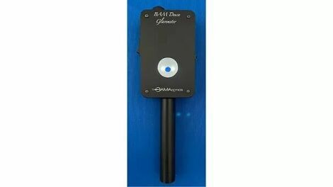
One of the primary benefits of the AMA BAM Deuce is its ability to deliver highly accurate results. By utilizing standardized lighting conditions and contrast levels, this device minimizes variability in measurements, ensuring consistent and reliable outcomes. This accuracy is invaluable in diagnosing eye conditions and monitoring changes in visual acuity over time.
Efficiency in Assessment:
Time is of the essence in any clinical setting, and the BAM Deuce excels in efficiency. With its automated testing procedures, this device streamlines the assessment process, allowing vision care professionals to conduct thorough examinations in a fraction of the time required by traditional methods. As a result, clinics can optimize their workflow and serve a larger number of patients without compromising the quality of care.
Objective Measurement:
Subjectivity can introduce errors and inconsistencies in vision assessments. The AMA Brightness Acuity Meter Deuce eliminates this factor by providing objective measurements based on scientific principles. By quantifying visual acuity in terms of brightness and contrast sensitivity, this device offers a standardized approach that transcends individual differences in perception. This objectivity enhances the reliability of diagnosis and treatment decisions, leading to better outcomes for patients.
Versatility in Applications:
The versatility of the AMA BAM Deuce extends its utility beyond routine vision testing. This device is invaluable in various clinical settings, including optometry, ophthalmology, and occupational therapy. Whether evaluating visual function in patients with eye diseases or assessing visual performance in occupational tasks, the BAM Deuce adapts to diverse applications with ease, making it a versatile tool for healthcare professionals.
Patient-Centric Benefits:
Ultimately, the true measure of any medical device lies in its impact on patient care. In this regard, the AMA BAM Deuce excels by prioritizing the needs and comfort of patients. Its non-invasive testing procedures and user-friendly interface ensure a positive experience for individuals undergoing vision assessments. Moreover, by delivering prompt and accurate results, this device empowers patients to make informed decisions about their eye health and treatment options. The AMA Brightness Acuity Meter Deuce represents a significant advancement in vision care technology. With its unparalleled accuracy, efficiency, and versatility, this device revolutionizes the way visual acuity is measured and assessed. By embracing the BAM Deuce, vision care professionals can enhance diagnostic precision, streamline clinical workflows, and improve patient outcomes.
0 notes
Text
Revolutionizing Ophthalmic Precision: Exploring the Zeiss IOL Master 700
In the realm of ophthalmology, precision is paramount. The Zeiss IOL Master 700 stands as a beacon of technological advancement, reshaping the landscape of intraocular lens (IOL) measurements. Let's delve into this cutting-edge device and unravel its myriad benefits for both practitioners and patients alike.
Unraveling the Zeiss IOL Master 700:
With precision at its core, the Zeiss IOL Master 700 epitomizes innovation in ophthalmic diagnostics. Let's dissect its key features and the benefits they offer.
Enhanced Accuracy:
At the heart of the Zeiss IOL Master 700 lies unparalleled accuracy. Utilizing swept-source optical coherence tomography (SS-OCT), this device captures precise biometric measurements of the eye with unprecedented detail. From axial length to anterior chamber depth, every parameter is meticulously analyzed, ensuring optimal IOL power calculation.
Streamlined Workflow:

Efficiency is synonymous with the Zeiss IOL Master 700. Its intuitive interface and automated processes streamline workflow, saving valuable time for both clinicians and patients. With swift measurements and seamless data integration, clinics can enhance productivity without compromising on accuracy.
Customized Solutions:
One size does not fit all, especially in the realm of ophthalmic care. The Zeiss IOL Master 700 recognizes this axiom and offers tailored solutions to meet diverse clinical needs. Whether it's optimizing IOL selection for cataract surgery or planning refractive procedures, this device empowers clinicians with customizable options, ensuring optimal outcomes for every patient.
Patient-Centric Care:
Behind every measurement lies a patient's vision and well-being. The Zeiss IOL Master 700 places patients at the forefront by prioritizing comfort and safety. Its non-contact measurements minimize discomfort, particularly for sensitive individuals. Moreover, the swift process reduces overall chair time, enhancing the patient experience and fostering satisfaction.
Future-Proof Technology:
Innovation knows no bounds, and neither does the Zeiss IOL Master 700. Built on a foundation of cutting-edge technology, this device evolves alongside the ever-changing landscape of ophthalmic advancements. With regular software updates and continuous refinement, practitioners can rest assured that they're equipped with the latest tools to deliver optimal care.
Clinical Versatility:
Versatility is a hallmark of excellence, and the Zeiss IOL Master 700 embodies this principle. Beyond cataract surgery, this device finds applications in diverse ophthalmic procedures, including refractive surgery and intraocular lens exchange. Its adaptability ensures that clinicians can confidently address a spectrum of visual impairments with precision and efficacy.
In the realm of ophthalmology, precision reigns supreme, and the Zeiss IOL Master 700 reigns as a beacon of technological prowess. From enhanced accuracy to patient-centric care, this device transcends conventional boundaries, redefining the standard of ophthalmic excellence. As we embrace the future of eye care, let us journey forward with confidence, guided by the transformative capabilities of the Zeiss IOL Master 700.
0 notes
Text
Streamlining Document Management with DGH Scanmate Flex
In the digital age, efficient document management is crucial for businesses to stay competitive. With the influx of paperwork and the need for accurate record-keeping, organizations are turning to innovative solutions like the DGH Scanmate Flex to streamline their document management processes. This cutting-edge technology offers a myriad of benefits, revolutionizing how businesses handle their documents from scanning to storage. Let's delve deeper into the advantages of integrating the DGH Scanmate Flex into your workflow.
Enhanced Productivity:
One of the primary benefits of the DGH Scanmate Flex is its ability to enhance productivity within your organization. With its high-speed scanning capabilities, this device can quickly convert paper documents into digital files, eliminating the need for manual data entry and reducing the time spent on document management tasks. This means your team can focus on more critical tasks, leading to increased efficiency and productivity across the board.
Improved Accuracy:
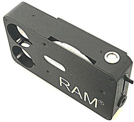
Manual data entry is not only time-consuming but also prone to errors. The DGH Scanmate Flex helps mitigate this risk by accurately capturing and digitizing documents with precision. Its advanced optical character recognition (OCR) technology ensures that even handwritten or poorly scanned documents are accurately converted into editable digital files. By minimizing errors in data entry, businesses can maintain the integrity of their records and make more informed decisions based on accurate information.
Cost Savings:
The DGH Scanmate Flex offers a cost-effective alternative by reducing the need for paper-based processes and streamlining document storage. With digital files stored securely in the cloud or on local servers, businesses can save on paper, ink, storage space, and other associated costs. Additionally, the time saved on document management tasks translates to cost savings in labor expenses.
Enhanced Accessibility:
In today's fast-paced business environment, accessibility to documents is paramount for collaboration and decision-making. The DGH Scanmate Flex enables businesses to access their documents anytime, anywhere, with secure cloud storage and remote access capabilities.
Seamless Integration:
Integrating new technologies into existing workflows can be daunting, but the DGH Scanmate Flex seamlessly integrates into your current document management system. Compatible with a wide range of software applications and document management platforms, this versatile device ensures a smooth transition without disrupting your business operations.
Enhanced Security:
Protecting sensitive information is a top priority for businesses, especially in industries with strict compliance regulations. The DGH Scanmate Flex prioritizes security with features like password protection, encryption, and user authentication, ensuring that only authorized personnel have access to confidential documents.
The DGH Scanmate Flex offers a comprehensive solution for businesses seeking to streamline their document management processes. From enhanced productivity and accuracy to cost savings and improved accessibility, this innovative technology empowers organizations to digitize their documents efficiently and securely. By investing in the DGH Scanmate Flex, businesses can stay ahead of the curve in today's digital landscape and unlock new opportunities for growth and success.
0 notes
Text
Exploring the Revolutionary Zeiss IOL Master 700: A Game-Changer in Ophthalmic Surgery
The Zeiss IOL Master 700 represents the pinnacle of innovation in ophthalmic surgery, revolutionizing the way cataract surgery and intraocular lens (IOL) calculations are performed. In this blog post, we'll delve into the features and benefits of the Zeiss IOL Master 700, highlighting its significance in modern eye care.
Unraveling the Zeiss IOL Master 700:
- The Zeiss IOL Master 700 is an advanced optical biometer designed to precisely measure the eye's axial length, corneal curvature, anterior chamber depth, and lens thickness.
- Utilizing swept-source optical coherence tomography (SS-OCT) technology, the IOL Master 700 captures high-resolution images of the eye's anatomy with unparalleled speed and accuracy.
- With its non-contact measurement technique, the Zeiss IOL Master 700 ensures patient comfort and minimizes the risk of intraoperative complications.
Key Features and Benefits:
1. Enhanced Precision and Accuracy:
- The Zeiss IOL Master 700 provides highly accurate measurements essential for successful cataract surgery and premium IOL selection.
- Its SS-OCT technology offers superior visualization of ocular structures, enabling precise calculations and optimal IOL power selection.
2. Streamlined Workflow:
- With its rapid measurement capabilities, the Zeiss IOL Master 700 streamlines the preoperative assessment process, reducing patient wait times and enhancing clinic efficiency.
- Automated alignment and focusing mechanisms further expedite measurements, allowing surgeons to perform more procedures with greater efficiency.
3. Comprehensive Biometry:
- The Zeiss IOL Master 700 offers comprehensive biometry of both the anterior and posterior segments of the eye, providing surgeons with essential data for accurate IOL power calculations.
- Its ability to measure parameters such as lens tilt and decentration enhances the predictability of refractive outcomes and reduces the need for postoperative adjustments.
4. Customized Surgical Planning:
- By integrating with the Zeiss Cataract Suite software, the IOL Master 700 enables surgeons to customize surgical plans based on individual patient characteristics and preferences.
- Advanced features such as toric and multifocal IOL planning tools facilitate precise astigmatism correction and optimal visual outcomes for patients with presbyopia.
5. Patient Satisfaction and Safety:
- The Zeiss IOL Master 700 enhances patient satisfaction by offering tailored treatment options and minimizing the risk of refractive surprises postoperatively.
- Its non-invasive measurement technique and user-friendly interface contribute to a positive patient experience and promote safety in ophthalmic surgery.
Advancing Ophthalmic Care with Zeiss IOL Master 700:
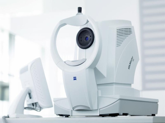
- The Zeiss IOL Master 700 represents a significant advancement in ophthalmic care, empowering surgeons with cutting-edge technology to achieve optimal outcomes for their patients.
- Its precision, efficiency, and versatility make it an indispensable tool in the modern cataract and refractive surgery practice.
- By embracing the Zeiss IOL Master 700, ophthalmic surgeons can elevate the standard of care and deliver superior visual outcomes to their patients.
0 notes
Text
A Guide to Smart Purchase of Refurbished Ophthalmic Equipment
Refurbished ophthalmic equipment presents an enticing opportunity for healthcare facilities, eye clinics, and practitioners to acquire high-quality devices at a fraction of the cost of new ones. However, making informed decisions in this arena requires careful consideration of various factors. This article aims to guide prospective buyers through the process of purchasing refurbished ophthalmic equipment, highlighting the best practices and identifying the ideal candidates for such acquisitions.
Understanding Refurbished Ophthalmic Equipment:
Refurbished Ophthalmic Equipment refers to devices that have been previously used but have undergone thorough inspection, repair, and testing to ensure they meet manufacturer specifications and function like new. These devices can include slit lamps, fundus cameras, phoropters, and more.

Best Practices for Purchasing Refurbished Equipment:
Research Reliable Suppliers: Identify reputable suppliers or dealers with a proven track record in refurbishing ophthalmic equipment. Look for certifications and customer reviews to gauge reliability and quality assurance.
Inspect and Test: Prioritize suppliers who offer transparency regarding the refurbishment process and provide detailed inspection reports. Request demonstrations or trials to assess the equipment's performance firsthand.
Warranty and Support: Choose suppliers that offer warranties and after-sales support. A comprehensive warranty offers protection against probable malfunctions or faults as well as peace of mind.
Evaluate Total Cost of Ownership: Consider not only the upfront cost of the equipment but also factors such as ongoing maintenance, calibration, and future upgrades. Calculating the total cost of ownership helps in determining the long-term value of the investment.
Compliance and Certification: Ensure that refurbished equipment adheres to industry standards and regulations, such as FDA approvals and ISO certifications. Compliance with these standards indicates the equipment's safety and reliability.
Who Should Consider Purchasing Refurbished Ophthalmic Equipment?
Refurbished Ophthalmic Equipment can be helpful for many, some examples are below:
Small Clinics and Practices: For small clinics or practices with budget constraints, refurbished equipment offers a cost-effective solution without compromising on quality. It allows them to access advanced technology that may have been otherwise financially prohibitive.
Medical Schools and Training Facilities: Educational institutions can benefit from refurbished equipment to provide hands-on training to students without overspending on new devices. It facilitates practical learning experiences while minimizing financial strain.
Emerging Markets and Developing Countries: In regions where healthcare infrastructure is still developing, refurbished ophthalmic equipment can bridge the gap by providing essential diagnostic and treatment tools at affordable prices. It enables healthcare providers to deliver quality eye care services to underserved populations.
Refurbished Ophthalmic Equipment presents a viable option for healthcare facilities and practitioners seeking to upgrade their diagnostic and treatment capabilities while managing costs effectively. By following best practices such as researching reliable suppliers, inspecting equipment thoroughly, and considering total cost of ownership, buyers can make informed decisions and acquire high-quality devices with confidence. Whether for small clinics, educational institutions, or healthcare initiatives in developing regions, refurbished equipment offers a pathway to enhanced eye care accessibility and affordability.
0 notes
Text
A Guide to Purchase Preowned Ophthalmic Equipment
In the realm of healthcare, obtaining quality equipment is crucial for providing optimal patient care. Ophthalmic practices, in particular, rely on advanced machinery to diagnose and treat various eye conditions. However, acquiring brand new ophthalmic equipment can be costly, prompting many practices to explore the option of purchasing preowned equipment. In this guide, we'll explore the best ways to procure preowned ophthalmic equipment while ensuring quality and reliability.
Assessing Your Needs:
Before diving into the market for Preowned Ophthalmic Equipment, it's essential to assess your practice's specific requirements. Consider factors such as the type of equipment needed, budget constraints, and compatibility with existing systems. This preliminary evaluation will help narrow down your search and ensure that you invest in equipment that aligns with your practice's goals.
Researching Reliable Suppliers:

When purchasing preowned ophthalmic equipment, choosing a reputable supplier is paramount. Look for suppliers with a track record of reliability, positive reviews from previous customers, and transparent policies regarding equipment condition and warranties. Online marketplaces, specialized medical equipment vendors, and industry forums can be valuable resources for finding trustworthy suppliers.
Quality Assurance and Certification:
To mitigate the risks associated with buying preowned equipment, prioritize vendors that offer quality assurance and certification processes. Reputable suppliers often conduct thorough inspections, refurbishments, and performance testing to ensure that the equipment meets industry standards and functions optimally. Additionally, inquire about warranty options to safeguard your investment against potential malfunctions or defects.
Requesting Detailed Product Information:
Before making a purchase of Preowned Ophthalmic Equipment, request comprehensive product information from the supplier. This should include specifications, maintenance history, usage statistics, and any upgrades or modifications performed on the equipment. Having a clear understanding of the equipment's condition and performance capabilities will enable you to make an informed decision and avoid unpleasant surprises post-purchase.
Negotiating Pricing and Terms:
While preowned equipment offers cost savings compared to new purchases, there's often room for negotiation when it comes to pricing and terms. Don't hesitate to discuss pricing, payment plans, shipping costs, and warranty coverage with the supplier. In some cases, bulk purchases or package deals may be available, further reducing the overall expenditure.
Ensuring Compatibility and Support:
Verify that the preowned equipment is compatible with your existing infrastructure and software systems. Compatibility issues can lead to operational disruptions and additional expenses down the line. Additionally, inquire about ongoing technical support, training resources, and maintenance services provided by the supplier to optimize the equipment's performance and longevity.
Purchasing Preowned Ophthalmic Equipment can be a cost-effective solution for healthcare practices seeking to upgrade their facilities without breaking the bank. By following these guidelines and exercising due diligence in the procurement process, practices can acquire high-quality equipment that enhances diagnostic accuracy, improves patient outcomes, and contributes to the overall efficiency of ophthalmic services. With trusted suppliers like Foresight International, practices can confidently invest in preowned equipment that meets their needs and exceeds expectations.
0 notes
Text
The Importance of Used Ophthalmic Equipment: Where to Find Quality Solutions
Ophthalmic equipment plays a crucial role in diagnosing and treating eye conditions, ensuring accurate assessments and effective treatments. However, the high cost of new equipment can pose challenges for many practices and healthcare facilities. This is where used ophthalmic equipment becomes invaluable, offering cost-effective solutions without compromising on quality.
Why Used Ophthalmic Equipment Matters:
Affordability:
Cost-saving is a significant advantage of purchasing Used Ophthalmic Equipment. By opting for pre-owned devices, practices can significantly reduce their initial investment, making essential equipment more accessible.
This affordability allows healthcare providers to allocate resources more efficiently, potentially expanding their services or investing in other areas of their practice.

Quality Assurance:
Contrary to common misconceptions, used ophthalmic equipment can offer excellent quality and performance. Reputable suppliers thoroughly inspect and refurbish equipment to ensure it meets industry standards before resale.
Advances in technology have led to durable and reliable equipment designs, meaning that used devices can still provide accurate results and long-term functionality.
Accessibility:
Access to specialized equipment is vital for ensuring comprehensive eye care services, especially in underserved areas or smaller practices with limited budgets.
By offering Used Ophthalmic Equipment, suppliers contribute to improving access to essential diagnostic and treatment tools, ultimately enhancing patient care outcomes.
Where to Purchase Quality Used Ophthalmic Equipment:
Certified Suppliers:
Opting for certified suppliers ensures that the used equipment has undergone rigorous testing and refurbishment processes to meet safety and performance standards.
Reputable suppliers often provide warranties and after-sales support, offering peace of mind to buyers regarding the reliability and longevity of the equipment.
Online Marketplaces:
Online platforms specializing in medical equipment resale offer a vast selection of used ophthalmic devices from various manufacturers.
Buyers can compare prices, specifications, and seller ratings to make informed purchasing decisions, often finding competitive deals and rare equipment options.
Industry Refurbishers:
Many companies specialize in refurbishing and reselling ophthalmic equipment, leveraging their expertise to ensure the quality and functionality of pre-owned devices.
These refurbishers often collaborate with manufacturers and adhere to strict refurbishment protocols, ensuring that the equipment meets or exceeds original performance standards.
Used Ophthalmic Equipment serves as a cost-effective solution for healthcare providers seeking to enhance their diagnostic and treatment capabilities without exceeding budgetary constraints. By prioritizing affordability, quality assurance, and accessibility, practices can acquire essential equipment that meets industry standards and improves patient care outcomes. Partnering with certified suppliers, exploring online marketplaces, or collaborating with industry refurbishers are effective strategies for sourcing quality used ophthalmic equipment, empowering healthcare providers to optimize their services and expand their reach.
0 notes
Text
Advancing Precision in Ophthalmic Surgery with Zeiss IOL Master 500
When it comes to ophthalmic surgery, accuracy is critical. As technology continues to evolve, so too do the tools and instruments at the disposal of surgeons. Among these advancements stands the Zeiss IOL Master 500, a revolutionary device that has transformed the landscape of cataract surgery and refractive lens exchange. In this article, we delve into the capabilities and significance of this cutting-edge equipment.
Introduction to Zeiss IOL Master 500
The Zeiss IOL Master 500 is a state-of-the-art device designed to measure the eye's parameters with unparalleled accuracy. Developed by the renowned optics company Carl Zeiss Meditec, this instrument utilizes partial coherence interferometry (PCI) to obtain precise measurements of axial length, corneal curvature, and anterior chamber depth. These measurements are crucial for determining the appropriate power of intraocular lenses (IOLs) to be implanted during surgery.

Unraveling the Technology: Partial Coherence Interferometry (PCI)
At the heart of the Zeiss IOL Master 500 lies the principle of partial coherence interferometry. This technology relies on the interference patterns created by splitting a light beam into two paths and recombining them. By analyzing these interference patterns, the device can accurately determine the distances within the eye, providing ophthalmic surgeons with essential data for surgical planning.
Enhancing Surgical Precision
One of the key advantages of the Zeiss IOL Master 500 is its ability to enhance surgical precision. By providing highly accurate measurements of ocular parameters, surgeons can better calculate the power of IOLs needed to achieve the desired refractive outcomes for patients. This precision is particularly critical in procedures such as cataract surgery, where the goal is to replace the clouded natural lens with an artificial one that restores clear vision.
Streamlining Workflow and Improving Efficiency
In addition to its precision, the Zeiss IOL Master 500 offers practical benefits in terms of workflow efficiency. With its user-friendly interface and rapid measurement capabilities, the device streamlines the preoperative assessment process, allowing surgeons to gather essential data quickly and accurately. This efficiency not only saves time but also contributes to better patient outcomes by ensuring thorough preoperative planning.
Elevating Patient Care and Satisfaction
Ultimately, the Zeiss IOL Master 500 plays a crucial role in elevating patient care and satisfaction. By optimizing the accuracy of preoperative measurements, surgeons can tailor their surgical approach to each individual patient, minimizing the risk of postoperative refractive errors and complications. The result is a higher likelihood of achieving the desired visual outcomes and improved overall satisfaction among patients undergoing cataract surgery or refractive lens exchange.
The Zeiss IOL Master 500 represents a significant advancement in the field of ophthalmic surgery. Its unparalleled precision, facilitated by partial coherence interferometry technology, enables surgeons to make informed decisions and deliver optimal visual outcomes for their patients. By streamlining workflow, enhancing surgical precision, and ultimately improving patient care and satisfaction, this innovative device continues to shape the future of ophthalmic surgery.
0 notes
Text
In the realm of ophthalmology and eye care, technological advancements continue to revolutionize the way professionals diagnose and treat various conditions. One such innovation that stands out is the Zeiss Visucam Fundus Camera. This cutting-edge device has become a cornerstone in the field, offering clinicians unparalleled insights into the health of their patients' eyes.
Unraveling the Benefits:
High-Quality Imaging: At the heart of the Zeiss Visucam Fundus Camera lies its ability to capture high-resolution images of the retina. This capability is crucial for detecting and monitoring a myriad of eye conditions, including diabetic retinopathy, glaucoma, and age-related macular degeneration. With its superior imaging quality, clinicians can make accurate diagnoses and develop personalized treatment plans with confidence.
Efficiency and Precision: Time is of the essence in any medical setting, and the Zeiss Visucam Fundus Camera excels in streamlining the diagnostic process. Its user-friendly interface and intuitive design allow clinicians to capture detailed images quickly and efficiently. Moreover, the camera's automated features ensure precise alignment and focus, minimizing the margin for error and reducing the need for retakes.
Early Detection of Pathologies: One of the most significant advantages of the Zeiss Visucam Fundus Camera is its ability to facilitate early detection of eye pathologies. By capturing detailed images of the retina, clinicians can identify subtle changes indicative of disease progression long before symptoms manifest. This proactive approach not only improves patient outcomes but also helps mitigate the risk of irreversible vision loss.
Patient Education and Engagement: Visual aids are invaluable tools for patient education, and the Zeiss Visucam Fundus Camera provides clinicians with a powerful means of engaging their patients. By visually demonstrating the condition of their eyes and explaining the significance of the images captured, clinicians can empower patients to take an active role in their eye health. This fosters better compliance with treatment regimens and encourages preventive measures to maintain optimal vision.
The Future of Eye Care
As technology continues to evolve, so too will the capabilities of devices like the Zeiss Visucam Fundus Camera. Future iterations may incorporate artificial intelligence algorithms for automated image analysis, further enhancing diagnostic accuracy and efficiency. Additionally, advancements in telemedicine may enable remote access to fundus imaging, expanding access to eye care services in underserved areas.

The Zeiss Visucam Fundus Camera represents a significant advancement in the field of ophthalmology, offering clinicians unparalleled insights into the health of their patients' eyes. From high-quality imaging to enhanced efficiency and precision, its benefits are undeniable. By leveraging this technology, clinicians can provide superior patient care, leading to improved outcomes and a brighter future for eye health worldwide.
0 notes
Text
Unlocking Efficiency and Precision with DGH Scanmate Flex: Revolutionizing Diagnostic Imaging
In the realm of diagnostic imaging, precision and efficiency are paramount. Whether it's detecting abnormalities or tracking disease progression, the accuracy of imaging tools can make all the difference in patient outcomes. One such groundbreaking innovation that's transforming the landscape of diagnostic ultrasound is the DGH Scanmate Flex. Let's delve into how this cutting-edge technology is reshaping the field and why it's an indispensable tool for healthcare professionals.
Understanding DGH Scanmate Flex:
The DGH Scanmate Flex is not just another ultrasound device; it's a game-changer. Engineered with advanced features and unparalleled flexibility, it offers a comprehensive solution for a wide range of diagnostic imaging needs. From routine screenings to complex examinations, this versatile tool excels in delivering high-quality images with exceptional clarity and detail.
Enhanced Imaging Capabilities:
At the core of its brilliance lies its superior imaging capabilities. Equipped with state-of-the-art technology, including advanced transducers and image processing algorithms, the DGH Scanmate Flex ensures unparalleled image resolution and contrast. This means healthcare providers can visualize anatomical structures with remarkable precision, enabling accurate diagnosis and treatment planning.
Versatility Redefined:
One of the standout features of the DGH Scanmate Flex is its versatility. Designed to adapt to diverse clinical settings and patient needs, it offers a multitude of scanning modes and customization options. Whether it's obstetrics, cardiology, musculoskeletal, or vascular imaging, this dynamic tool delivers consistent results across various specialties, streamlining workflow and optimizing resource utilization.
Streamlined Workflow:
Efficiency is at the heart of the DGH Scanmate Flex. With intuitive controls and user-friendly interface, it empowers healthcare professionals to perform scans with ease and efficiency. Its seamless integration with existing healthcare systems further enhances workflow efficiency, allowing for seamless data management and sharing. This not only saves valuable time but also ensures continuity of care for patients.
Precision-guided Interventions:
In diagnostic imaging, precision is non-negotiable, especially when it comes to guiding interventions. The DGH Scanmate Flex excels in this aspect, offering real-time imaging capabilities that enable precise needle guidance during procedures such as biopsies and injections. This not only enhances procedural accuracy but also minimizes patient discomfort and reduces the risk of complications.
Empowering Patient-centered Care:
Ultimately, the true measure of any diagnostic tool's importance lies in its ability to improve patient outcomes. In this regard, the DGH Scanmate Flex shines brightly. Providing healthcare providers with the tools they need to make accurate diagnoses and informed treatment decisions, plays a pivotal role in delivering patient-centered care. From early detection of diseases to monitoring treatment response, its impact on patient lives is profound and far-reaching.
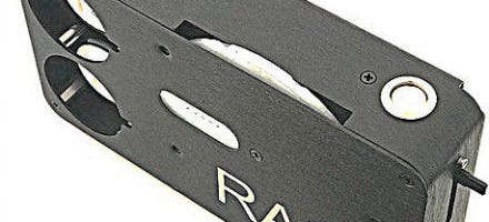
In the ever-evolving landscape of healthcare, innovation is key to driving progress. The DGH Scanmate Flex stands as a testament to this ethos, revolutionizing diagnostic imaging with its unparalleled precision, versatility, and efficiency.
0 notes
Text
Enhancing Eye Health with Zeiss Clarus 500: A Revolutionary Diagnostic Tool
In the realm of eye care, technological advancements continue to revolutionize diagnosis and treatment. Among these innovations stands the Zeiss Clarus 500, a state-of-the-art imaging system that offers unparalleled clarity and precision in ocular diagnostics. Designed by the renowned optics company Zeiss, the Clarus 500 is redefining the way eye health professionals detect and monitor various ocular conditions.
Unrivaled Clarity and Detail
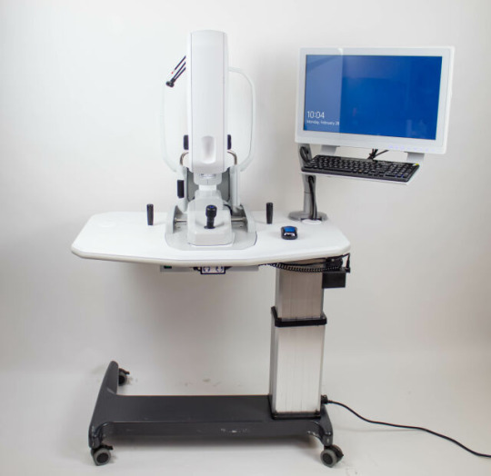
At the heart of the Zeiss Clarus 500 lies its advanced imaging capabilities, providing eye care professionals with unprecedented clarity and detail. Utilizing ultra-widefield retinal imaging technology, this system captures high-resolution images of the retina, optic nerve, and anterior segment of the eye. With a field of view up to 200 degrees, the Clarus 500 ensures comprehensive visualization of the entire retina, enabling early detection of subtle abnormalities that may indicate sight-threatening conditions.
Enhanced Efficiency and Patient Comfort
In addition to its superior imaging capabilities, the Zeiss Clarus 500 offers enhanced efficiency and patient comfort. Unlike traditional imaging methods that may require dilation or prolonged examination times, the Clarus 500 captures widefield images quickly and non-invasively. Its intuitive design and user-friendly interface streamline the diagnostic process, allowing eye care professionals to assess retinal health with ease. Moreover, patients benefit from reduced discomfort and shorter examination times, making the experience more pleasant and convenient.
Early Detection and Monitoring of Ocular Pathologies
One of the most significant benefits of the Zeiss Clarus 500 is its ability to facilitate early detection and monitoring of ocular pathologies. By providing detailed images of the retina and optic nerve, this system enables eye care professionals to identify signs of diabetic retinopathy, age-related macular degeneration, glaucoma, and other sight-threatening conditions at their earliest stages. Early intervention is key to preserving vision and preventing irreversible damage, making the Clarus 500 an invaluable tool in the fight against ocular disease.
Precision Treatment Planning
Furthermore, the Zeiss Clarus 500 assists in precision treatment planning for patients with ocular conditions. By accurately capturing the size, location, and severity of retinal abnormalities, eye care professionals can develop personalized treatment strategies tailored to each patient's needs. Whether it involves laser therapy, intravitreal injections, or surgical intervention, the detailed images provided by the Clarus 500 empower clinicians to deliver targeted and effective treatments, ultimately optimizing patient outcomes and preserving vision.
The Zeiss Clarus 500 represents a significant advancement in ocular diagnostics, offering unrivaled clarity, efficiency, and precision. From comprehensive retinal imaging to early detection of ocular pathologies, this revolutionary system has transformed the way eye care professionals assess and monitor eye health. With its ability to facilitate early intervention and personalized treatment planning, the Clarus 500 plays a vital role in preserving vision and enhancing the quality of life for patients worldwide. As technology continues to evolve, innovations like the Zeiss Clarus 500 pave the way for a brighter future in eye care.
0 notes