Don't wanna be here? Send us removal request.
Photo

What is this strange animal?
Is it a jellyfish? Nope, it’s a doliolid, closely related to sea squirts that you might see in a tide pool! Pseudusa bostigrinus was discovered by MBARI researchers and was the first example of a carnivorous doliolid. Unlike other doliolids, its bell-shape is more like a jellyfish. This adaptation allows it to collect sinking particles by simply directing its large opening upward. The jellyfish-like velum allows it to trap zooplankton prey and to propel itself with considerable force and control. You can read more about this animal here MBARI - Pseudusa.
(via: Monterey Bay Aquarium Research Institute)
232 notes
·
View notes
Photo

Giant stinky Corpse Flower, blossoming
“The Huntington, a wonderful destination here in Los Angeles, has posted GIFs of the rare Corpse Flower blossoming. If you’re in Southern California, you gotta see it in person. The plant’s latin name, Amorphophallus titanum comes from Ancient Greek amorphos, “without form, misshapen” + phallos, “phallus”, and titan, “giant.”
(via BoingBoing)
3K notes
·
View notes
Photo

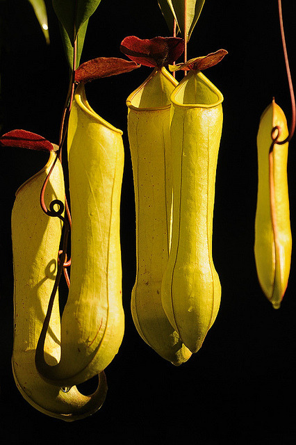
Pitcher plant (Nepenthes distillatoria), Sri Lanka by KSberg on Flickr.
471 notes
·
View notes
Photo


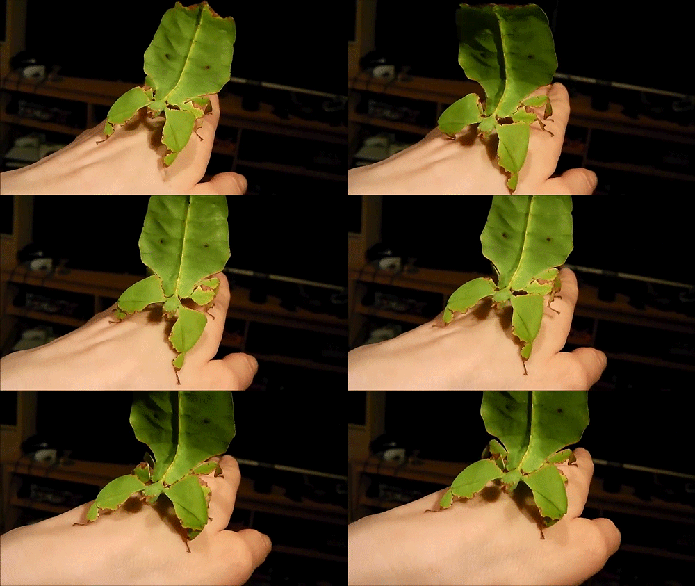
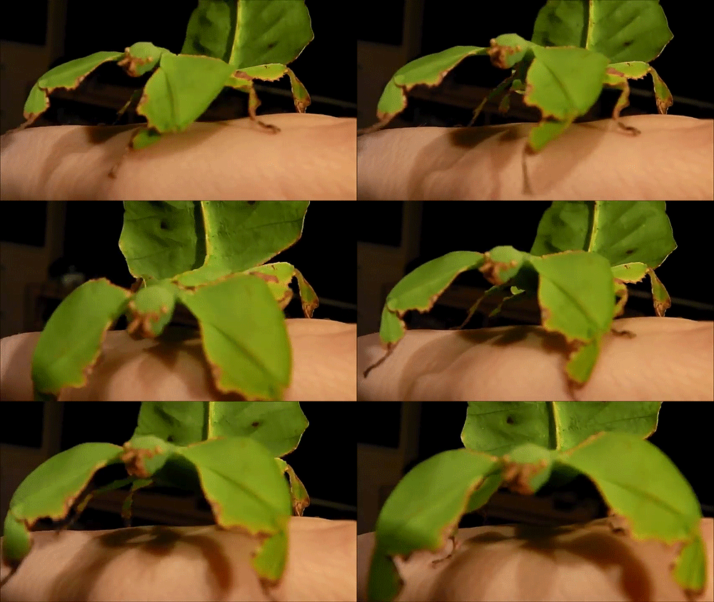
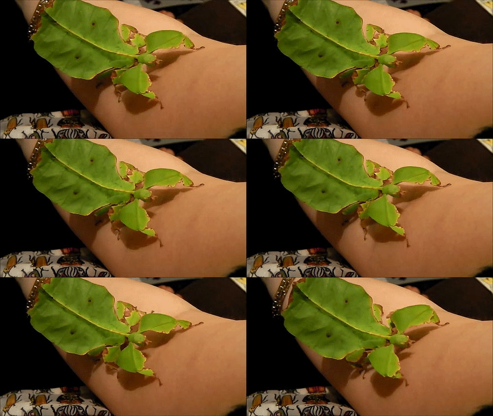
Leaf bug (Phyllium giganteum)
198K notes
·
View notes
Photo

Ebola has a nasty reputation for damaging the body, especially its blood vessels. But when you look at the nitty-gritty details of what happens after a person is infected, a surprising fact surfaces.
How Ebola Kills You: It’s Not The Virus
Illustration credit: Lisa Brown for NPR
671 notes
·
View notes
Photo

Placental Vasculature of a transgenic mouse embryo.
Want to see more beautiful and significant life science images, captured through a light microscope? Then visit our Olympus BioScapes Digital Imaging Competition Exhibit. #Microscope #Science (at New York Hall of Science)
101 notes
·
View notes
Photo


New Species of Venomous Jellies Discovered in Australia By Megan Gannon, News Editor, Live Science | August 13, 2014
Two new species of jellyfish (Keesingia gigas and Malo bella) have been discovered off the coast of Western Australia. One is surprisingly large. The other is tiny. Both are extremely venomous.
These two newfound creatures are thought to pack painful stings that cause Irukandji syndrome, a constellation of symptoms that includes lower back pain, vomiting, difficulty breathing, cramps and spasms. Though Irukandji syndrome usually isn’t life threatening, two people who were stung in the Great Barrier Reef in 2002 died from severe Irukandji-related hypertension.
Research scientist Lisa-ann Gershwin, who is director of the Australian Marine Stinger Advisory Services, described the new box jellyfish, or cubozoans, last month in the Records of the Western Australian Museum [available as a PDF].
Gershwin said that in all of the photos the jellyfish did not appear to have tentacles and that the specimen was also captured without them.
“Jellyfish always have tentacles ... that’s how they catch their food,” she said. “The tentacles are where they concentrate their stinging cells. ... Some of the people working with it through the years actually got stung by it and experienced rather distressing Irukandji syndrome.”
IMAGES: [1] An example of the Keesingia gigas jellyfish. Photograph: John Totterdell/MIRG Australia. Via The Guardian [2] Keesingia gigas in bloom of sea tomatoes, Crambione mastigophora. Image credit: John Totterdell / MIRG Australia.. Via Sci-News [3] That’s not a plastic bag. That’s a newly described species of box jellyfish, Keesingia gigas. Image Credit: Lisa-ann Gershwin
#new species#box jellyfish#cubozoan#Keesingia gigas#marine biology#Irukandji syndrome#australia#jbwii#jbwau
91 notes
·
View notes
Photo

What does Ebola actually do? By Kelly Servick, 13 August 2014 || Science/AAAS | News
Behind the unprecedented Ebola outbreak in West Africa lies a species with an incredible power to overtake its host.
Zaire ebolavirus and the family of filoviruses to which it belongs owe their virulence to mechanisms that first disarm the immune response and then dismantle the vascular system.
The virus progresses so quickly that researchers have struggled to tease out the precise sequence of events, particularly in the midst of an outbreak. Much is still unknown, including the role of some of the seven proteins that the virus’s RNA makes by hijacking the machinery of host cells and the type of immune response necessary to defeat the virus before it spreads throughout the body. But researchers can test how the live virus attacks different cells in culture and can observe the disease’s progression in nonhuman primates—a nearly identical model to humans.
Continue reading to find out some of the basic things we understand about how Ebola and humans interact ...
IMAGE: The Ebola virus THOMAS W. GEISBERT
251 notes
·
View notes
Photo


Indian Giant Flying Squirrel - Petaurista philippensis
Indian Giant Flying Squirrels, Petaurista philippensis (Rodentia - Sciuridae) vary in color from grey to coffee-brown with a mottled back, a grey tail and pale undersides. They feed on fruits and leaves and have a fondness for fig fruits.
They nest mostly in tree cavities and are found in a mosaic of forests of tall trees and plantations. When dusk falls, they leave their nests and return only before dawn.
Glides of flying squirrels are a thrill to watch. Gliding has been the subject of much research, as some scientists believe it may be a precursor to powered flight. Two membranes, one between the wrist and the ankle and another from their wrist to neck are what help them achieve the feat of gliding. Large flying squirrels like the Indian Giant Flying Squirrel have an additional membrane between their tail and hind limbs.
To glide, the flying squirrel climbs high up a tree to the upper canopy and jumps down. It then stretches its gliding membranes taut with the help of a cartilaginous wrist bone so that aided by the force of lift, it glides spread-eagle along an oblong trajectory. While air borne, it uses its tail to manoeuvre and then billows its gliding membranes outwards to decelerate so that it can land lightly. A vertical trunk is usually picked for landing when glides are long.
Indian Giant Flying Squirrels have been known to cover distances as great as 90m in a single glide, although they prefer shorter glides. They also prefer climbing across tree branches when distances between trees are short.
Petaurista philippensis is widely distributed in South Asia, southern and central China, and mainland Southeast Asia.
References: [1] - [2]
Photo credit: ©Gopi Krishnamurthy | Locality: Dandeli, Uttara Kannada, Karnataka, India
318 notes
·
View notes
Photo

Cabbage Coral, Milne Bay, New Guinea 1997 | ©Russell Gilbert
153 notes
·
View notes
Photo

Mouse embryo
“Image courtesy Celeste Nelson and Joe Tien, Princeton Art of Science
Fluorescent light illuminates an embryo’s inner workings in “Baby Mouse.” The unborn mouse’s vascular system, usually red with blood, is rendered here in green. The blue coloring reveals its DNA.”
http://tinyurl.com/opsckl8
78 notes
·
View notes
Photo

Unexpected stem cell factories found inside teeth by Sarah C. P. Williams || 30 July 2014 || Science/AAAS | News
Development is typically thought to be a one-way street. Stem cells produce cells that mature into specific types, such as the neurons and glia that compose nervous systems, but the reverse isn't supposed to happen.
Yet researchers have now discovered nervous system cells transforming back into stem cells in a very surprising place: inside teeth. This unexpected source of stem cells potentially offers scientists a new starting point from which to grow human tissues for therapeutic or research purposes without using embryos.
Continue reading ...
131 notes
·
View notes
Photo

01 August 2014
Gut Bugs Attack
Diarrhoea, sickness, cramps, fever – just some of the symptoms of food poisoning. Some of us will be unlucky enough to experience these as a result of infection by a nasty gut bug known as Campylobacter jejuni, found on raw poultry such as chicken. It’s one of the leading global causes of food-related illness, yet we know little about how it infects us. To find out more, researchers are studying mice carrying a faulty version of a gene called Sigirr that are particularly prone to Campylobacter infection. Pictured is a highly magnified section of gut from one of these animals showing the bacteria (highlighted in red) aggressively attacking and invading the gut tissue (blue and green). Figuring out how cells in the gut and the wider immune system respond to this onslaught reveals more about what’s going on, and opens new approaches for more effective prevention and treatment.
Written by Kat Arney
—
Adapted from an image by Bruce Vallance and colleagues Child and Family Research Institute, Canada Originally published under a Creative Commons Licence (BY 4.0) Research published in PLOS Pathogens, July 2014
—
You can also follow BPoD on Twitter and Facebook
40 notes
·
View notes
Photo


Largest aquatic insect in the world found in China By Bec Crew | July 22, 2014 | Scientific American Blog Network
New species of Megaloptera insect, shown with a chicken’s egg for scale. Credit: China News Service/Zhong Xin
The largest aquatic insect ever found. Credit: China News Service/Zhong Xin
Images have surfaced of a newly discovered insect reported to be the largest aquatic insect in the world. Found in the mountains of Chengdu in China’s Sichuan province, the specimen boasts a wingspan of 21 cm. While very little is known about the specimen at this point, it’s been identified as belonging to the order Megaloptera, which includes about 300 described species of winged alderflies, dobsonflies and fishflies.
Continue reading ...
33 notes
·
View notes
Photo

Keeping Viral Load Low
By Thomas Deerinck, NCMIR, USCD
Over the past 30 years, the combined efforts of scientists and clinicians have delivered remarkable successes in HIV therapeutics. Since 1987, the FDA has approved more than 30 antiviral drugs, including 12 HIV protease inhibitors and one integrase inhibitor. These drugs stop ~99% of viral replication, essentially transforming HIV infection from a deadly disease to a chronic one. What will the next 30 years bring?
Image: Here numerous HIV-1 particles leave a cultured HeLa cell. These viruses lack their vpu gene and thus can’t detach from the cell’s tethering factor, BST2. Each viron particle is ~120nm in diameter. The image was captured with a Zeiss Merlin ultra high-resolution scanning electron microscope. The cells were fixed, dehydrated, critical-point dried, and lightly sputter-coated with gold/palladium.
through Cell.com
94 notes
·
View notes

