
he/him | I post random stuff, whatever has to do with my hyperfixations | Current hyperfixations: mycology, marine biology, dungeons and dragons.
Don't wanna be here? Send us removal request.
Statistics
We looked inside some of the posts by theprinceofmycologia and here's what we found interesting.
Average Info
Notes Per Post
28K
Likes Per Post
16K
Reblog Per Post
12K
Reply Per Post
287
Time Between Posts
14 hours ago
Number of Posts By Type
Text
17
Last Seen Tumblr Blogs


Fun Fact
28.6 is the average number of monthly visits per US mobile user.
Text
Thank you for tagging me ! ♡

A lot has changed, but that is okay, life is dynamic .
Tagging: @squidsandthings @lameotello @edukincon @belis86 @pissass11 @emmakapla @poishroom @strobilomyces-confusus @jasontoddlovr @pinkished @dinonugasaurus + anyone else who wants to join :)
Picrew tag game!- Create yourself now vs how you looked when you were a kid
Link
I was tagged by @cutebisexualmess for this but the chain was too long so I'm restarting!

If only that little girl could see me now (she'd probably think I was cool tbh)
uhm tagging: @b3achfagz (ik you dont do tag games so u can just ignore this but i though u might find it cool) @cassiecryptic @viktheviking1 @depressedgremlinbitch @ramencat12 @inkyslimee @the-horrifying-digital-circus @patipati @cute--thing @musicalsiphonophore @tastetherainbow290 @disenchantedwarlock @bookishcatcafe and anyone else who sees this and thinks it looks cool!!
12K notes
·
View notes
Text
Thank you for tagging me ! ♡ Tadaaaaaa

Tagging : @squidsandthings @lameotello @edukincon @belis86 @pissass11 @emmakapla @poishroom @strobilomyces-confusus @jasontoddlovr @pinkished @dinonugasaurus + anyone else who wants to join :)
No pressure btw ! :)
fuck it. worm on a string picrew chain. let's fucking go

happy worm creation my friends
tagging @areyoudoingthis @cursed-coat-of-homosexuality @peanutbutterex @tfemteach @piratecaptainscaptainpirates (no pressure 💛)
12K notes
·
View notes
Text
You are totally right !
I have a new life motto :
"Laziness will sometimes help you achieve great things."
- One of my best friends
3 notes
·
View notes
Text
I have a new life motto :
"Laziness will sometimes help you achieve great things."
- One of my best friends
3 notes
·
View notes
Text
This is my favourite Fallout : New Vegas fanart EVER ! ! ! It is drop dead gorgeous !
And I just feel the need to reblog it, because it is so awesome !
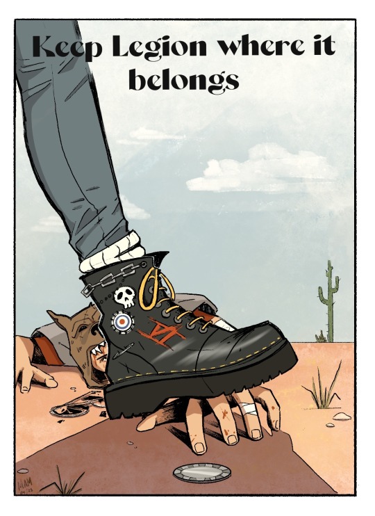
These boots stomp fascists
Based off of the extremely misogynistic 1974 Weyenberg Shoes ad. Image ID under the cut.
[Image ID: A picture of a heavy combat boot crushing the hand of Vulpes Inculta. The text across the top reads “Keep Legion where it belongs.” The combat boot is decorated with skulls and chains and the Roman numeral for six. Vulpes’ face is bloody and his glasses are lying broken in front of him. He is reaching for a platinum chip on the ground, just out of reach. The scene takes place in an empty desert.]
#fallout new vegas#fnv#fnv art#fallout art#fallout fan art#courier six#vulpes inculta#caesar’s legion#legionary#courier oc#fnv oc#fallout oc#this art is drop dead gorgeous!!#so awesome!#special interest
4K notes
·
View notes
Text
This is a Schlumbergera, specifically the Thanksgiving (which refers to the time of the year it blooms) variant, it is named Kal / Cal . . .


I named it Cal, after the Dutch word for turkey because it is a thanksgiving cactus. The Dutch word for turkey is kalkoen, I decided Kal / Cal would be a nice name :)
I bought this Cereus Forbesii Spiralis today !
I named it Polyphemus, like the one-eyed cyclops from Greek Mythology. I did that, because I put it on the pot I painted with pine chafers a.k.a. Polyphylla fullo on it, and I thought *poly* . . . Polyphemus and I thought it was a really nice name so yea . . .
Long story short, I named it Polyphemus ♡


I love it to so much ! ! !
#hyperfixation#botany#house plants#cactus#cacti#spiral cactus#cereus forbesii spiralis#pine chafers#polyphylla fullo#plant collection#schlumbergera#thanksgiving cactus
31 notes
·
View notes
Text
This is my Opuntia microdasys, I named it Deniz . . .


Deniz is Turkish for sea, I named my Opuntia this because, at least to my knowledge and experience, this species of Opuntia grows near the West coast of Turkey and so near the sea :)
It is also growing two new leaves ! ! !
I bought this Cereus Forbesii Spiralis today !
I named it Polyphemus, like the one-eyed cyclops from Greek Mythology. I did that, because I put it on the pot I painted with pine chafers a.k.a. Polyphylla fullo on it, and I thought *poly* . . . Polyphemus and I thought it was a really nice name so yea . . .
Long story short, I named it Polyphemus ♡
I love it to so much ! ! !
#hyperfixation#botany#house plants#cactus#cacti#spiral cactus#cereus forbesii spiralis#pine chafers#polyphylla fullo#plant collection#opuntia#opuntia microdasys#bunny ears cactus#deniz
31 notes
·
View notes
Text
The cactus in the far corner is a Cleistocactus winterii a.k.a. a rat's tail cactus . . .

I named it Argyle, just because I like the name ♡
I already described the other two cacti in previous posts, but the bulbous one on the left is called Gar / Gargouille and the one with really long spikes on the right is called Gnoom :)
I bought this Cereus Forbesii Spiralis today !
I named it Polyphemus, like the one-eyed cyclops from Greek Mythology. I did that, because I put it on the pot I painted with pine chafers a.k.a. Polyphylla fullo on it, and I thought *poly* . . . Polyphemus and I thought it was a really nice name so yea . . .
Long story short, I named it Polyphemus ♡
I love it to so much ! ! !
#hyperfixation#botany#house plants#cactus#cacti#spiral cactus#cereus forbesii spiralis#pine chafers#polyphylla fullo#plant collection#rat's tail cactus#cleistocactus winterii
31 notes
·
View notes
Text
This my peyote, specifically Lophophora Williamsii caespitosa . . .


I named it Dēlos, after 'del' in 'psychedelic', it means 'to reveal' / 'to make visible' / 'to manifest'. I thought it would be fitting for a psychedelic cactus, so yeaa that . . .
I bought this Cereus Forbesii Spiralis today !
I named it Polyphemus, like the one-eyed cyclops from Greek Mythology. I did that, because I put it on the pot I painted with pine chafers a.k.a. Polyphylla fullo on it, and I thought *poly* . . . Polyphemus and I thought it was a really nice name so yea . . .
Long story short, I named it Polyphemus ♡
I love it to so much ! ! !
#hyperfixation#botany#house plants#cactus#cacti#spiral cactus#cereus forbesii spiralis#pine chafers#polyphylla fullo#plant collection#peyote#lophophora williamsii caespitosa
31 notes
·
View notes
Text
This is my Trichocereus bridgesii monstrosa . . .


I named it Gnoom ( Dutch for Gnome, a variant of Gnoom in Dutch is Kabouter and in older Dutch Kaboyter ) , I got to Gnoom because the 'thorns' of the cactus reminded my of nails, nail in Dutch is 'spijker' which in certain dialects / accents is pronounced as 'spieker' , which reminded me of a gnome from a childhood fairytale I cannot quite remember . . . so yea, basically that :)
I bought this Cereus Forbesii Spiralis today !
I named it Polyphemus, like the one-eyed cyclops from Greek Mythology. I did that, because I put it on the pot I painted with pine chafers a.k.a. Polyphylla fullo on it, and I thought *poly* . . . Polyphemus and I thought it was a really nice name so yea . . .
Long story short, I named it Polyphemus ♡
I love it to so much ! ! !
#hyperfixation#botany#house plants#cactus#cacti#spiral cactus#cereus forbesii spiralis#pine chafers#polyphylla fullo#plant collection#trichocereus bridgesii monstrosa#penis cactus#gnome#kabouter
31 notes
·
View notes
Text
This cactus is a Gymnocalycium gibbosum, I named it Gar / Gargouille . . .


Gargouille is French for gargoyle, I do not have much reasons for naming this specific cactus Gargouille besides that I LOVE gargoyles ♡
I bought this Cereus Forbesii Spiralis today !
I named it Polyphemus, like the one-eyed cyclops from Greek Mythology. I did that, because I put it on the pot I painted with pine chafers a.k.a. Polyphylla fullo on it, and I thought *poly* . . . Polyphemus and I thought it was a really nice name so yea . . .
Long story short, I named it Polyphemus ♡
I love it to so much ! ! !
#hyperfixation#botany#house plants#cactus#cacti#spiral cactus#cereus forbesii spiralis#pine chafers#polyphylla fullo#plant collection#gymnocalycium gibbosum#gargoyles#gargouille
31 notes
·
View notes
Text
Okayyy, so this succulent is named Bula . . .


I started out with 'bulbasaur' because . . . well, the way I have the plant potted makes it kind of look like a bulbasaur and at some point I got to the name Bula, which literally means life, I liked it so yea . . . that's that :)
I bought this Cereus Forbesii Spiralis today !
I named it Polyphemus, like the one-eyed cyclops from Greek Mythology. I did that, because I put it on the pot I painted with pine chafers a.k.a. Polyphylla fullo on it, and I thought *poly* . . . Polyphemus and I thought it was a really nice name so yea . . .
Long story short, I named it Polyphemus ♡
I love it to so much ! ! !
#hyperfixation#botany#house plants#cactus#cacti#spiral cactus#cereus forbesii spiralis#polyphylla fullo#pine chafers#plant collection#plant pots#succulents#frogs#bulbasaur
31 notes
·
View notes
Text
I bought this Cereus Forbesii Spiralis today !
I named it Polyphemus, like the one-eyed cyclops from Greek Mythology. I did that, because I put it on the pot I painted with pine chafers a.k.a. Polyphylla fullo on it, and I thought *poly* . . . Polyphemus and I thought it was a really nice name so yea . . .
Long story short, I named it Polyphemus ♡
I love it to so much ! ! !
#hyperfixation#botany#house plants#cactus#cacti#spiral cactus#cereus forbesii spiralis#pine chafers#polyphylla fullo#plant collection
31 notes
·
View notes
Text
Honestly that's fair ! I LOVE LOVE LOVE Polyphemus from EPIC ! ! !
I'm legit so happy you introduced me to EPIC because it's soo good ! ! ! :)
I bought this Cereus Forbesii Spiralis today !
I named it Polyphemus, like the one-eyed cyclops from Greek Mythology. I did that, because I put it on the pot I painted with pine chafers a.k.a. Polyphylla fullo on it, and I thought *poly* . . . Polyphemus and I thought it was a really nice name so yea . . .
Long story short, I named it Polyphemus ♡
I love it to so much ! ! !
31 notes
·
View notes
Text
Thank you so much ! !
EPIC the musical did cross my mind, but my first thought was the original Odyssey ( one of my friends has been translating and analysing parts of it, so I get to hear about it, which is awesome ! )
I bought this Cereus Forbesii Spiralis today !
I named it Polyphemus, like the one-eyed cyclops from Greek Mythology. I did that, because I put it on the pot I painted with pine chafers a.k.a. Polyphylla fullo on it, and I thought *poly* . . . Polyphemus and I thought it was a really nice name so yea . . .
Long story short, I named it Polyphemus ♡
I love it to so much ! ! !
31 notes
·
View notes
Text
I bought this Cereus Forbesii Spiralis today !
I named it Polyphemus, like the one-eyed cyclops from Greek Mythology. I did that, because I put it on the pot I painted with pine chafers a.k.a. Polyphylla fullo on it, and I thought *poly* . . . Polyphemus and I thought it was a really nice name so yea . . .
Long story short, I named it Polyphemus ♡
I love it to so much ! ! !
#hyperfixation#botany#house plants#cactus#cacti#spiral cactus#cereus forbesii spiralis#pine chafers#polyphylla fullo
31 notes
·
View notes
Text
I made cookies!! They are soo good! So I am gonna share the recipe :)
They are chai cookies, I got the inspiration from my moot @jasontoddlovr, thank you so much! <3
I also gotta thank my son for helping me in the kitchen again <3


Chai Cookies
Ingredients
150 grams flour
75 grams white sugar
100 grams butter
a pinch of salt
Some lemon zest ( or a little bit of lemon curd, I did not have lemon zest)
Some instant chai powder ( I used Drink Me Chai Latte Spiced, because @jasontoddlovr recommended it ) , just use as much as you want to. I eyeballed it and just improvised.
Optional : a little bit of honey
Recipe
Throw all the ingredients in a bowl and knead them together, when kneaded into a dough wrap it in plastic foil and put it in the fridge for 20 minutes to let the dough cool.
Preheat the oven to 170 ℃ / 338 ℉.
Take the dough out of the fridge and, roll it out on a flour-covered counter, cut the cookies out and put them on a baking paper - covered baking sheet.
Bake the cookies for 15 minutes and let them cool down.
Enjoyy! :)
4 notes
·
View notes