#radionuclide
Explore tagged Tumblr posts
Note
Hi! What's your favorite radioactive isotope?
oh man!! the short answer: yes, probably all of them since there's so many! a lot of elements can become radioactive and start to decay!!
long answer:
for the most part my favorites are gonna the ones that are able to have fission reactions, because my love for this stemmed from learning about nuclear energy! so the Big Three are Uranium-233, Uranium-235, and plutonium-239.
i fucking love uranium-235 specifically because it was the one from the natural fission reactor that was discovered in 1972 at Oklo! it's primordial (which means it was around before the earth was formed!) so it makes sense for it to be the one to have made a natural fission reactor. this was billions of years ago, so sadly we don't have anything "recent" that would have made a natural reactor to observe!
there are tons of radiosotopes though! bismuth-209 is so fun to know about, since that was a 2003 discovery that it was decaying. and its half life was confirmed in 2012! (it's 20.1 quintillion years btw) but it's not very radioactive with an alpha decay!
honestly this took me on a fun wiki dive i havent done in a while, and im sorry for the pretty boring answers but, theres a reason i have the nuclide chart is one of my laptop screen backgrounds!

look at all of those!! not all of them are unstable which is what we categorize is radioactive outside of it's "natural" state basically, such are the middle ones, but still!
i love learning about all of them, from the bottom of my heart, we have found and discovered and made such cool radioactive materials! a lot of fission byproducts, which are typically still very radioactive, have been put to use in other things! this is where a lot of our nuclear medicine comes from, and i think thats so fucking cool that humans are trying to STILL to this day, despite the reputation nuclear has gotten, to find ways to help us!
this is also now my petition to please learn about nuclear energy and how its the best energy producer we have and CLEANER than anything else we have!
thank you for the question!! this was a lot of fun c:
#dead or alive youre asking me#i literally stared at several wiki pages having a fun time learning about other radionuclide and radioisotopes#so many to learn and so little time!
5 notes
·
View notes
Text
PRRT is a molecular technique in which a radioisotope which is labeled with a small body that actually targets a particular receptor which is known as the somatostatin receptors is used to treat a specific kind of tumor known as a Neuroendocrine Tumor.
#prrt therapy#Actinium Ac 225 Alpha PRRT#PRRT in India#Peptide Receptor Radionuclide Therapy in India#Nuclear Medicine Expert in India#PRRT Treatment for Neuroendocrine Tumors#PRRT Therapy Side Effects#PRRT Therapy#PRRT Treatment#PRRT in Neuroendocrine Tumors#Peptide Receptor Radionuclide Therapy#PRRT
0 notes
Text
Everything you need to know about subspecialties of Radiology
Radiology is the art of interpreting visual information by using very complex equipment creating very complex images. The field of radiology is a lot more complicated than reading an x-ray and determining a problem. Undeniably, radiology is complex and includes several subspecialties to choose from.
The Basics
After completing high school studies, on average, it will take 13 years to become a Radiologist. The 13 years includes completing an undergraduate degree, which usually takes four years, followed by four years of Medical school, then a one-year internship, followed by four years of residency training in Diagnostic Radiology. Additionally, more than 95% of physicians who complete residency always prefer to pursue a Fellowship in radiology sub-specialty. In addition, a minimum of one year of additional training.
Radiology Categories:
Radiology generally falls into 2 categories: Diagnostic and Interventional Radiology. Diagnostic radiologists use imaging technologies, such as ultrasounds, CT scans, MRIs, and PET scans to diagnose a patient's health condition. Interventional radiology uses image-guided procedures (angioplasty, ablation, and stent placements) to diagnose and treat various patients' conditions.
Both diagnostic radiology and interventional radiology have further subspecialties. Most of the radiology subspecialties require a one to two-year fellowship completion.
Radiology subspecialty options include the following:
Diagnostic Radiology
A diagnostic radiologist uses x-rays, ultrasound, radionuclides, and electromagnetic radiation to diagnose and treat a patient’s health problems. The required training for diagnostic radiology is five years: one year of clinical training, followed by four years of radiology training.
Interventional Radiology
Interventional radiology (IR) is the most procedure-oriented and fast-paced radiology subspecialty. An incumbent can directly go into an integrated interventional radiology residency after medical school, which is a 6-year path. After completing your radiology residency, you can also do this as a 2-year fellowship.
Procedures in interventional radiology are minimally invasive and can be performed using wires and catheters. Interventional radiology helps you cure cancers, salvage critical limbs, stop life-threatening hemorrhage, and reverse disabling genitourinary conditions through an incision merely centimeters long.
Neuroradiology
Neuroradiology is for those who are highly intellectual, driven by curiosity, and have a great passion for learning. The main job is to diagnose pathologies involving the brain and spinal cord and guide clinical decision-making. Neurological radiology is an interdisciplinary approach to apply radiology in the diagnosis and cure of neurological challenges.
Diagnosing strokes is one such condition, where neuroradiologist deploys his radiology related acumen to diagnose the patient’s condition. If you are interested in more procedural interventions with the brain and spinal cord, you can pursue a neuro-interventional fellowship afterward.
Radiology for Diagnosis and treating Breast cancers
Radiology is applied under the clinical settings to perform mammograms and biopsies on female patients. Radiologists that specialize in treating breast cancers may lead a comfortable career path with occasional night shifts, as there would not be many emergency conditions while treating breast cancer cases.
Pediatric Radiology
Pediatric radiology is strictly for those who enjoy the pathology of pediatrics but not necessarily the clinical aspect. Pediatric radiologists use imaging and interventional procedures related to diagnosing, caring, and managing congenital abnormalities and diseases particular to infants and children. A pediatric radiologist also treats diseases that begin in early childhood and cause impairments in adulthood.
Musculoskeletal Radiology
Musculoskeletal radiologists’ responsibilities revolve around orthopedics and sports medicine in diagnosis and management. As a result, musculoskeletal radiologists will be working side by side with the orthopedic specialists. It’s a highly procedural field, with joint aspirations for diagnosis, joint injections for pain, and kyphoplasties to treat vertebral fractures.
Body Imaging & Body MRI
Body imaging and MRI is the work-horse of radiology and the backbone of the clinic. Radiation oncologists use ionizing radiation and other modalities to treat malignant and benign diseases. Radiation oncologists may also use computed tomography (CT) scans, ultrasound, magnetic resonance imaging (MRI), and hyperthermia (heat) as additional interventions to aid in treatment planning and delivery.
Summary
Radiology has been a fast-growing field, and it plays a critical role in the health system. The demand for professional radiologists in clinics, hospitals, and physicians' offices is at an all-time high.
With the advent of technology, the demand for radiology will continue to increase and extend its impact on inpatient care – from prevention and diagnosis to treatment and recovery of chronic and acute cancerous diseases. Significance of radiologists will continue to increase in the future. So, choosing a profession as a radiologist will be the best decision ever for all upcoming medical professionals.
#Radiology#Subspecialties#Diagnostic Radiology#Interventional Radiology#X-rays#Ultrasound#Radionuclides#Neuroradiology#Pediatric Radiology#Radiologists#Musculoskeletal Radiology#Body imaging and MRI#Healthcare#Medical Profession
1 note
·
View note
Text
Radionuclide therapy, also known as targeted radiotherapy, is a specialized medical treatment that employs radioactive substances (radionuclides) to selectively deliver radiation to specific diseased tissues within the body. In Rohtak, this advanced form of therapy is offered as part of a comprehensive approach to managing various medical conditions, particularly cancer.
0 notes
Link
In this –this episode of the Onco’Zine Brief, Peter Hofland is talking with Dr. Matthias Bucerius. Dr. Bucerius is Vice President and General Manager at MilliporeSigma. He is responsible for Contract Development and Manufacturing Organsation (CDMO) business of the company, leading a fully integrated global team with Manufacturing Operations, Commercial, Marketing & Strategy, Technology & Innovation organizations. The company is helping its clients in developing and manufacturing a variety of products, including antibody-drug conjugates. Antibody-drug conjugates or ADCs are targeted therapies that have opened new ways in targeting diseases like cancer and hematological malignancies. What is unique about ADCs is that they leverage the specific targetability benefits offered by antibodies and combine that with the high potency of small-molecule drugs. This combination makes these agents uniquely targetable therapies. And unlike traditional chemotherapy, these ADCs target tumors by delivering the attached payload to destroy cancer cells while sparing the healthy or normal cells, thereby potentially reducing negative side effects for patients.
#adc#adc-express#antibody-drug-conjugates#bispecifics#cdmo#chetosensar™#dolastatins#dolcore™#hpapi#linker/payload#maycore™#maytansinoids#milliporesigma#oligonucleotides#pbdcore™#pharmaceutical#pyrrolobenzodiazepines#radionuclides#safebridge®-certification#therapeutic
0 notes
Text
"Radionuclides". That's a new thing to be afraid of!
you know a report is about to get bad when they mention a technicians mass
1 note
·
View note
Text
It would be intentionally dishonest to say that the Chornobyl Disaster of 1986 was an accident, as official party line stated. According to the nuclear scientists who analyzed the event, not only was it inevitable, but "it was just a matter of time and which power unit that would not withstand the first". The problems were present on every single level - starting from the materials used for the plant, and ending with the work protocols.
The higher-ups at Moscow not only knew that the Chornobyl Nuclear Plant was one of the most dangerous NP in ussr, and that "the radioactive danger of a potential disaster is 60 times than that of Hirosima and Nagasaki" - according to the results of the official KGB investigation; at the moment of the disaster the project managers had reports of at least 29 emergency shutdowns, 9 accidents and 68 key equipment failures that had already happened on the Chornobyl NP. The real number could be much higher but is currently unknown due to many KGB archives remaining classified.
For example:
On September th 9th 1982 at 18:18 during a trial run of the reactor of the first power unit there was a significant release of radioactive substances into the environment. The total activity of beta-emitting radionuclides exceeded natural levels by dozens of times, and in the area of Chystohalivka village, located 5 kilometres from the Chornobyl power plant, the figure was exceeded by hundreds of times. The investigation team found about 20 gross violations in the operation of the power unit. Instead of following the protocol of alerting the civillians and declaring the village a "temporarily contaminated territory", KGB implemented measures to hide the fact of an accident ever happening.
Every report of the KGB investagative teams that we have access to ended the same: taking measures to conceal the very fact of the accident occurence. It came to the absurd situations when the workers were unaware of the fact that the previous shift's team had encountered an emergency situation.
------
Source: The KGB dossier on Chornobyl - from construction to accident
271 notes
·
View notes
Text
Excerpt from this story from Nation of Change:
The United States is grappling with significant water challenges, according to a new report from the U.S. Geological Survey (USGS), which reveals that nearly 30 million Americans live in areas with limited water supplies. The report, based on data from 2010 to 2020, highlights growing concerns over water availability, pollution, and climate change.
“Water availability is an issue everywhere in our country and beyond,” Lori Sprague, USGS national program manager for the water availability assessment, said in a webinar presenting the report. “It raises the question – do we have enough water to sustain our nation’s economy, ecosystems, and drinking water supplies?”
The findings show that approximately 27 million people lived in regions with “a high degree of local water stress” during the assessment period. The report also notes that socially vulnerable populations face a disproportionately higher risk of limited water supplies. Additionally, widespread contamination in aquifers and waterways exacerbates the issue, posing health risks to millions.
“Substantial areas of aquifers that provide about one-third of public water supplies have elevated concentrations of contaminants,” the report noted. These include arsenic, manganese, radionuclides, and nitrate, which disproportionately impact low-income and minority communities reliant on domestic wells for drinking water.
The report underscores the role of climate change in exacerbating water-related challenges. “The amount of water stored within and moving between vapor, liquid, and frozen components of the water cycle is shifting, with substantial consequences for water availability,” the agency warned. Climate impacts such as extreme heat, drought, reduced snow cover, and saltwater intrusion threaten water quality and availability nationwide.
“Climate change impacts water quality as well, with threats posed by rising water temperatures, flooding, and saltwater intrusion in coastal areas,” the report states. These changes also harm aquatic ecosystems, with species like the Arkansas River shiner losing more than 50% of their habitat due to supply and demand imbalances.
4 notes
·
View notes
Text
Neuroendocrine Tumors often present with large volume Liver Metastases and because these are relatively Indolent Tumors the patient often does not know that he has a Neuroendocrine Tumors specially those who have non-functioning Neuroendocrine Tumors.
#Transarterial Radioembolisation#Transarterial Radioembolisation in India#Side Effects of TARE#TACE vs TARE#PRRT#PRRT in India#PRRT in Neuroendocrine Tumors#PRRT Therapy#PRRT Therapy Side Effects#PRRT Treatment#PRRT Treatment Cost in India#PRRT Treatment for Neuroendocrine Tumors#PRRT Treatment in India#Peptide Receptor Radionuclide Therapy#Peptide Receptor Radionuclide Therapy in India#Nuclear Medicine Expert in India#Nuclear Medicine Therapy#Nuclear Medicine Therapy in India#Dr. Ishita B. Sen
0 notes
Text
The Search for the Fuel: The Elephant’s Foot
From the first days of the accident, locating and monitoring the 190 tons of nuclear fuel that had been in the fourth reactor at the time of the explosion was a top priority for the commission overseeing the cleanup. They wanted to ensure that no further disasters would unfold at Chernobyl; with the Soviet Union's international prestige already significantly battered, it was critical that they felt in control of the situation once more. After the initial plume of radionuclides from the burning reactor declined significantly in the early days of May 1986, it was established by the scientists assisting the Commission that the nuclear fuel had three distinct hazards that it could present. These were a radioactive hazard, a nuclear hazard, and a thermal hazard.
Perhaps the most obvious, the radioactive hazard was that of the aforementioned radioactive cloud rising from reactor 4. Although it had decreased significantly, it was still a danger and could potentially flare up again unless measures were taken to prevent it.
The nuclear hazard was the fear of a new uncontrolled nuclear chain reaction like the one that had initially destroyed the reactor. The state of the core was unknown at this time, and scientists had to determine if any of the reactor assembly was still in place and if it or any other mass of fuel had the necessary elements to sustain another catastrophic reaction. Basically, it was a possibility that the fuel could gather in such a way that a new nuclear chain reaction would start.
Finally, the thermal hazard was that of the hot nuclear fuel melting through the concrete of the unit block and into the Earth below. This is known as the “China Syndrome” after a movie of the same name. It was also feared at the time that the fuel could melt down into the bubbler tanks below the reactor, which stored a large reservoir of cooling water, and cause a significant steam explosion. This was the main concern of the government commission and the most effort was put in place to reduce this hazard first.
Having established the potential dangers of the fuel the Commission wanted absolute assurance that the hazard was not an immediate threat to the safety of the workers at Chernobyl and the world at large.
This undertaking was assigned to the team of experts assembled by the Kurchatov Institute, a scientific institute for the study of nuclear physics. It was established early on that most fuel was somewhere within the ruins of the fourth unit, since very little was ejected by the explosion. The building itself was enormous, with winding passages known only to those who worked for years in its labyrinthine walls.
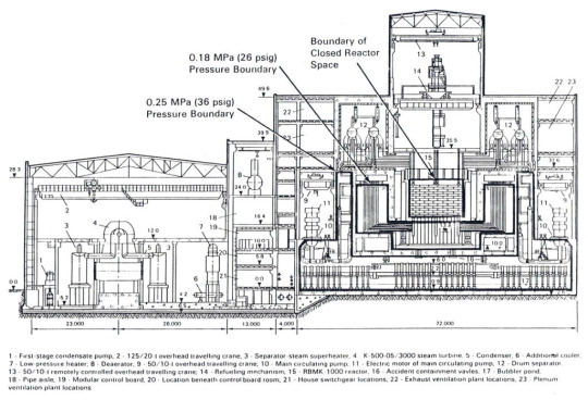
Below: A schematic diagram of the fourth unit block seen from the west. The dimensions of the building are marked in meters. Note the enormous region of rooms located below the reactor core (Closed Reactor Space on this diagram). The area at the bottom of the building with the large vertical pipes are the bubbler tanks that held emergency cooling water for the reactor. This was the area scientists feared a steam explosion if the fuel lava gained access.
Adding to the problem of the scale of the search area was the fact that the building was potentially unstable due to the explosion of the fourth reactor. Rubble filled hallways and walls and ceilings sagged dangerously. Radiation levels fluctuated wildly within the building, with some areas almost entirely safe and others able to cause sickness and death in minutes. Even more pressing was that starting in the spring of 1986, the lower levels of the fourth unit began to slowly fill with fresh concrete. The Ministry of Medium Machine Building unit US-605, who were building the Sarcophagus to cover the radioactive remains of the unit, poured concrete into the structures of the Sarcophagus 24 hours a day. However, huge gaps and sinkholes existed in their work area, and a good portion of the concrete pumped into the Sarcophagus ended up deep in the lower levels of the block. This concrete blocked hallways, doors, and even (as they would later come to learn) covered up some of the melted fuel.
It was not until late spring of 1986 that exploration of the fourth unit block began. In June of 1986, two men were probing a steam distribution corridor in the southeast corner of the block from another corridor just below it using a powerful dosimeter which could detect radiation levels of up to 3,000 roentgens per hour. Since radiation levels in the stairway up to the corridor they were probing were already quite high (~25 roentgens per hour) they decided to send the detection head of the device up the stairs ahead of them via an assembly of metal rods. As soon as the device entered the corridor above, it went off the scale and burnt out. From this result the team was able to pinpoint a source of extreme gamma radiation.
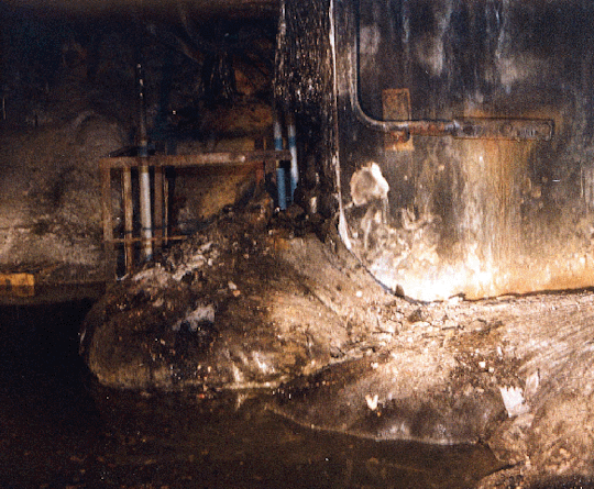
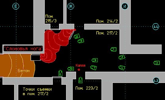
In December of 1986, and expedition was mounted to room 217/2 to make visual contact with the suspected fuel concentration. Moving along the steam corridor this time, the team spotted a large metallic gray mass sitting neatly within the corner of the room. This formation was dubbed the "Elephant's Foot" (though some source translate it as"Elephant's Leg") due to its similarity to the leg of an elephant. The black glassy mass emitted over 8,000 roentgen an hour, deadly after just one and a half minutes (this is the maximum recorded emission, the levels would decrease significantly in the months after the disaster). For the first time, the theory of fuel lava was visually confirmed. The team branded the materiel "lava-like fuel containing masses" (LFCM).
Below: A picture of the Elephant's Foot from the direction which it was first observed. The railings just behind the main formation (Label 1) is the railing around the metal stairs from which the formation was first detected via dosimeter. Note the streaks of fuel above the formation showing where the fuel had dripped down from above. Label 4 denotes the "fresh" concrete that made its way into the building during the construction of the Sarcophagus.

Below: a view of room 217/2 from above. The red is the Elephant’s Foot, the orange is the fresh concrete, and the gray are the walls of the block. The Foot itself is the accumulation closest to the bottom left of the photograph. You can see there is more lava in an unnamed formation next to it.
After locating the fuel and taking some pictures, the team was tasked with analyzing what the lava was composed of. This may seem kind of obvious, but really it was not known what was in the LFCM. Presumably the fuel of course, but what else? It had been debated from the early days of the accident if the efforts to douse the fire and melting fuel with lead, boron, and sand had been effective (I refer here to the April 26th-May 12th aerial bombardment campaign of the reactor via helicopter- I will make a post on this effort at a later time and link it here). The contents of the fuel is an interesting topic which I will not go over more in this post. You can find more info on the quest to get a piece of the LFCM here.
After procuring a sample, one issue remained. They had found some of the fuel, but nowhere near the total amount. The grand majority of the fuel had yet to be located. By the summer of 1987 the fresh concrete that had run into the lower levels of the block began to cause real obstacles to the scientists. Many rooms suspected to contain fuel were inaccessible or could not be reached safely. A new approach was needed.
At the end of 1987 the Kurchatov team was reassigned to be part of the adventurously named Chernobyl Complex Expedition (CCE), an enormous liquidation effort that was tasked with exploring the interior of the Sarcophagus and locating the rest of the fuel in the years after the disaster. The CCE was composed to representatives from all the major Soviet scientific institutions, as well as builders and engineers. At its peak over 3,000 people worked as part of the Expedition. Through it all, the main core of this group was the scientists from the Kurchatov Institute.
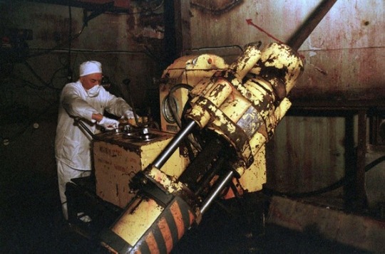
Backed by resources of the entire CCE, the scientific team sought new approaches for searching for the fuel. After extensive discussion, the scientists came up with a rather ingenious solution. They would use coring drills like those used in oil drilling exploration set up in specially decontaminated rooms in the fourth unit block to drill so called ‘wells’ into inaccessible rooms. Through these they would send specially built monitoring equipment. These included thermometers, periscopes, and radiation sensors. Not only could they monitor the LFCM with this equipment, they could also obtain information about the building and the LFCM via analysis of the cores made by the drill. This allowed them to remotely locate, monitor, and sample the LFCM with minimal risk to personnel.
Below: A schematic of the wells drilled at the 9 meter mark below the reactor containment vessel. The purple lines are the wells themselves, with the gray being the concrete walls of the block. The large blue cross is the metal support Scheme S. I provided this as an example of how the wells were drilled and laid out. For more info, feel free to contact me.

Below: a member of the CCE operates a drill in the bowels of the fourth unit block. Note the protective clothing he is wearing to prevent the spread of airborne radiological contamination.
Between 1988 and 1992 a total of 134 wells were drilled between the 9 and 16 meter marks (the method by which levels of the plant are identified) and at the 20, 21, and 25 meter marks. It was eventually determined that over 180 of the 190 tons of fuel remained in the reactor block. The missing ten tons had either been blown out of the active zone and into the area around the reactor by the explosion, or had vaporized into the radioactive column that had emerged from the reactor in 1986. The remaining fuel within the block took the form of the LFCM, as well as dust. The dust created by the fuel is the main radiological enemy post 1986 and continues to be an issue to this day. Primarily composed of plutonium, the dust has thankfully remains mostly within the Sarcophagus. The LFCM, initially almost indestructible, has started to crumble and decay. As time goes on this creates even more dust, and the formations slowly erode away. This has contributed to a significant drop in gamma radiation emissions from the fuel masses and allowed for further study of the premises of the fourth unit block.
In 1994, the Complex Expedition used data collected from these wells to compile an official report on the status of the fourth unit block. With their findings published (and the Soviet Union dissolved) the CCE disbanded. Many of the scientists who worked on the expedition (and even some who were on the original Kurchatov Institute team) continued to work at Chernobyl for years. Expeditions are still sometimes mounted into the Sarcophagus, but they have not been carried out with any regularity since the early 2000s. However, visual inspection remains the only way to accurately monitor the condition of the LFCM.
You may be left wondering: how exactly did these men navigate such a radioactive environment without adverse effects? Once again I shall make a post on this in the future, but the main answer is: speed! Defense against radiation (with some exceptions) is as simple as not lingering in high radiation areas. To facilitate safe movement through the block, the scientists located and marked safe areas of reduced radiation levels (such as the room in this post) as well as dangerous areas to avoid. In the end, only a select few “Stalkers” actually set foot within the ruins of Reactor 4.
This post serves as the (admittedly lose) historical context for the exploration of the fourth unit block and the location of the LFCM. I will be making another post about the fuel rest of the fuel soon. I always feel bad for the other fuel formations because the Elephants Foot gets all of the attention. This will be a lot more technical, with locations and diagrams (joy of joys!) of the fuel as well as more context to its identification.
#chernobyl#elephants foot#radiation#history#autism#nuclear#nuclear power#reactor#china syndrome#chernobyl complex expedition#accidents and disasters#kurchatov institute#LFCM#chnpp#soviet union
106 notes
·
View notes
Text
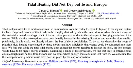
This is a fun paper - I'd always kind of assumed the reason Io and Europa are relatively* dry is because of their high tidal heating having desiccated their surfaces. Seems this is plausibly not actually the case! More likely that they just formed inwards of the ice-line of the jovian circumplanetary disk, similarly to how the inner planets in the Solar System are rocky and the outer planets and moons are volatile-rich.
Particularly interesting to me for worldbuilding was this bit:
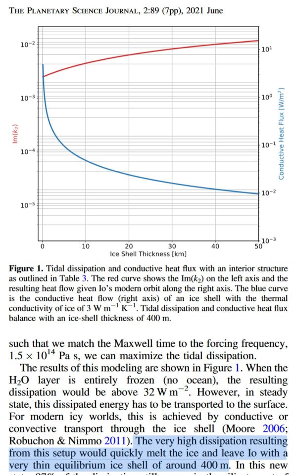
Actual numbers for the correlation between tidal heating and ice shell thickness! I've always wondered just how much internal heat flux you'd need for a super-thin ice shell that could let some visible light through (like you see under the thin sea ice in the Arctic and Antarctic oceans) in areas with locally thinned ice above hydrothermal upwellings. Turns out the answer is "quite a bit, probably a bit more than 10x the heat flow of present-day Io", but plausibly not completely unachievable**, especially with the mention of periods of high-eccentricity - and concomitant high tidal heating - in the histories of moon systems that exhibit Laplace resonance. The more exciting thing is that this implies that a moon with a higher water mass fraction could migrate into an Io-like orbit and not be desiccated, which could yield some interesting cryovolcanism from the thinned out ice shell - plausibly even temporary melt lakes like the ones which we've seen frozen-out remnants of on Triton!
(*) Europa is of course famous for being a wet moon with an ocean, but notably it only has a water mass fraction of about 5-9 percent. While being about three times the mass of all the water in all the oceans on Earth, this is teeny-tiny in comparison to Ganymede's water mass fraction of 50 to 30 percent - tens of times the total surface water inventory of the Earth! Io, on the other hand, is very very dry to the best of our knowledge. Unpleasant place.
(**) This becomes far more reasonable with a larger icy moon in a younger star system like Epsilon Eridani (age cca 400-800 million years), where the residual heat of formation and radionuclide decay in the hearts of these worlds has had less time to diminish.
11 notes
·
View notes
Text
Radionuclide Therapy in Rohtak
Radionuclide therapy, also known as targeted radiotherapy, is a specialized medical treatment that employs radioactive substances (radionuclides) to selectively deliver radiation to specific diseased tissues within the body. In Rohtak, this advanced form of therapy is offered as part of a comprehensive approach to managing various medical conditions, particularly cancer.
0 notes
Text
loved one got medical test results back and it's not great. but it'll likely be okay. thank god for radionuclide therapy. would love to put them in front of a clover detector
7 notes
·
View notes
Text
On a spring day in 1978, a fisherman caught a tiger shark in the lagoon surrounding Enewetak Atoll, part of the Marshall Islands in the north Pacific. That shark, along with the remains of a green sea turtle it had swallowed, wound up in a natural history museum. Today, scientists are realizing that this turtle holds clues to the lagoon’s nuclear past—and could help us understand how nuclear research, energy production, and warfare will affect the environment in the future.
In 1952, the world’s first hydrogen bomb test had obliterated a neighboring island—one of 43 nuclear bombs detonated at Enewetak in the early years of the Cold War. Recently, Cyler Conrad, an archeologist at Pacific Northwest National Laboratory, began investigating whether radioactive signatures of those explosions had been archived by some particularly good environmental historians: turtles.
“Anywhere that nuclear events have occurred throughout the globe, there are turtles,” Conrad says. It’s not because turtles—including sea turtles, tortoises, and freshwater terrapins—are drawn to nuclear testing sites. They’re just everywhere. They have been mainstays of mythology and popular culture since the dawn of recorded history. “Our human story on the planet is really closely tied to turtles,” Conrad says. And, he adds, because they are famously long-lived, they are uniquely equipped to document the human story within their tough, slow-growing shells.
Collaborating with researchers at Los Alamos National Laboratory, which was once directed by J. Robert Oppenheimer, Conrad was able to use some of the world’s most advanced tools for detecting radioactive elements. Last week, his team’s study in PNAS Nexus reported that this turtle, and others that had lived near nuclear development sites, carried highly enriched uranium—a telltale sign of nuclear weapons testing—in their shells.
Turtle shells are covered by scutes, plates made of keratin, the same material in fingernails. Scutes grow in layers like tree rings, forming beautiful swirls that preserve a chemical record of the turtle’s environment in each sheet. If any animal takes in more of a chemical than it’s able to excrete, whether through eating it, breathing it in, or touching it, that chemical will linger in its body.
Once chemical contaminants—including radionuclides, the unstable radioactive alter egos of chemical elements—make their way into scute, they’re basically stuck there. While these can get smeared across layers in tree rings or soft animal tissues, they get locked into each scute layer at the time the turtle was exposed. The growth pattern on each turtle’s shell depends on its species. Box turtles, for example, grow their scute outward over time, like how humans grow fingernails. Desert tortoise scutes also grow sequentially, but new layers grow underneath older layers, overlapping to create a tree ring-like profile.
Because they are so sensitive to environmental changes, turtles have long been considered sentinels of ecosystem health—a different kind of canary in the coal mine. “They’ll show us things that are emergent problems,” says Wallace J. Nichols, a marine biologist who was not involved in this study. But Conrad’s new findings reveal that turtles are also “showing us things that are distinct problems from the past.”
Conrad’s team at Los Alamos handpicked five turtles from museum archives, with each one representing a different nuclear event in history. One was the Enewetak Atoll green sea turtle, borrowed from the Bernice Pauahi Bishop Museum in Honolulu, Hawaii. Others included a Mojave desert tortoise collected within range of fallout from the former Nevada Test Site; a river cooter from the Savannah River Site, which manufactured fuel for nuclear weapons; and an eastern box turtle from Oak Ridge, which once produced parts for nuclear weapons. A Sonoran desert tortoise, collected far from any nuclear testing or manufacturing sites, served as a natural control.
While working at Los Alamos, Conrad met isotope geochemist and soon-to-be coauthor Jeremy Inglis, who knew how to spot even the most subtle signs of nuclear exposure in a turtle shell. They chose to look for uranium. To a geochemist, this might initially feel like an odd choice. Uranium is found everywhere in nature, and doesn’t necessarily flag anything historically significant. But with sensitive-enough gear, uranium can reveal a lot about isotope composition, or the ratio of its atoms containing different configurations of protons, electrons, and neutrons. Natural uranium, which is in most rocks, is configured very differently from the highly enriched uranium found in nuclear labs and weapons.
To find the highly enriched uranium hidden among the normal stuff in each turtle shell sample, Inglis wore a full-body protective suit in a clean room to keep his uranium from getting in the way. (“There’s enough uranium in my hair to contaminate a picogram of a sample,” he says.) Inglis describes the samples like a gin and tonic: “The tonic is the natural uranium. If you add lots of natural uranium tonic into your highly enriched uranium gin, you ruin it. If we contaminate our samples with natural uranium, the isotope ratio changes, and we can’t see the signal that we’re looking for.”
The team concluded that all four turtles that came from historic nuclear testing or manufacturing sites carried traces of highly enriched uranium. The Sonoran desert tortoise that had never been exposed to nuclear activity was the only one without it.
They collected bulk scute samples from three of their turtles, meaning that they could determine whether the turtle took in uranium at some point in its life, but not exactly when. But the researchers took things a step further with the Oak Ridge box turtle, looking at changes in uranium isotope concentrations across seven scute layers, marking the seven years of the turtle’s life between 1955 and 1962. Changes in the scutes corresponded with fluctuations in documented uranium contamination levels in the area, suggesting that the Oak Ridge turtle’s shell was time-stamped by historic nuclear events. Even the neonatal scute, a layer that grew before the turtle hatched, had signs of nuclear history passed down from its mother.
It’s unclear what this contamination meant for the turtles’ health. All of these shells were from long-dead animals preserved in museum archives. The best time to assess the effects of radionuclides on their health would have been while they were alive, says Kristin Berry, a wildlife biologist specializing in desert tortoises at the Western Ecological Research Center, who was not involved in this study. Berry adds that further research, using controlled experiments in captivity, may help figure out exactly how these animals are taking in nuclear contaminants. Is it from their food? The soil? The air?
Because turtles are nearly omnipresent, tracing nuclear contamination in shells from animals living at various distances from sites of nuclear activity may also help us understand the long-term environmental effects of weapons testing and energy production. Conrad is currently analyzing desert tortoise samples from southwestern Utah, collected by Berry, to better relate exposure to radionuclides (like uranium) to their diets over the course of their lives. He also hopes that these findings will inspire others to study plants and animals with tissues that grow sequentially—like mollusks, which are also found in nearly all aquatic environments.
The incredible migratory patterns of sea turtles, which sometimes span the entire ocean (as anyone familiar with Finding Nemo may recall), open up additional opportunities. For example, sea turtles forage off the Japanese coast, where in 2011 the most powerful earthquake in Japan’s history caused a tsunami that led to a chain reaction of failures at the Fukushima Daiichi Nuclear Power Plant. With lifespans of up to 100 years, many of those turtles are likely still alive today, carrying traces of the disaster on their backs.
Recently, the Japanese government started slowly releasing treated radioactive water from the Fukushima Daiichi plant into the Pacific Ocean. Scientists and policymakers seem to hesitantly agree that this is the least bad option for disposing of the waste, but others are more concerned. (The Chinese government, for instance, banned aquatic imports from Japan in late August.) Through turtle shells, we may better understand how the plant’s failure, and the following cleanup efforts, affect the surrounding ocean.
The bodies of these creatures have been keeping score for millennia. “For better or for worse, they get hit by everything we do,” Nichols says. Maybe, he adds, “the lesson is: Pay more attention to turtles.”
50 notes
·
View notes
Text
Anomaly in the deep sea
Extraordinary accumulation of rare atoms could improve geological dating methods
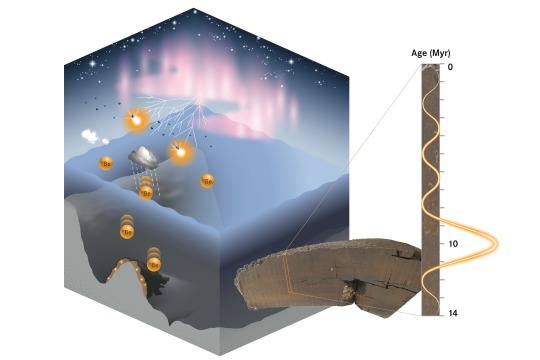
Beryllium-10, a rare radioactive isotope produced by cosmic rays in the atmosphere, provides valuable insights into the Earth's geological history. A research team from the Helmholtz-Zentrum Dresden-Rossendorf (HZDR), in collaboration with the TUD Dresden University of Technology and the Australian National University (ANU), has discovered an unexpected accumulation of this isotope in samples taken from the Pacific seabed. Such an anomaly may be attributed to shifts in ocean currents or astrophysical events that occurred approximately 10 million years ago. The findings hold the potential to serve as a global time marker, representing a promising advancement in the dating of geological archives spanning millions of years. The team presents its results in the scientific journal Nature Communications (DOI: 10.1038/s41467-024-55662-4).
Radionuclides are types of atomic nuclei (isotopes) that decay into other elements over time. They are used to date archaeological and geological samples, with radiocarbon dating being one of the most well-known methods. In principle, radiocarbon dating is based on the fact that living organisms continuously absorb the radioactive isotope carbon-14 (14C) during their lifetime. Once an organism dies, the absorption ceases, and the 14C content starts to decrease through radioactive decay with a half-life of approximately 5,700 years. By comparing the ratio of unstable 14C to stable carbon-12 (12C), researchers can determine the date of the organism's death.
Archaeological finds, such as bones or remnants of wood, can be dated quite accurately in this way. “However, the radiocarbon method is limited to dating samples no more than 50,000 years old,” explains HZDR physicist Dr. Dominik Koll. “To date older samples, we need to use other isotopes, such as cosmogenic beryllium-10 (10Be).” This isotope is created when cosmic rays interact with oxygen and nitrogen in the upper atmosphere. It reaches the Earth through precipitation and can accumulate on the seabed. With a half-life of 1.4 million years, 10Be decays into boron, allowing geological dating that can extend back over 10 million years.
Conspicuous accumulation of beryllium
Some time ago, Koll's research group examined unique geological samples retrieved from the Pacific Ocean at a depth of several kilometers. The samples consisted of ferromanganese crusts, primarily composed of iron and manganese, which had formed slowly but steadily over millions of years. To date the samples, the team analyzed the 10Be content using a highly sensitive method – Accelerator Mass Spectrometry (AMS) at HZDR. In this process, the sample is chemically purified before undergoing analysis for trace isotopes. Individual atoms from the sample are accelerated by high voltage, deflected by magnets, and then registered by specialized detectors. This method allows for the precise identification of 10Be, distinguishing it from other beryllium isotopes as well as molecules and isotopes with the same mass, such as boron-10.
When the research group evaluated the collected data, they were in for a surprise. “At around 10 million years, we found almost twice as much 10Be as we had anticipated,” reports Koll. “We had stumbled upon a previously undiscovered anomaly.” To eliminate any possibility of contamination, the experts analyzed additional samples from the Pacific, which also exhibited the same anomaly. This consistency allows the team to conclude that it is indeed a real phenomenon.
Ocean currents, stellar explosion or interstellar collision?
But how did such a striking increase in concentration come about 10 million years ago? Koll, who completed his doctorate at the TU Dresden and the ANU, proposes two possible explanations. One is related to the ocean circulation near Antarctica, which is thought to have changed drastically 10 to 12 million years ago. “This could have caused 10Be to be unevenly distributed across the Earth for a period of time due to the altered ocean currents,” explains the physicist. ”As a result, 10Be could have become particularly concentrated in the Pacific Ocean.”
The second hypothesis is astrophysical in nature. It suggests that the after-effects of a near-Earth supernova could have caused cosmic radiation to become temporarily more intense 10 million years ago. Alternatively, the Earth might have temporarily lost its protective solar shield – the heliosphere – due to a collision with a dense interstellar cloud, making it more vulnerable to cosmic radiation. ”Only new measurements can indicate whether the beryllium anomaly was caused by changes in ocean currents or has astrophysical reasons,” says Koll. ”That is why we plan to analyze more samples in the future and hope that other research groups will do the same.” If the anomaly were found all over the globe, the astrophysics hypothesis would be supported. On the other hand, if it were detected only in specific regions, the explanation involving altered ocean currents would be considered more plausible.
The anomaly could be extremely useful for geological beryllium dating. When comparing different archives for dating, one fundamental problem arises. Common time markers must be identified in all data sets so they can be properly synchronized with each other. Dominik Koll explains, “For periods spanning millions of years, such cosmogenic time markers do not yet exist. However, this beryllium anomaly has the potential to serve as such a marker.”
Publication: D. Koll, J. Lachner, S. Beutner, S. Fichter, S. Merchel, G. Rugel, Z. Slavkovská, C. Vivo-Vilches, S. Winkler, A. Wallner: A cosmogenic 10Be anomaly during the late Miocene as independent time marker for marine archives, in Nature Communications, 2025 (DOI: 10.1038/s41467-024-55662-4)
Further information: Dr. Dominik Koll | Institute of Ion Beam Physics and Materials Research at HZDR Phone: +49 351 260 3804 | Email: [email protected]
Media contact: Simon Schmitt | Head Communications and Media Relations at HZDR Phone: +49 351 260 3400 | Mobile: +49 175 874 2865 | Email: [email protected]
The Helmholtz-Zentrum Dresden-Rossendorf (HZDR) performs – as an independent German research center – research in the fields of energy, health, and matter. We focus on answering the following questions:
How can energy and resources be utilized in an efficient, safe, and sustainable way?
How can malignant tumors be more precisely visualized, characterized, and more effectively treated?
How do matter and materials behave under the influence of strong fields and in smallest dimensions?
To help answer these research questions, HZDR operates large-scale facilities, which are also used by visiting researchers: the Ion Beam Center, the Dresden High Magnetic Field Laboratory and the ELBE Center for High-Power Radiation Sources. HZDR is a member of the Helmholtz Association and has six sites (Dresden, Freiberg, Görlitz, Grenoble, Leipzig, Schenefeld near Hamburg) with almost 1,500 members of staff, of whom about 680 are scientists, including 200 Ph.D. candidates.
IMAGE: Schematic depiction of production and incorporation of cosmogenic 10Be into ferromanganese crusts. Credit HZDR / blrck.de
3 notes
·
View notes
Text
@silverjetsystm | caelesis.

Minutes fold toward the past, happening just like the steady, nacre thermosphere when Videntis pulls a new body in.
They scrubbed at his fingers and feet for hours, intermittently, leeching trace radionuclides and toxins from his nails, his cuts. The chafing should have whittled him raw. But the Sisters and their peons have their ways of leeching evidence, too.
Most especially from mind.
To keep and study, like the rest of what they've siphoned─they'll find clever ways to know him before he can even wake to lie.
There are silver cups, and some gold, sitting quietly on surfaces around the closeted sanctuary of his featureless sepulcher; a body-length rock risen from the blue-dark floor. They've stale water in them. Water mixed with blood.
But in the corner of the small crypt-likeness, only a youth observes him. She keeps to a liquid shadow on the secret turn of the wall; but she knows he is awakening. She has felt that something in him is.
The way she'd felt his crash, before its inevitable destruction, before his slip into the black unconscious. The way she knows none of these things were accident.
“You woke me,” she tells him, a crystal voice and head of snow in the shadow.
2 notes
·
View notes