#minutum
Explore tagged Tumblr posts
Text

Craterium minutum by Barry Webb
#craterium minutum#barry webb#crateria#myxomycota#slime mold#slime mould#macro photography#myxomycetes#nature#forest floor#microbiology#microbiota#microorganisms
196 notes
·
View notes
Text
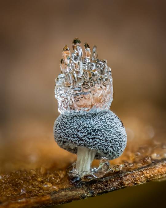
Didymium squamulosum with ice crown.
In Macro Photos, Barry Webb Captures the Fleeting, Otherworldly Characteristics of Slime Molds and Fungi

Metatrichia floriformis and physarum
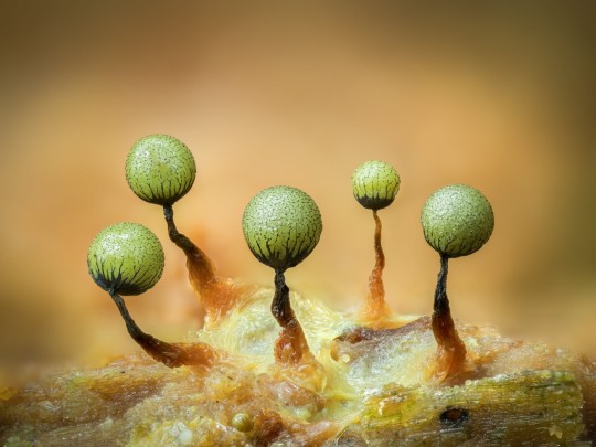
Cribraria

Craterium minutum
#barry webb#photographer#fungus#mushroom#fungi#didymium squamulosum with ice crown#macro photography#slime molds#metatrichia floriformis and physarum#cribraria#craterium minutum
206 notes
·
View notes
Text
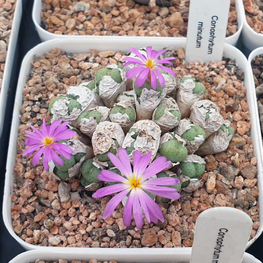
Conophytum minutum. September 2022.
32 notes
·
View notes
Text
#2184 - Tapinoma sp.

From the Greek ταπείνωμα for 'low position', but I don't know why that applies to this species - for one thing many of the 74 species are arboreal, often living in myrmecophilous plants.
Generalists, as far as diet goes.
The Ghost Ant Tapinoma melanocephalum and the Odorous House Ant Tapinoma sessile are both house pests where they occur, but the later still hasn't made it to Austrslia as far as anyone knows, and the Ghost Ant is limited to the coastlines of Queensland and the Northern Territory. Tapinoma minutum, the Dwarf Pedicel Ant, is found in Sydney but has pale shins.
Mascot, Sydney, New South Wales
2 notes
·
View notes
Text
In Macro Photos, Barry Webb Captures the Fleeting, Otherworldly Characteristics of Slime Molds and Fungi
August 26, 2023
ThisIsColossal.com
Grace Ebert
All images © Barry Webb, licensed
Photographer Barry Webb (previously) continues his hunt for the speckled, glimmering, and ice-crested organisms that pop up near his home in South Buckinghamshire, U.K. Armed with a 90-millimeter macro lens, Webb ventures into woodlands and other natural areas where slime molds and fungi thrive. There, he zeroes in on their microscopic features, documenting their wildly diverse characteristics that often last for just a brief moment in time. Recent shots include a tuft of Muppet-like fuzz topping Metatrichia floriformis, a water droplet suspended between two cup-like Craterium minutum, and a cluster of Pink stemonitis filaments propped on spindly black legs.
Webb has won several awards in recent months, including from the Royal Photographic Society and Close-Up Photographer of the Year. Four of his photos will be featured at the Vienna Mushroom Festival next month, prints are available on his site, and you can find more of his work on Instagram.
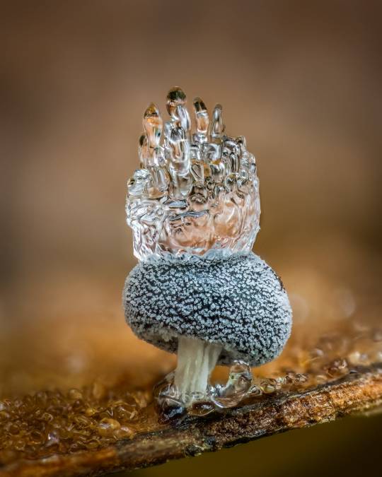
Didymium Squamulosum with ice crown
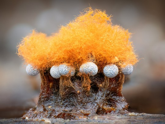
Metatrichia floriformis and physarum

Cribraria
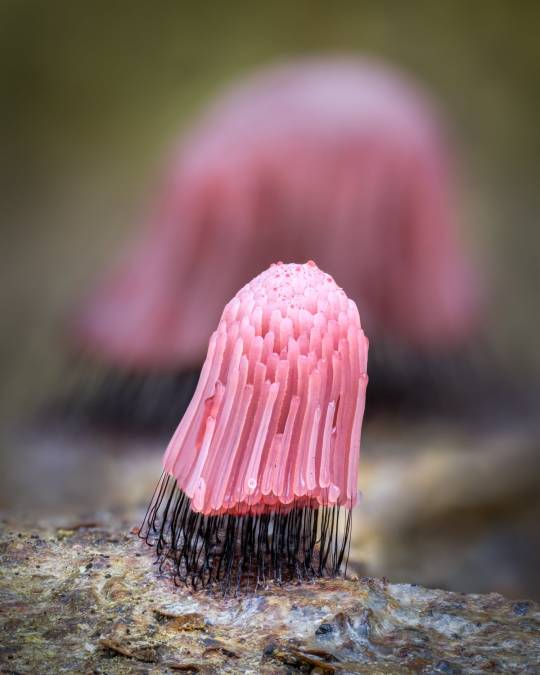
Pink Stemonitis
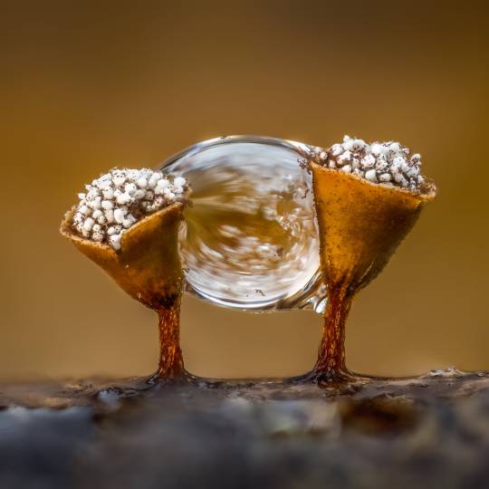
Craterium minutum
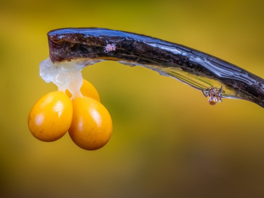
Leocarpus fragilis
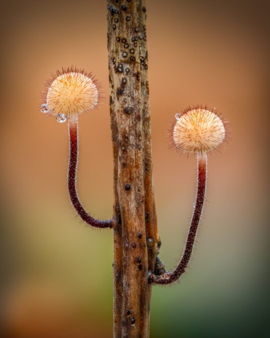
Holly Parachute fungus Marasmius hudsonii
0 notes
Text
Részeges álmok, józan gondolatok.
Józan álmok,részeges gondolatok.
Mindez csupán 1 minutum műve.
0 notes
Text
Cellule di Acropora tenuis che si mangiano le alghe, ci avreste mai creduto?
Cellule di Acropora tenuis che si mangiano le alghe, ci avreste mai creduto? - Non stiamo scherzando, è quello che hanno scoperto per la prima volta degli scienziati giapponesi, cellule di corallo che si mangiano le alghe simbionti.
Un nuovo articolo su http://www.danireef.com/2021/07/16/cellule-di-acropora-tenuis-che-si-mangiano-le-alghe-ci-avreste-mai-creduto/
Cellule di Acropora tenuis che si mangiano le alghe, ci avreste mai creduto?
#AcroporaTenuis, #AlgheSimbionti, #Biologia, #BreviolumMinutum, #Dinoflagellati, #KazKawamura, #Ricerca, #SatokoSekida, #Sbiancamento, #Simbiosi, #Università, #UniversityOfKochi, #Zooxantellae, #Zooxantelle, #Zooxanthellae
- by Danilo Ronchi
#acropora tenuis#alghe simbionti#biologia#Breviolum minutum#dinoflagellati#Kaz Kawamura#ricerca#Satoko Sekida#Sbiancamento#simbiosi#università#University of Kochi#zooxantellae#zooxantelle#zooxanthellae#NEWS
0 notes
Photo

Conophytum minutum
5 notes
·
View notes
Text
Slime mold of Tasmania. Photographed by Sarah Lloyd.


Elaeomyxa cerifera

Ceratrimeria feeding on slime mold (Lycogala epidendron)

Species: ?

Species: ?

Lamproderma echinulatum

Craterium minutum

Arcyria dendudata

Elaeomyxa reticulospora


Ceratrimeria feeding on slime mold

Plasmodium and sporangia


Lamproderma cf. muscorum
More photos and description of habitat, ecology at Sarah Lloyd’s site: sarahlloydmyxos at word/press

218 notes
·
View notes
Note
re: thing in the garage: do you have those samples on any sort of medium? all i see is paper towels. what's the game plan here?
i have a few different sizes of containers in there: 1 large (2 inches across, 2 inches deep) one, 2 medium (two inches across, one inch deep) ones, and 8 small (one inch across, one inch deep) ones. the large one just has a wet paper towel in it, but with the medium containers i did one with just a paper towel and one with mulch from next to the garage in it along with a paper towel. for the small ones, i did 4 with just a paper towel and 4 with a paper towel and mulch. i didn’t have many containers left, but i tried to do controls by putting just paper towels in a 1.5 across, one inch deep container, and paper towels + mulch inside a medium container (no spores in either). unfortunately i dont have petri dishes with actual agar in them to provide for these dudes, or even like, sterile containers i know are sterile, but for the purposes of just seeing what grows through all of them, i don’t think it needs to be as sterile as it would be otherwise (this is like....way too many variables for an actual science experiment too lmao but i’m not really testing for anything inparticular).
now, the general idea of this experiment comes from the book ‘Myxomycetes: A Handbook of Slime Molds’ by Stephen L. Stephenson and Henry Stempen (side note: this is a cool slime mold book in general). they describe how to go about finding and collecting slime molds in the wild by just like, kinda getting a ton of bark from your local forest and shoving them in wet containers. because i directly put spores from the thing on the paper towels in each with q-tips, i didn’t follow this tutorial exactly, but here’s the full quote (pages 41-44, so putting it under a cut for those interested):
“PREPARING MOIST CHAMBERS
The next step in setting up a moist chamber involves preparing the culture chamber itself. Petri dishes (Figure 3-2) have been widely used as culture chambers, but any similar container or shallow bowl that can be covered with a top or lid will work just as well.
Many modern workers use plastic Petri dishes, which are simply discarded when no longer needed. Plastic containers have another major advantage over glass containers. In a moist chamber, the fruiting bodies of myxomycetes sometimes develop on the side of the container and on the lower surface of the top or lid. When this happens, the only way to harvest the fruiting bodies is to cut away the portion of the container upon which they developed. This is rather difficult to do with a glass container, but relatively easy with plastic.
Once a suitable container has been selected, it should be fitted with a filter paper disk or a piece of absorbent paper towel trimmed to the appropriate size. For experiments designed to study the differences that exist in the species of myxomycetes obtained from bark samples from different tree species or trees growing in different habitats, care should be taken to prevent the potential contamination of one culture with spores from another by sterilizing all the equipment used in preparing moist chamber cultures, including all the paper used to cover the bottom of the culture chambers. If the only objective of preparing cultures is simply to obtain a variety of different myxomycetes, sterilization is not necessary.
After the filter paper or paper towel is in place, it should be covered with samples of the organic debris (bark, leaves, twigs, or dung) to be cultured. The samples should not overlap with each other to any great extent, and bark samples should be placed in the container with their cut surfaces facing downward. Enough distilled water (tap water can be used if distilled water is not available) is then added to completely cover the samples. The container is covered and set aside to allow the sample to soak up the moisture overnight.
Each container should be labeled, either with a piece of tape applied to the side so as not to obscure any portion of the bottom when the container is viewed from above or by writing directly on the lid with a wax pencil. If the latter method is used, it sometimes becomes necessary to rotate the lid of the dish when making observations of the samples to view portions of the bottom otherwise obscured by the writing. The next day, excess water in the container must be poured off and the cultures set aside and disturbed as little as possible. If Petri dishes are used, they may be stacked to save space. Cultures should be incubated at ordinary room temperatures under diffuse light.
CHECKING THE CULTURES
Cultures should be observed a day or two after they have been established and thereafter on a regular basis (that is, at least once every few days) for at least two weeks. After this time, cultures may be checked at less frequent intervals for as long as they are maintained. It is possible to maintain cultures for several months or more. In fact, some species of myxomycetes which appear in moist chamber cultures typically require weeks or even months to develop. This is almost always the case for species associated with dung. In contrast, many of the species that produce very small fruiting bodies, such as species of Echinostelium (including the exceedingly common E. minutum), often appear rather quickly, sometimes within 24 to 48 hours. Since the cultures slowly dry out, it may be necessary to add more water from time to time to keep cultures moist throughout the incubation period.
It is important that all cultures be checked as carefully as possible to detect all the fruiting bodies that may be present. Sometimes only one or a few fruiting bodies of a given species develop; in other cases hundreds of fruiting bodies are produced. Moreover, it is not unusual for a single culture to yield the fruiting bodies of several different species.
Cultures are most easily checked using a dissecting microscope, but a good hand lens (at least x10) may be used if a microscope is not available. In some instances, fruiting bodies are clearly visible to the naked eye. Since removing the lid can interfere with the development of fruiting bodies not yet completely mature, a culture should be examined by viewing through the lid whenever possible.
When fruiting bodies are detected in a culture, they should be harvested as soon as circumstances permit. If this is not done, the fruiting bodies soon become moldy. Cultures should be left covered until the fruiting bodies are completely mature, though often it is helpful to raise the lid on one side to allow gradual drying of the fruiting bodies. When dry, the fruiting bodies can be harvested by removing both the fruiting bodies and portions of the substrate upon which they developed. Once the fruiting bodies have been removed from the culture, they should be allowed to air-dry. Then each individual collection should be placed in a small box for permanent storage, as described earlier in this chapter.
The question has been raised whether the species of myxomycetes appearing on various substrates placed in moist chamber cultures necessarily produce fruiting bodies on the same substrates in nature, since their appearance in such cultures may reflect nothing more than the presence of spores, microcysts, or sclerotia on these substrates (Brooks 1967; Keller and Brooks 1973; Blackwell and Gilbertson 1984). Although the data available on myxomycete distribution patterns are still too limited to permit a definitive answer to this question, there is mounting evidence that with diligent collecting many (and perhaps most) of the species obtained from moist chamber cultures prepared with material from a particular microhabitat occur as natural fruiting within the same microhabitat (Whitney 1982; Blackwell and Gilbertson 1984; Stephenson 1988, 1989). It does not always follow, however, that these species are necessarily restricted to the particular habitat, although this sometimes seems to be the case.”
#indigo-adult#i just realized OH SHIT I NEED CONTROLS and went and did them lmao#also idk why it put the keep reading thing at the top when i put it in the middle?#weird.#plont asks#slime molds#mycology#thing on the garage
93 notes
·
View notes
Text
“Red was generally used by scribes in ancient and medieval times when they wished to decorate their manuscripts. The word "miniature" comes from the word minium, the Latin name for "red lead." The pictures that they incorporated with this red lead were quite small, and so the word "miniature" got somehow mixed up in people's minds with the Low Latin word minutum, meaning "a small portion." Hence "miniature" is now used to signify a little painting, and by extension, anything on a reduced scale.”
—Alexander Theroux, The Primary Colours: Three Essays
#quotes#words#color#colours#colour theory#primary colours#red#alexander theroux#etymology#word origins#linguistics#language#latin#mine#art#mim#not blue but hey colours!
424 notes
·
View notes
Text
The Five Medicine that is essential to be carried while travelling... It addresses the ailments of the head, organs, stomach and constipation. It protects the user from catching diseases and boosts the immune system. This has been a prescription for travellers.
1.) Botebotekoro - Ageratum conyzoides
2.) Totowiwi - Oxalis Corniculata
3.) Sinu - Excoecaria agallocha L.
4.) Mulokaka - Trifolium Vitex
5.) Sasaqilu - micromelum minutum
2 notes
·
View notes
Photo
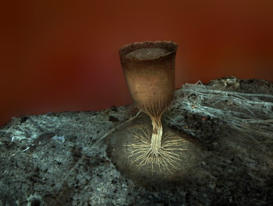
Cheers! Image of the Week – June 15, 2020
CIL:41635 - http://www.cellimagelibrary.org/images/41635
Description: Fluorescent image of the sporangium, an enclosure in which spores are formed, of the slime mold Craterium minutum. Honorable Mention, 2011 Olympus BioScapes Digital Imaging Competition®.
Authors: Dalibor Matýsek and 2011 Olympus BioScapes Digital Imaging Competition®
Licensing: Attribution Non-Commercial No Derivatives: This image is licensed under a Creative Commons Attribution, Non-Commercial, No Derivatives License
15 notes
·
View notes
Photo

マメツブウミコチョウ Gastropteron minutum (Ong & Gosliner, 2017)
45 notes
·
View notes
Photo

Craterium minutum
[@sonjas_shrooms / instagram]
20 notes
·
View notes