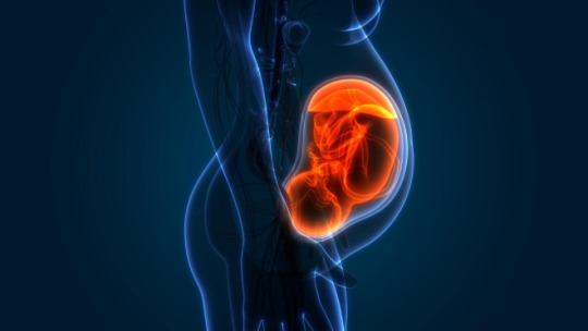
#inhibin alpha
Explore tagged Tumblr posts
Visit Tumblr Blog
Explore Tumblr blogs with no restrictions, modern design and the best experience.
Last Seen Tumblr Blogs


Fun Fact
Celebrities use Tumblr as well.
Text
Các chỉ số Xét nghiệm sàng lọc trước sinh: Ý nghĩa và cách đọc
Xét nghiệm sàng lọc trước sinh là một phần quan trọng trong chăm sóc thai kỳ, giúp phát hiện sớm các bất thường di truyền và bệnh lý ở thai nhi. Bài viết này sẽ giải thích ý nghĩa của các chỉ số trong xét nghiệm sàng lọc trước sinh và cung cấp hướng dẫn chi tiết về cách đọc kết quả để hiểu rõ hơn về sức khỏe của mẹ và bé.

Các xét nghiệm sàng lọc trước sinh phổ biến
Trong xét nghiệm sàng lọc trước sinh, có một số xét nghiệm phổ biến mà các bà m��� cần biết để hiểu rõ hơn về sức khỏe của thai nhi.
Double Test

Triple Test

Non-Invasive Prenatal Testing (NIPT)

>>> Dịch vụ Xét Nghiệm NIPT
Ý nghĩa của các chỉ số trong xét nghiệm sàng lọc trước sinh
Hiểu rõ ý nghĩa của các chỉ số trong xét nghiệm sàng lọc trước sinh giúp các bà mẹ và bác sĩ đánh giá chính xác tình trạng của thai nhi.
hCG (Human Chorionic Gonadotropin)
hCG là một hormone quan trọng được tiết ra trong quá trình mang thai. Nồng độ hCG cao có thể liên quan đến nguy cơ cao mắc hội chứng Down, trong khi nồng độ thấp có thể gợi ý đến nguy cơ thai nhi mắc hội chứng Edwards hoặc Patau. Việc theo dõi chỉ số này giúp bác sĩ đưa ra đánh giá chính xác hơn về nguy cơ dị tật bẩm sinh.
Ngưỡng an toàn:
Trong tam cá nguyệt đầu tiên, nồng độ hCG thông thường nằm trong khoảng 25,700 – 288,000 mIU/mL.
Ngưỡng nguy cơ:
Nồng độ hCG quá cao (trên ngưỡng thông thường) có thể liên quan đến nguy cơ mắc hội chứng Down.
Nồng độ hCG quá thấp có thể gợi ý nguy cơ thai chết lưu hoặc hội chứng Edwards.
PAPP-A (Pregnancy-Associated Plasma Protein A)
PAPP-A là một protein liên quan đến thai kỳ và được đo lường trong Double Test. Nồng độ PAPP-A thấp có thể là dấu hiệu của nguy cơ hội chứng Down hoặc các bất thường nhiễm sắc thể khác. Chỉ số này thường được kết hợp với hCG và độ mờ da gáy để đưa ra đánh giá tổng thể về sức khỏe của thai nhi.
Ngưỡng an toàn:
Nồng độ PAPP-A thường trên 0.5 MoM (Multiples of the Median).
Ngưỡng nguy cơ:
Nồng độ PAPP-A thấp dưới 0.4 MoM có thể liên quan đến nguy cơ hội chứng Down và các bất thường nhiễm sắc thể khác.
AFP (Alpha-Fetoprotein)
AFP là một protein được sản xuất bởi gan thai nhi và có thể được đo lường trong Triple Test. Nồng độ AFP cao có thể gợi ý đến nguy cơ dị tật ống thần kinh, chẳng hạn như tật nứt đốt sống, trong khi nồng độ thấp có thể liên quan đến hội chứng Down. Việc đo lường AFP giúp đánh giá nguy cơ của các bất thường này một cách chính xác hơn.
Ngưỡng an toàn:
Nồng độ AFP trong tam cá nguyệt thứ hai thường nằm trong khoảng 10 – 150 ng/mL.
Ngưỡng nguy cơ:
Nồng độ AFP cao trên 2.5 MoM có thể gợi ý đến nguy cơ dị tật ống thần kinh, chẳng hạn như tật nứt đốt sống.
Nồng độ AFP thấp có thể liên quan đến nguy cơ hội chứng Down.
Estriol (uE3)
Estriol là một hormone estrogen được sản xuất bởi nhau thai và thai nhi. Nồng độ estriol thấp có thể liên quan đến nguy cơ các dị tật bẩm sinh hoặc suy thai. Việc theo dõi chỉ số estriol giúp đảm bảo thai nhi phát triển bình thường và không gặp các vấn đề sức khỏe nghiêm trọng.
Ngưỡng an toàn:
Nồng độ Estriol thường nằm trong khoảng 0.5 – 2.5 MoM trong tam cá nguyệt thứ hai.
Ngưỡng nguy cơ:
Nồng độ Estriol thấp dưới 0.5 MoM có thể liên quan đến nguy cơ hội chứng Down hoặc các bất thường di truyền khác.
Inhibin-A
Inhibin-A là một protein được sản xuất bởi nhau thai và thường được đo lường trong Triple Test. Nồng độ Inhibin-A cao có thể là dấu hiệu của nguy cơ hội chứng Down. Chỉ số này, khi kết hợp với các chỉ số khác như hCG và AFP, giúp đưa ra đánh giá chính xác hơn về nguy cơ các bất thường di truyền.
Ngưỡng an toàn:
Nồng độ Inhibin-A thường dưới 2 MoM.
Ngưỡng nguy cơ:
Nồng độ Inhibin-A cao trên 2 MoM có thể là dấu hiệu của nguy cơ hội chứng Down.

>>> Giải Đáp Về Xét Nghiệm NIPT: Tất Cả Những Gì Bạn Cần Biết
Cách đọc kết quả xét nghiệm sàng lọc trước sinh
Đọc và hiểu kết quả xét nghiệm sàng lọc trước sinh là điều quan trọng để có thể đưa ra quyết định đúng đắn trong việc chăm sóc thai kỳ.
Hiểu về nguy cơ cao và nguy cơ thấp
Kết quả xét nghiệm sàng lọc trước sinh thường được đưa ra dưới dạng nguy cơ cao hoặc nguy cơ thấp. Nguy cơ cao không có nghĩa là thai nhi chắc chắn có vấn đề, mà chỉ là khả năng xảy ra bất thường cao hơn so với bình thường. Ngược lại, nguy cơ thấp cho thấy khả năng thai nhi mắc các bất thường là rất nhỏ, nhưng không hoàn toàn loại trừ.
Kết quả dương tính giả và âm tính giả
Xét nghiệm sàng lọc trước sinh có thể cho ra các kết quả dương tính giả hoặc âm tính giả. Điều này có nghĩa là một số xét nghiệm có thể cho kết quả nguy cơ cao trong khi thai nhi không thực sự mắc các dị tật, hoặc ngược lại. Vì vậy, nếu có kết quả nguy cơ cao, bạn nên tham khảo ý kiến bác sĩ để quyết định xem có cần thực hiện thêm các xét nghiệm chẩn đoán xâm lấn như chọc ối hoặc sinh thiết gai nhau hay không.
Khi nào cần làm thêm các xét nghiệm chẩn đoán?

Những điều cần lưu ý khi đọc và hiểu kết quả xét nghiệm sàng lọc trước sinh
Việc hiểu rõ các kết quả xét nghiệm sàng lọc trước sinh đòi hỏi sự cẩn trọng và tham khảo ý kiến chuyên gia.
Tham khảo ý kiến bác sĩ

Tính toàn diện của xét nghiệm
Các xét nghiệm sàng lọc trước sinh chỉ cung cấp đánh giá nguy cơ chứ không phải chẩn đoán chính xác. Kết quả sàng lọc cần được kết hợp với các yếu tố khác như tuổi của mẹ, tiền sử gia đình và các kết quả siêu âm để đưa ra đánh giá toàn diện nhất về sức khỏe thai nhi.
Cách quản lý lo lắng và căng thẳng khi nhận kết quả

Viện Nghiên Cứu Vietcare – địa chỉ Xét nghiệm NIPT uy tín
Xét nghiệm sàng lọc trước sinh là một công cụ quan trọng giúp phát hiện sớm các bất thường di truyền và bệnh lý ở thai nhi. Việc hiểu rõ các chỉ số trong xét nghiệm và cách đọc kết quả không chỉ giúp bạn theo dõi sức khỏe của thai nhi mà còn đưa ra những quyết định chính xác và kịp thời trong quá trình mang thai.

Tại sao nên chọn Viện Nghiên Cứu Vietcare??
Kết quả xét nghiệm chính xác tuyệt đối, trên 99,96%.
Thai phụ được tư vấn và đồng hành cùng Bác sĩ Di truyền trong suốt thai kỳ.
Đã có hơn 8+ năm kinh nghiệm trong lĩnh vực tư vấn di truyền.
Quy trình xét nghiệm chuẩn quốc tế, được kiểm duyệt bởi một tiến sĩ khoa học trước khi đưa ra kết luận cuối cùng.
Giá xét nghiệm cực kỳ cạnh tranh so với thị trường.
Hỗ trợ lấy mẫu tận nơi 24/7.
Hãy để lại thông tin, chúng tôi sẽ trực tiếp gọi điện tư vấn sớm nhất trong 24h.
https://vietcarelab.vn/cac-chi-so-xet-nghiem-sang-loc-truoc-sinh-y-nghia-va-cach-doc/
0 notes
Text
Quadruple Marker Test- Purpose, Risks, Treatment and Results
Quadruple Marker Test: Unveiling Insights for a Healthier Tomorrow
The Quadruple Marker Test, also known as the Quad Marker or Quad Screen, is a prenatal screening test designed to provide valuable insights into the health of both the fetus and the expecting mother. This comprehensive test is typically performed between the 15th and 20th weeks of pregnancy and assesses specific markers to identify potential risks and conditions.
Purpose: The primary purpose of the Quadruple Marker Test is to assess the risk of certain chromosomal abnormalities and neural tube defects in the developing fetus. The test evaluates four specific markers:
Alpha-fetoprotein (AFP): Produced by the fetal liver, AFP levels are measured to assess the risk of neural tube defects, such as spina bifida.
Human Chorionic Gonadotropin (hCG): This hormone is crucial for maintaining pregnancy and abnormal levels may indicate chromosomal abnormalities.
Estriol: Produced by both the fetus and the placenta, low estriol levels may suggest potential chromosomal abnormalities.
Inhibin-A: Elevated levels of inhibin-A can be associated with an increased risk of Down syndrome.
Quadruple Marker Test Time In pregnancy, a quadruple marker test is performed during the second trimester which ideally means sometime between 15 weeks and 20 weeks of pregnancy. If an individual has any of the following risk factors, the doctor might recommend them to go for a quadruple marker test:
Is above the age of 35 or older when the baby is due.
Has had a viral infection during pregnancy.
Has a family history of birth defects or congenital disabilities.
Is diagnosed with type 1 diabetes during pregnancy.
On harmful medication during pregnancy.
Has been exposed to high levels of radiation.
Risks: It’s important to note that the Quadruple Marker Test is a screening test, not a diagnostic one. While it provides valuable information, it does not guarantee a definitive diagnosis. False positives and false negatives are possible, necessitating further diagnostic tests for confirmation.
Treatment: The Quadruple Marker Test itself does not offer treatment; instead, it serves as an informative tool to identify potential risks. In the case of abnormal results, healthcare providers may recommend additional tests such as amniocentesis or chorionic villus sampling (CVS) for a more accurate diagnosis. These diagnostic tests can provide detailed information about the fetus’s chromosomal makeup.
Results: Interpreting the results requires the expertise of healthcare professionals. Normal results indicate a lower likelihood of the assessed conditions, providing reassurance to the expecting parents. Abnormal results, however, may prompt further testing and discussions about potential courses of action.
Conclusion
the Quadruple Marker Test is a valuable tool in prenatal care, offering insights that can guide healthcare decisions for both the mother and the developing fetus. Expecting parents are encouraged to consult with their healthcare providers to understand the implications of the results and make informed decisions regarding their pregnancy journey.
Navigate the path to parenthood with confidence at DMS Diagnostics! Our Quadruple Marker Test unravels the intricacies of prenatal health, providing a thorough understanding of potential risks and conditions. With precision and care, we guide you through the purpose, risks, treatments, and results, ensuring a journey to parenthood rooted in knowledge and support.
#2d echo test services in kothrud#best radiology doctor in kothrud#best radiologists in baner#sonography center in kothrud#2d echo test services near me#2d echo test near me#2d echo test in baner#sonography center in baner#best pathology lab in kothrud
0 notes
Text
Quadruple Marker Test
The quadruple marker test in pregnancy is a screening device used to survey the probability of specific chromosomal irregularities and brain tube defects in a creating baby. It's regularly suggested between the 15th and 20th week of pregnancy. This test clearly assesses four substances in the mother's blood.
Alpha-fetoprotein (AFP): Raised levels could show issues with the child's spine or mind, for example, brain tube issues like spina bifida or anencephaly.
Human chorionic gonadotropin (hCG): Unusual levels could flag potential chromosomal differences like Down condition.
Estriol: Lower-than-typical usual could recommend specific chromosomal abnormalities.
Inhibin-A: Raised levels may be related to an increased gamble of down situations.
The quadruple marker test isn't inspection yet rather, it surveys the likelihood of specific circumstances, taking into account further assessment if necessary.
1 note
·
View note
Text
Lecture from today:
It’s recommended to start ASA for prevention of pre-eclampsia. Start ASA 81 mg qd at 12 weeks gestation for pts at risk for pre-eclampsia (high risk: hx of pre-eclampsia, chronic HTN, DM, renal disease, autoimmune disease; moderate risk: nulliparous, obesity, family hx of pre-eclampsia, low SES, minority race, 35 y/o or older; low risk: previous, uncomplicated full term delivery).
Second trimester women at risk for preterm birth should be offered IM or vaginal progesterone.
Cell free DNA can be done from 10 weeks until term to screen for down syndrome
First trimester (10-13 weeks) screening: nuchal translucency (sign of neural tube defects), PAPPA, free beta hCG
Quad screen (15-22 weeks): beta hCG, estriol, inhibin A, alpha fetoprotein
Integrated screen: 10-13 - NT and PAPPA; or 15-22 weeks - quadruple screen
Quad screen is 15 to 22 weeks (16 to 18 weeks is ideal): Alpha fetoprotein made by baby’s liver (can indicate NTD, chromosomal disorders; elevated in twins or triplets); beta hCG, estriol, inhibin A
Trisomy 21: AFP decreased, uE3 decreased, hCG increased, DIA (dimeric inhibin A) increased
Trisomy 18: ADP decreased, uE3 decreased, hCG decreased, DIA decreased
Quad screens have high false positive rates
2% of women with high AFP have fetuses with NTDs
Anatomy scan of fetus is done in second trimester; evaluates the whole fetus.
Cell free DNA is the most sensitive and specific test for Down Syndrome screening.
1 note
·
View note
Text
Fetal Medicine Specialist - A Glimpse Into A Fascinating Field Of Medicine

Pregnancy is also referred to as gestation. It is a period in which a fetus or baby grows inside the uterus of a woman. A missed menstruation cycle or period is the most common indication of a pregnancy. A few women show other indications such as weakness, back pain, and nausea. A maternal fetal medicine specialist or a fetal medicine expert is a doctor who has the expertise in helping and taking care of the women with high-risk pregnancies.
These doctors are also known as obstetricians who have also completed three additional years of training in high-risk pregnancy. Specialists for fetal medicine are also referred to as a perinatologist.
No matter if it is your very 1st pregnancy or 3rd one, hearing your obstetrician, midwife or nurse practitioner say that your pregnancy is high risk can feel concerning. High-risk pregnancy is a team that can be a symbol of a wide range of common conditions. A lot of them are associated with pre-existing conditions you may have had prior to turning out to be pregnant or conditions that you may have developed at the time of being pregnant or during delivery.
A high-risk pregnancy does not essentially signify that your pregnancy will be more challenging or difficult as compared to the pregnancy with low risks. However, at times, it does signify that you will need to have a word with a reputed and experienced maternal fetal medicine doctor in Alain and go through more monitoring as compared to a person with low-risk pregnancy.
Roles and responsibilities of a fetal medicine doctor
One will carry out routine pregnancy, non-invasive tests such as routine screening tests and ultrasound, and invasive tests such as amniocentesis and CVS (chorionic villus sampling)
One will help keep an eye on a pregnant woman who may fall at high risk of developing pre-eclampsia
One will help manage the prevailing conditions of a pregnant mom such as hypertension or diabetes
One will provide routine antenatal care for the ones with high-risk pregnancies
Tests that a pregnant woman has to go through in routine during pregnancy
The very 1st test is to confirm a pregnancy. It is simple urine pregnancy test that can be carried out with the help of a general kit at home, or it can even be performed at a lab. You will also need to go through checking the Beta HCG levels in a laboratory. After this, a woman goes through a transvaginal scan, which is also referred to as a dating scan, to check for the position of the embryo.
The doctor can find out whether the implantation has taken place and check for the fetal heartbeat by means of a transvaginal scan. In general, this scan takes place between four to six weeks of gestation.
The following are the two pregnancy tests:
Screening Tests
Confirmatory Tests
Screening tests help to find out the risk of recognizing a genetic abnormality or chromosomal abnormality with the help of ultrasound tests or blood tests. Screening tests are non-invasive tests, at the same time as confirmatory tests are invasive tests.
Let us begin with screening tests that are carried out as per the gestation period.
Combined 1st Trimester Screening (NT Scan and Double Marker)
In order to measure the biochemical approximation of two parameters – Beta HCG and pregnancy-associated plasma protein-A (PAPP-A), combined 1st trimester screening is carried out. This screening is performed along with an ultrasound examination of Nuchal Translucency (NT). Depending on the age and level of PAPP-A, Beta HCG, and NT, the risk is projected with the help of making use of Astria or Lifecycle platforms. It is carried out from 11 - 13.6 weeks of gestation. The sensitivity and specificity for this testing are 85% - 90%.
Quadruple Marker Test
In general, a quadruple marker test is carried out between fifteen to eighteen weeks of gestation. The specialist for maternal fetal medicine normally checks four biomarkers:
Inhibin A,
Estriol (uE3),
Human Chorionic Gonadotropin (HCG), and
Alpha-fetoprotein (AFP)
Depending on the levels of these biomarkers, the test can help evaluate the risk for Trisomy 13, 18, 21, and NTD (Neural Tube Defects).
Non-Invasive Prenatal Testing
Also known as NIPS (non-invasive prenatal screening) non-invasive prenatal testing helps to screen chromosomal aneuploidies. It is a plain blood test that a pregnant woman can go through from the tenth weeks of gestation. The specificity and sensitivity of NIPS for Trisomy21 are more than 99% at the same time as the specificity and sensitivity for Trisomy 13 and 18 are 93% - 95%. For sex chromosomes, the specificity and sensitivity are 85%.
TIFFA(Targeted Imaging for Fetal Anomalies)
Every pregnant woman goes through a thorough head-to-toe checkup of the fetus around the eighteenth week of gestation. An expert for fetal medicine checks for structural abnormality in a fetus by making use of an ultrasound. TIFFA scan is normally carried out from 18 - 23 weeks of gestation. Every major organ is imagined and inspected in this scan. This scan is also used to find out congenital anomalies such as movement of the fetus, septum defects, clubfoot, and a lot more.
If any of the test mentioned above are high risk, invasive testing such as amniocentesis or CVS (chorionic villus sampling) is recommended to a pregnant woman.
Amniocentesis - It is an ultrasound-guided invasive procedure. A fetal medicine expert initially checks for the position of the fetus by making use of an ultrasound, and about 20 ml of amniotic fluid is gathered with the help of a sterile syringe. Amniocentesis is carried out from sixteen to eighteen weeks of gestation. There is a risk for miscarriage from one to two percent.
CVS (Chorionic Villus Sampling) - It is an ultrasound-guided invasive procedure. An expert for fetal medicine collects a sample either from the abdomen or from the cervix, known as transcervical, known as transabdominal. It is carried out from ten to twelve weeks gestation. Chorionic villus sampling can be performed in order to find out any type of chromosomal aneuploidies or for any single-gene disorders such as Thalassemia. There is a risk for miscarriage from one to two percent. This is for the reason that this is an invasive procedure.
Will all my future pregnancies be high risk?
Having a high-risk pregnancy does not signify that all your future pregnancies will be deemed high risk as well. You may have a fetal complication to take place in one pregnancy that would not be in another, and certain health conditions may change in the fullness of time.
However, if you have had a pregnancy that ended in preterm delivery, you are at greater risk or having preterm labor at the time of your next pregnancy. If this takes place, your obstetric provider will manage the pregnancy with the help of medication, and a specialist for maternal fetal medicine will keep an eye on the cervical length with ultrasound surveillance.
Ultimately, the most important thing to keep in mind regarding having a high-risk pregnancy is that your specialists for maternal fetal medicine and OB/GYN have the experience and knowledge necessitated to keep you and your baby as healthy as possible. If you are also in search of a specialist for maternal fetal medicine near me, look no further than visiting Mothers and Fetuses!
0 notes
Link
0 notes
Text
Second Trimester Tests In Pregnancy
Overview
The second trimester lasts from week 14 to 26 and the most comfortable period during pregnancy.
During the second trimester, a woman experiences decreased nausea, better sleep patterns, and an increased energy level. However, regular visits to the doctor are still essential to receive valuable information about a woman’s health, monitor development, and assess any kind of potential threats to the baby.
Let us go over different types of second trimester prenatal tests recommended by the doctor in the next sections.
What happens during the Second Trimester?
Most of the early pregnancy symptoms such as morning sickness, extreme fatigue usually ease up and even disappear during the second trimester. By the start of the trimester, the fetus has developed all its organs and systems and will now begin to grow in length and weight. The second trimester is when most women can feel their baby move for the first time, usually by 20 weeks.
What are the important Second Trimester Tests?
The doctor may recommend various tests at certain intervals during the second trimester based on the woman’s age, history, and health conditions. Some of these important tests are listed below.
● Glucose Screening: This test is used to detect gestational diabetes in pregnant women. It is usually done between the 24th and 28th week of pregnancy. Gestational Diabetes occurs due to the hormonal changes of pregnancy that make it more difficult for the body to effectively use insulin resulting in high blood sugar levels. It also increases the risk of cesarean delivery, birth injuries, and stillbirth.
● Urine Examination Routine: This test is used to detect a range of medical conditions such as kidney infection, diabetes, and urinary tract infection. It also detects protein, which could indicate the presence of preeclampsia, a pregnancy-induced disease that is accompanied by high blood pressure. If left untreated, preeclampsia can lead to eclampsia or seizures, kidney failure, and even death in the mother and the unborn baby.
● Quadruple Test: This test is one of the most common prenatal screening that measures levels of four substances in a woman’s blood:
○ Alpha-fetoprotein (AFP), a protein produced by the growing baby
○ Human chorionic gonadotropin (hCG), a hormone produced in the placenta
○ Unconjugated Estriol (uE3), a form of the hormone estrogen produced in the fetus and the placenta
○ Inhibin A, another hormone released by the placenta
The Quadruple screening test is typically performed between the 15th and 20th weeks of the pregnancy and evaluates the chances of carrying a baby who may have any of the following disorders:
○ Down syndrome (Trisomy 21): A chromosomal disorder that causes lifelong intellectual disability and developmental delays.
○ Edward’s syndrome (Trisomy 18): A chromosomal disorder that is often fatal and causes severe developmental delays and abnormalities in the structure of the body.
○ Spina bifida: A birth defect that occurs when a portion of the neural tube fails to develop or close properly, causing defects in the spinal cord and in the bones of the spine.
○ Abdominal wall defects: In these birth defects, the baby's intestines or other abdominal organs stick through the belly button.
● CBC (Complete Blood Count): This blood test is done to determine any health issues a woman may have developed. It monitors the red blood cells that carry oxygen throughout the body and also determines the count of red blood cells, white blood cells, and platelets.
● Amniocentesis: This particular test is done between the 15th and the 18th week of pregnancy, especially in the case of women above the age of 35 years, who pose a higher risk of genetic disorders and other related issues. Its analysis aids in the detection of neural tube defects and genetic disorders.
● Fetal Doppler Ultrasound: This test makes use of sound waves to produce images of blood flow and determines whether or not the flow of blood to the placenta and fetus is normal.
● Fetoscopy - Fetoscopy is an endoscopic procedure during pregnancy to allow viewing of the fetus. It can help detect some kind of diseases and defects that other tests may fail to identify.
● Ultrasound: Ultrasounds are done throughout the entire period of pregnancy. Generally, the first ultrasound is ordered in the 20th week of pregnancy and helps in verifying the due date of delivery, look for multiple fetuses, investigate complications, and even detect malformations inside the womb.
● Chorionic Villus Sampling: This diagnostic procedure may be recommended to women above the age of 35 years and who have a family history of some specific pregnancy-related diseases. CVS has the ability to diagnose a broad range of genetic defects, including Down Syndrome, muscular dysentery, sickle cell anemia, hemophilia, and cystic fibrosis.
0 notes
Text
What is Alpha FetoProtein Test and Why is it done AFP Blood Test

What is Alpha FetoProtein Test

What is Alpha FetoProtein Test AFP, which we call Alpha-Fetoprotein, is made by the liver of the fetus. It is the main source of protein during a baby's first three months. It decreases as the age of the children increases and becomes less in one-year-old children. It is found in very small amounts in adults. AFP is also a marker of many cancers, its amount increases greatly in the blood in liver cancer, liver cirrhosis, hepatitis, testicle cancer and cancer of the ovaries. Although AFP is not an accurate test to diagnose a tumour or cancer, it does provide a lot of information.
Where is the AFP Test Most Used?
The AFP test is most commonly used by gastro doctors. This test is most commonly used to monitor liver cirrhosis and hepatitis. Apart from this, it is also used a lot in monitoring cancer. Many times female doctors also use the AFP test to find out if there is any defect in the unborn child of pregnant women.
What the AFP Test
- Result Says Alpha-Fetoprotein Levels Normal Range. The normal value of AFP ranges from 10 ng/mL to 20 ng/mL. - If this value comes above 400 then it means that you may have liver cancer. - If your AFP value is increasing, it means that your cancer is spreading. - If the value of your AFP is decreasing, it means that your treatment is going well. - If the value of your AFP does not increase or decrease, it means that your cancer has stopped. - If your AFP value first decreases and then starts increasing, it means that your cancer is coming back.
What other tests are done with the AFP test?
Since AFP is not able to give complete information, many more tests are also done with it like 1) Liver Function Test 2) Kidney Function Test 3) HCG Test 4) LDH Test 5) Hepatitis B and Hepatitis C Tests 6) PTINR Test
What disease does AFP test positive indicate?
The AFP test is mainly used for liver patients and in some cases pregnant women. This test gives the following information 1) Hepatitis 2) Liver cirrhosis 3) Liver Cancer 4) Testicle Cancer
Whereas in pregnant women it tells
1) Down syndrome 2) Ovaries cancer 3) Birth defects Doctors do this test on pregnant women with Triple Marker (AFP, Estriol & hCG) and Quadruple Marker (AFP, Estriol, hCG and Inhibin A). To eliminate any possibility of birth defects.
It has different meanings in pregnant women
If the mother's AFP level is high, the baby may develop birth defects such as spina bifida. This value mainly occurs when it is 2.5 times more than the normal value. On the other hand, if the value of AFP is low in pregnant women, it means that the child may have Down syndrome or Edward's syndrome. To confirm this, the doctor then does an ultrasound.
AFP Blood Test Cost
The cost of an AFP test is close to 1000. This price may increase or decrease in each city and lab. Follow us on Google News, Twitter and Facebook for the latest tv health news health. Read all the TV Health, and Get news straight to your email through our Newsletter Read the full article
0 notes
Text
HCG ‘The Pregnancy Hormone’- Everything You Need to Know About It
HCG is a hormone that is completely produced at the time of pregnancy.
Not only this, but it also plays a vital role in supporting an embryo until and unless the placenta is formed.
Also, known as the “Pregnancy Hormone”, Human Chorionic Gonadotropin (HCG) is a hormone that is produced in large during pregnancy.
During the first 8-11 weeks of pregnancy, the levels of HCG can be found peaking in the urine of pregnant women.
HCG helps the pregnancy go smoothly and even has an effect on the development of a baby.
This hormone is produced by pregnant women and also men & women who are suffering from certain cancers or medical conditions.
The Role of HCG in Pregnancy
HCG in its regular form is usually produced by pregnant women by special cells that later become part of the placenta.
This is the reason why we see HCG at high levels in women who are pregnant.
The main role of HCG is to support the production of the progesterone hormone by the ovaries until the placenta is formed appropriately.
It offers support till the time acceptable amounts of progesterone on its own are produced, usually by 10 weeks gestation.
This is necessary because progesterone is really essential to ensure healthy reproduction.
A higher level of HCG than normal can show the presence of twins or triplets.
On the other hand, a lower level than normal indicates an ectopic pregnancy.
Once you conceive, the body starts producing HCG the moment fertilized egg implants into the uterine wall.
It further takes another 8 to 14 days before the HCG levels rise to be detected through an at-home pregnancy test.
Most urine pregnancy tests are going to detect a pregnancy when you miss your first period.
HCG Also Detects Possible Birth Defects
In order to check possible birth defects like Downs Syndrome, the levels of HCG in your blood are tested.
The tests are going to check either three or four different substance levels present in your blood.
In the case of triple tests, a type of estrogen is included called uE3 and a check for the levels of alpha-fetoprotein.
The quadruple test is also going to check the levels of a hormone known as inhibin A.
Other factors are also considered by the doctor like the age of the mother to determine the chances of baby birth defects.
Testing for HCG can be done easily with the help of a blood sample or urine sample.
To determine whether pregnancy exists or not, the testing of HCG is done with the first urine of the day.
This is done because the first urine of the day contains the highest levels of HCG.
After the urination, a test can be done in less than four hours.
The levels of HCG in your blood start increasing during the first 14-16 weeks of pregnancy.
You need to know that the first week of gestation can be somewhere between 5 and 50 IU/L.
While during the four weeks of gestation these levels increase and reach 1,100 and 30,000 IU/L.
By the 12th week of gestation, these figures range between 12,000 to 2,70,000 IU/L.
You can find the best brand like Ovidac 5000IU HCG which is made by Indian company zydus health care. Price is very cheap as compared to other HCG injections.
Remember in a pregnant woman very high levels of HCG use indicate the presence of multiple pregnancies like twins or triplets.
On the other hand, it can also show the chances of Downs Syndrome or molar pregnancy.
A molar pregnancy is usually a huge amount of abnormal placental cell growth in the uterus that can fill it partially.
In this case, the symptoms of pregnancy include serious morning sickness along with increased blood pressure.
A molar pregnancy is treated by removing all the tissue from the uterus by force through the cervix and the vagina.
Hucog 5000 IU Premix is also contained human chorionic gonadotrophin as an active ingredient. It works in the body as similar to the sex hormone known as (chorionic gonadotropin).
HCG in men and women who are not pregnant or expectant
In non-pregnant women or men, HCG levels are extremely low and do not play a vital role in everyday hormone function.
But there are some conditions that might lead to moderate to high HCG levels in non-pregnant women or man including:
Response to chemotherapy
Perimenopause
Familial HCG, a very rare genetic condition
A gestational trophoblastic disease that is linked with high levels of HCG
All these conditions need to be discussed with and managed by your doctor.
You must know that certain types of cancers are capable of producing higher than normal levels of HCG in men & women.
Below-stated is some types of cancers that can increase HCG levels:
Testicular
Lung
Ovarian
Stomach
Liver
By measuring HCG levels in the above types of cancers can easily identify the presence of tumors in your body.
Therefore, after reading this article, you must have attained solid knowledge about HCG.
0 notes
Text
IHC stain without interpretation Inhibin Alpha Test
IHC stain without interpretation Inhibin Alpha Test
Rs. 1100
View On WordPress
0 notes
Text
Quadruple Marker Test- Purpose, Risks, Treatment and Results
Quadruple Marker Test: Unveiling Insights for a Healthier Tomorrow

The Quadruple Marker Test, also known as the Quad Marker or Quad Screen, is a prenatal screening test designed to provide valuable insights into the health of both the fetus and the expecting mother. This comprehensive test is typically performed between the 15th and 20th weeks of pregnancy and assesses specific markers to identify potential risks and conditions.
Purpose: The primary purpose of the Quadruple Marker Test is to assess the risk of certain chromosomal abnormalities and neural tube defects in the developing fetus. The test evaluates four specific markers:
Alpha-fetoprotein (AFP): Produced by the fetal liver, AFP levels are measured to assess the risk of neural tube defects, such as spina bifida.
Human Chorionic Gonadotropin (hCG): This hormone is crucial for maintaining pregnancy and abnormal levels may indicate chromosomal abnormalities.
Estriol: Produced by both the fetus and the placenta, low estriol levels may suggest potential chromosomal abnormalities.
Inhibin-A: Elevated levels of inhibin-A can be associated with an increased risk of Down syndrome.
Quadruple Marker Test Time In pregnancy, a quadruple marker test is performed during the second trimester which ideally means sometime between 15 weeks and 20 weeks of pregnancy. If an individual has any of the following risk factors, the doctor might recommend them to go for a quadruple marker test:
Is above the age of 35 or older when the baby is due.
Has had a viral infection during pregnancy.
Has a family history of birth defects or congenital disabilities.
Is diagnosed with type 1 diabetes during pregnancy.
On harmful medication during pregnancy.
Has been exposed to high levels of radiation.
Risks: It’s important to note that the Quadruple Marker Test is a screening test, not a diagnostic one. While it provides valuable information, it does not guarantee a definitive diagnosis. False positives and false negatives are possible, necessitating further diagnostic tests for confirmation.
Treatment: The Quadruple Marker Test itself does not offer treatment; instead, it serves as an informative tool to identify potential risks. In the case of abnormal results, healthcare providers may recommend additional tests such as amniocentesis or chorionic villus sampling (CVS) for a more accurate diagnosis. These diagnostic tests can provide detailed information about the fetus’s chromosomal makeup.
Results: Interpreting the results requires the expertise of healthcare professionals. Normal results indicate a lower likelihood of the assessed conditions, providing reassurance to the expecting parents. Abnormal results, however, may prompt further testing and discussions about potential courses of action.
Conclusion
the Quadruple Marker Test is a valuable tool in prenatal care, offering insights that can guide healthcare decisions for both the mother and the developing fetus. Expecting parents are encouraged to consult with their healthcare providers to understand the implications of the results and make informed decisions regarding their pregnancy journey.
Navigate the path to parenthood with confidence at DMS Diagnostics! Our Quadruple Marker Test unravels the intricacies of prenatal health, providing a thorough understanding of potential risks and conditions. With precision and care, we guide you through the purpose, risks, treatments, and results, ensuring a journey to parenthood rooted in knowledge and support.
#2d echo test services in kothrud#best radiology doctor in kothrud#best radiologists in baner#sonography center in kothrud#2d echo test services near me#2d echo test near me#2d echo test in baner#sonography center in baner#best pathology lab in kothrud
0 notes
Text
Wow. I should've gotten this question right! I chose "yolk sac tumor," but the yolk sac doesn't make beta hCG, the placenta does. The chorion contributes to the formation of the placenta, so it secretes beta hCG and the pt had choriocarcinoma.
Choriocarcinoma typically affects younger men in the age range of 15 to 35-years-old. These tumors are highly malignant and can be associated with gynecomastia or testicular enlargement. This is because this tumor secretes beta human chorionic gonadotropin, which has an analogue similar to luteinizing hormone. Thus, patients can present with precocious puberty, gynecomastia, impotence, or loss of libido. On physical examination, testicular tumors are firm, nontender masses that do not transilluminate. Histological evaluation of a choriocarcinoma shows the presence of both syncytiotrophoblastic cells and cytotrophoblastic cells.
Yolk sac tumor, also known as endodermal sinus tumor, is the most common testicular neoplasm in infants and children. Yolk sac tumors have a median age of onset of 1 to 2-years-old. Children with yolk sac tumors usually present with a painless and bulky testicular mass. Histologically, a honeycomb pattern is seen in which a reticular network is formed by the vacuolated cytoplasm of tumor cells. The presence of Schiller-Duvall bodies is pathognomonic of yolk sac tumors. Schiller-Duvall bodies take the appearance of a central vessel that is rimmed by fibrous tissue and surrounded by malignant epithelial cells in a cystic space. The tumor marker for yolk sac tumor is alpha-fetoprotein.
Bottom Line: The tumor marker for choriocarcinoma is beta human chorionic gonadotropin. The tumor marker for yolk sac tumors is alpha fetoprotein. Testicular sex cord stromal tumors such as leydig cell tumors and sertoli cell tumors have inhibin-alpha as its tumor marker.
0 notes
Text
Testicular Masses in Long Standing Congenital Adrenal Hyperplasia Boy: A Diagnosis Dilemma by Randa I *

Abstract
Congenital adrenal hyperplasia (CAH) refers to group of inherited diseases resulting from impaired adrenal steroidogenesis, and the most common cause is 21-hydroxylase enzyme deficiency. Genital examination must be done to all patients with CAH at every follow up visit. Testicular masses as testicular adrenal rest tumors (TART) and leydig cell hyperplasia or tumors may appear occasionally in both adequately treated and untreated cases. We report a case of bilateral leydig cell tumor in a 6-year-old male patient with lately diagnosed simple virilizing form of CAH. He presented with gradually increasing asymmetric bilateral testicular masses. Testosterone levels were high despite receiving appropriate dose of glucocorticoids and controlling the puberty advancement. Testicular biopsy showed bilateral leydig cell tumor. Testis sparing surgery was done after which testosterone normalized.
Keywords: CAH; Testicular Masses; Leydig Cell Tumors
Abbreviations: CAH: Congenital Adrenal Hyperplasia; TART: Testicular Adrenal Rest Tumors; ACTH: Adrenocorticotropic Hormone; LCTs: Leydig Cell Tumors; SD: Standard Deviation; Na : Sodium; K : Potassium; DHEA : Dehydroepiandrosterone; FSH: Follicle Stimulating Hormone; LH: Luteinizing Hormone; LHRH: Lutenizing Hormone Releasing Hormone; HCG: Human Chorionic Gonadotropin; CD: Cluster of Differentiation; MRI: Magnestic Resonance Imaging
Introduction
Testicular masses in cases of congenital adrenal hyperplasia (CAH) were originally described in 1940s. They are known as testicular tumors of the adrenogenital syndrome or testicular adrenal rest tumours (TARTs) arising in almost 94% of CAH adults and may appear in children [1]. TARTs originate due to prolonged adrenocorticotropic hormone (ACTH) hypersecretion from old adrenal cells present in the testes during embryogenesis. They are often small and may be overlooked in genital examination but become evident when they increase in size becoming palpable or causing dragging pain in the scrotum. The etiology of leydig cell tumors (LCTs) remains unknown. They are not associated with cryptorchidism. The disruption of the hypothalamic-pituitary testicular axis by excess luteinizing hormone may induce Leydig cells oncogenesis [2]. Due their completely different therapeutic approaches, endocrinologists face a clinical challenge to differentiate between them with the help of imaging and histopathological studies.
Case Presentation
A 6-year-old male patient presented with progressively enlarging genitalia over a period of 1 year. On examination, his height was 104.5 cm (+3SD) and bone age was 8.5 years, Tanner staging: P3, penile length was 8.5cm and testes were 4ml bilaterally.
The results of hormonal profile as shown in (Table 2) were consistent with the diagnosis of isosexual peripheral precocious puberty complicating untreated lately diagnosed simple virilizing form of CAH. The patient was started on regular daily oral hydrocortisone (12.5mg/m2). Height and puberty were advancing very rapidly despite of proper compliance to hydrocortisone so Lutenizing hormone releasing hormone (LHRH) analogue was started monthly after which testosterone level decreased to 0.47pg/ml. Six months later both testes increased to 12 ml and testosterone level increased to 5.8 ng/ml. At this point the dose of hydrocortisone was modified to 15mg/m2 and testosterone levels went down to 0.34 pg/ml. 6 months later despite giving the stronger suppressive dose of hydrocortisone, the patient presented with further enlarged testes size (20 ml) with multiple palpable scrotal swellings more on the right side and serum testosterone was 5.8 ng/ml. (Table 2) (Figure 2)
Scrotal ultrasound revealed bilateral soft lesions above the testes with homogenous echogenicity and no focal lesions confirming the presence of TARTs. Alpha-feto protein and human chorionic gonadotropin (HCG) levels were normal. Scrotal magnetic resonance imaging (MRI) showed the presence of lobulated exophytic soft tissue masses replacing most of testes parenchyma with thin mantle of testicular tissue left around. The masses extended to epididymis & spermatic cord with hydrocele and varicocele concluding the diagnosis of leydig cell tumors and excluding TARTs. After the contradicting results of the two imaging techniques, fine needle aspiration and cytology was done. The specimen showed the proliferation of leydig cells with eosinophilic cytoplasm and prominent nuclei arranged in nodules with prominent fibrous tissue septae. There were no reinke crystals seen. The cells showed negative staining to synaptophysin and positively stained with inhibin. This was consistent with the diagnosis of bilateral Leydig cell tumor and not TARTs. The patient was referred for a testis sparing surgery. Both testes grossly showed varicocele, hydrocele and the appearance of multiple well-defined rounded brown masses. Under microscopy they showed the same previously mentioned criteria confirming again the presence of bilateral leydig cell tumor. The testosterone dropped to 0.22 ng / ml after the successful surgical resection of the masses. Last follow up of the patient at the age of 8 years shows that his height is at 143.8 cm (+ 3.2 SD) with height velocity of 4.8 cm per year, testes were 4ml and 17 OH progesterone was 0.95 ng/ml on 15 mg/m2 hydrocortisone.
Discussion
Late diagnosis of simple virilizing form of CAH may predispose the patient to precocious puberty and the development of testicular adrenal rest tumors due to the prolonged exposure to adrenal androgens [3]. It has been shown that most of those cases present with the CAH before the appearance of the testicular masses. Differential diagnosis of testicular masses arising in CAH cases may include TARTs and leydig cell hyperplasia or tumors [4]. (Figure 3)
Despite of the benign nature of TARTs, they must be differentiated from the leydig cell hyperplasia and tumors to avoid unnecessary surgical procedures. They can be often misdiagnosed as the more dangerous leydig cell tumor especially when the lesions are resistant and do not subside even after proper control of the CAH [5].
Figure 3: Frozen sections: The lesion was composed of sheets and nests separated by dense fibrous tissue. The individual cells were large round and polygonal cells with defined cell borders, abundant eosinophilic cytoplasm and round central nuclei (H and E, ×400)
Scrotal ultrasonography can be useful in differentiating leydig cell hyperplasia from leydig cell tumor by the lesions’ echogenicity. In leydig cell hyperplasia it is a mix of hypo and hyperechoic lesions while in leydig cell tumor it is either homogeneously hypoechoic, or of mixed echogenicity. The more the hyperechogenic lesions in the testes the more necessary to do testicular excisional biopsy with frozen- section analysis [6].
Grossly TARTs are often bilateral while leydig cell tumors are unilateral. Both are brown in color due to lipofuscin pigment. Histopathologically, TARTs are confined to testicular hilum in most cases while leydig cell tumor are located in testicular interstitium. TARTs appear as large polygonal cells with abundant eosinophilic cytoplasm arranged in strands or cords separated by fibrous septae. Features such as lack of cytological atypia, low mitotic activity, dense fibrous septae, lymphoid aggregates are present in TARTs. Reinke crystals are confirmatory of leydig cell tumors, yet present in only 25–40% of cases and absent in TARTs. Immunohistochemically, strong positivity for CD56 as well as strong reactivity for synaptophysin are present in TARTs. Negative reactivity for CD56, and synaptophysin are seen in leydig cell tumors [7].
It is also important to differentiate between leydig cell hyperplasia and leydig cell tumor. Leydig cell tumor is solitary, whereas leydig cell hyperplasia is characteristically multifocal. In addition, leydig cell tumor is larger (more than 0.5cm in diameter), whereas the foci of leydig cell hyperplasia are smaller (less than 0.5cm in diameter). Leydig cell tumor is more likely to be diagnosed with the presence of the distinctive immunopanel (inhibin, calretinin, and Melan-A) also with the identification of Reinke crystals (in some cases) and lipofuscin pigment [8].
In our patient, the clinical examination and the gross appearance of the testes confirmed the multifocality of the lesions favoring the dianosis of leydig cell hyperplasia. The scrotal ultrasound showed that the lesions were multiple and not invasive suggesting again the diagnosis of leydig cell hyperplasia although the report confirmed the presence of TARTs. The scrotal MRI showed features highly suggestive of invasive leydig cell tumor, in agreement to the more confirmative histopathological results that showed the exact features of leydig cell tumor.
Conclusion
This case highlights on the importance of undergoing scrotal ultrasonography, scrotal MRI and histopathological examination for any testicular masses in lately diagnosed congenital adrenal hyperplasia patients.
For more information about Journal : https://ijclinmedcasereports.com/
https://ijclinmedcasereports.com/pdf/IJCMCR.SC.ID.00034.pdf https://ijclinmedcasereports.com/ijcmcr-sc-id-00034/
#CAH#Congenital Adrenal Hyperplasia#Testicular Masses#Leydig Cell Tumors#ijcmcr#clinical studies#Randa I *
0 notes
Text
Sex Cord Gonadal Stromal Tumor Market Trends, Growth, Sales, Cost and Profit Till 2027
Market Highlights
Owing to the increasing cases of ovarian and testicular cancer, technological advancements, increasing healthcare expenditure, growing awareness about sex cord-gonadal stromal tumor, unmet medical needs, enhancing regulatory framework, increasing government assistance, and rising funding and reimbursement, the sex cord-gonadal stromal tumor market has evolved significantly.
However, huge capital investments with low-profit margins, strict FDA regulations, and poor healthcare system in low and middle-income countries are likely to hamper the growth of the sex cord-gonadal stromal tumor market over the forecast period.
It is estimated that the sex cord-gonadal stromal tumor market is expected to grow at a CAGR of 6.5% during the forecast period of 2017–2023.
Regional Analysis
The Americas dominates the sex cord-gonadal stromal tumor market owing to the increasing prevalence of ovarian cancer in this region and high healthcare expenditure. According to the American Cancer Society, around 22,240 women are likely to be diagnosed with ovarian cancer, and 14,070 women are likely to die from ovarian cancer, in 2018.
Europe holds the second position in the sex cord-gonadal stromal tumor market. The financial support provided by the government towards R&D and technological advancements are expected to drive the European sex cord-gonadal stromal tumor market. The increasing healthcare expenditure is also boosting the European sex cord-gonadal stromal tumor market.
According to the data suggested by Eurostat, it is observed that among the EU Member States, the highest value of healthcare expenditure was recorded in Germany in 2014, i.e., EUR 321 billion which is further followed by France with EUR 237 billion and United Kingdom with EUR 223 billion. In 2016, the R&D expenditure in the pharmaceutical industry was EUR 35000 million, suggested by the European Federation of Pharmaceutical Industries and Association.
Asia Pacific is the fastest growing sex cord-gonadal stromal tumor market owing to a huge patient pool and development in the pharmaceutical sector. Healthcare expenditure is found to be boosting in various regions of Asia Pacific. As per the data suggested by the Australian Institute of Health and Welfare during the year 2015–2016, the total health expenditure was nearly USD 170.4 billion, i.e., 3.6% higher than the expenditure of 2014–2015.
The Middle East and Africa holds the lowest share of the global market due to slow development, lack of technical knowledge, and poor medical facilities.
Access Report Details @ https://www.marketresearchfuture.com/reports/sex-cord-gonadal-stromal-tumor-market-5912
Segmentation
The global sex cord-gonadal stromal tumor market is segmented on the basis of type, diagnosis, treatment, and end-user.
On the basis of tumor type, the sex cord-gonadal stromal tumor market is classified as granulosa cell tumor, Sertoli cell tumor, thecoma, Leydig cell tumor, Sertoli Leydig cell tumor, gynandroblastoma, and Sex Cord Tumor with Annular Tubules (SCTAT). The granulosa cell tumor is further classified as functioning tumors and non-functioning tumors.
On the basis of diagnosis, the sex cord-gonadal stromal tumor market is classified as microscopy immunohistochemistry, tumor marker, ultrasound, Magnetic Resonance Imaging (MRI), and others. The tumor marker segment is further classified as inhibin-alpha, calretinin, Melan-A, and others.
On the basis of treatment, the sex cord-gonadal stromal tumor market is classified as chemotherapy, radiotherapy, surgery, and others. The surgery segment is further classified as salpingo-oophorectomy, abdominal hysterectomy, Retroperitoneal Lymph Node Dissection (RPLND), and radical inguinal orchiectomy. The sub-segment of salpingo-oophorectomy includes unilateral salpingo-oophorectomy and bilateral salpingo-oophorectomy.
On the basis of end-user, the sex cord-gonadal stromal tumor market is segmented into hospitals and clinics, cancer research centers, research and academic institutes, and others.
Key Players
Some of the key players in the global sex cord–gonadal stromal tumor market are Abbott, Abcam plc, Affymetrix, Inc., Agilent Technologies, Inc., Beckman, Dickinson and Company (BD), Bio SB Inc., BioCurex, BioModa, Inc., Bio-Rad Laboratories, Inc., Boston Scientific Corporation, Cell Signaling Technology, Inc., Clarient, Inc., CooperSurgical Inc., Correlogic Systems, Inc., Danaher Corporation, Epigenomics AG, F. Hoffmann-La Roche AG, Gen-Probe, Inc., Hologic, Inc., Johnson & Johnson Services, Inc., Karl Storz GmbH & Co. Kg, LiNA Medical USA, Merck Millipore, PerkinElmer, Inc., Qiagen, Radient Pharmaceuticals, Thermo Fisher Scientific Inc., Veridex LLC, and others.
Browse Related Reports at:
Chronic Lymphocytic Leukemia Treatment Market Size and Share | Forecast to 2027
Global Coronary Stents Market Demand, Size, Share, Growth, Forecast to 2027 | MRFR
ADME Toxicology Testing Market Size, Share, Trend, Growth, Industry Analysis - Global Forecast to 2027 | MRFR
0 notes
Link
Congrats! Getting good results in your pregnancy tests. Stay put, keep up the spirits and get good grades. The exams aren’t leaving you till the end.
Your whole pregnancy period from the starting till the end is accompanied by a lot of tests, diagnoses, screenings, and hospital visits. To make sure that your baby is healthy and enjoying its days, your doctor recommends some routine and diagnostic tests every trimester to check upon your unborn child’s growth and development. These tests not only give you an idea how well your baby is doing currently right there in your womb but also helps in finding the risk of many threatening disorders which can affect your baby’s mental and physical growth.
If you are from a middle-income family then, the cost will probably bother you. But nothing is more important than your baby’s health. So, here we are going to talk about the quadruple marker test and its price, which will help you make the right decisions.
A quadruple marker test (also called Quad Marker Screening) is one of the important tests that you may undergo during your pregnancy. It is a screening test carried out in the second trimester of pregnancy. Though it is recommended for all pregnant women, you can choose, if you want the test to be done or not.
The test is conducted mostly for women above the age of 35 or who have a family history of birth defects, type 1 diabetes, or other major complications.
It is a simple blood test in which your doctor is interested to know about four elements found in your blood which is why it is called a quadruple marker test.
The four elements are Alpha-fetoprotein (AFP), Unconjugated Estriol (UE), Human chorionic gonadotropin HCG, and Inhibin-A. These four substances are the key to determine your baby’s health. The test tells your doctor about the chances of your baby having or not having any birth defect, genetic disorder, or any other abnormality.
The test helps to screen out conditions like down’s syndrome, trisomy, or other chromosomal abnormalities.
Click Here, to know more about Quad Marker Test.
So, the question is, how much does it cost?
Huh! This is a tricky question to answer. While the cost for the test varies from hospital to hospital. It can range between Rs. 1,890 to 4,000/-.
So, you can easily choose a place for the test at the price that better suits you.
Now you are ready to go for the test.
To know more about Quad Marker Test and inquire about best offers at Remedo, Click Here
REFERENCE
American College of Obstetrics and Gynecologists. Your Pregnancy and Birth. 4th ed. Washington, DC: ACOG; 2005.
https://www.acog.org/womens-health/faqs/prenatal-genetic-screening-tests
https://www.fortishealthcare.com/india/key-medical-procedures/quad-screen-test-72#:~:text=Quad%20Screen%20or%20Quadruple%20marker,women's%20blood%20as%20mentioned%20below.
0 notes
Text
IHC single marker with reporting Inhibin Alpha Test
IHC single marker with reporting Inhibin Alpha Test
Rs. 1900
View On WordPress
0 notes